

 PDF
PDF ePub
ePub Citation
 Print
Print


Abstract
Purpose
Lower respiratory tract infection (LRTI) is one of the most common causes of hospitalization in the pediatric population. In this study, we investigated the clinical characteristics of LRTI, particularly in low birth weight children.
Methods
We reviewed medical records of children at ages 0–6 years with LRTI in Korea University Anam Hospital between January and December of 2014. Clinical data including age, sex, birth history, viral pathogens, blood test results, and clinical courses were collected.
Results
In the 828 eligible cases, 617 (74.5%) were pneumonia and followed by bronchiolitis 180 (21.7%) and bronchitis 31 (3.7%). The median age of the subjects was 17 months (interquartile range [IQR], 7–28 months), the median gestational age was 39.0 weeks (IQR, 38.0–40.0 weeks) and the median birth weight was 3,200 g (IQR, 2,900–3,480 g). Sixty-four children (7.7%) were low birth weight (<2,500 g) and their median gestational age and birth weight were 33.0 weeks (IQR, 30.0–36.0 weeks) and 2,045 g (IQR, 1,565–2,300 g), respectively. The rates of oxygen supplement (17.2% vs. 4.6%, P<0.001) and systemic steroid use (20.3% vs. 4.7%, P<0.001) were significantly higher in low birth weight children than normal birth weight children. Respiratory viruses were identified in 82.6% (519 of 628 subjects); RSV was detected in 240 subjects (38.2%), followed by rhinovirus 168 (26.8%) and adenoviruses 75 (11.9%). The distribution of respiratory viruses was not different between normal birth weight children and low birth weight children.
Conclusion
Low birth weight children show more severe clinical manifestations than normal birth weight children during hospitalization for LRTI, although respiratory viral pathogens were not different. Clinicians should be aware that the severity may be increased when low birth weight children were hospitalized due to low respiratory tract infection.
REFERENCES
1. Kim HJ, Jung YM, Park SK, Park HJ, Shin MJ, Kang SC. The statistical observations for pediatric inpatients(1971-1990). J Korean Pediatr Soc. 1993; 36:615–25.
2. Shin YS, Kang DS, Lee KS, Kim JK, Chung EH. Clinical characteristics of respiratory virus infection in children admitted to an intensive care unit. Allergy Asthma Respir Dis. 2013; 1:370–6.

3. Liu L, Oza S, Hogan D, Perin J, Rudan I, Lawn JE, et al. Global, regional, and national causes of child mortality in 2000-13, with projections to inform post-2015 priorities: an updated systematic analysis. Lancet. 2015; 385:430–40.
4. Jain S, Williams DJ, Arnold SR, Ampofo K, Bramley AM, Reed C, et al. Community-acquired pneumonia requiring hospitalization among U.S. children. N Engl J Med. 2015; 372:835–45.
5. Choi EH, Lee HJ, Kim SJ, Eun BW, Kim NH, Lee JA, et al. The association of newly identified respiratory viruses with lower respiratory tract infections in Korean children, 2000-2005. Clin Infect Dis. 2006; 43:585–92.
6. Ham H, Jang J, Choi S, Oh S, Jo S, Choi S, et al. Epidemiological characterization of respiratory viruses detected from acute respiratory patients in Seoul. Ann Clin Microbiol. 2013; 16:188–95.
7. Hall CB, Weinberg GA, Iwane MK, Blumkin AK, Edwards KM, Staat MA, et al. The burden of respiratory syncytial virus infection in young children. N Engl J Med. 2009; 360:588–98.
8. Lim JS, Woo SI, Kwon HI, Baek YH, Choi YK, Hahn YS. Clinical characteristics of acute lower respiratory tract infections due to 13 respiratory viruses detected by multiplex PCR in children. Korean J Pediatr. 2010; 53:373–9.
9. Kim HY, Moon CS. Integrated care center for high risk pregnancy and neonate: an analysis of process and problems in obstetrics. Korean J Peri-natol. 2014; 25:140–52.
10. National Health Medical Survey. Census statics survey, total birth rate. Daejeon (Korea): Statistics Korea;2014.
11. Walter EC, Ehlenbach WJ, Hotchkin DL, Chien JW, Koepsell TD. Low birth weight and respiratory disease in adulthood: a population-based case-control study. Am J Respir Crit Care Med. 2009; 180:176–80.
12. Chan KN, Noble-Jamieson CM, Elliman A, Bryan EM, Silverman M. Lung function in children of low birth weight. Arch Dis Child. 1989; 64:1284–93.
13. Chan KN, Elliman A, Bryan E, Silverman M. Respiratory symptoms in children of low birth weight. Arch Dis Child. 1989; 64:1294–304.
14. Lee NH, Kim SJ, Choi HJ. Clinical characteristics of lower respiratory infections in preterm children with bronchopulmonary dysplasia. Allergy Asthma Respir Dis. 2017; 5:92–8.
15. Palta M, Sadek-Badawi M, Sheehy M, Albanese A, Weinstein M, McGuin-ness G, et al. Respiratory symptoms at age 8 years in a cohort of very low birth weight children. Am J Epidemiol. 2001; 154:521–9.
16. Park KH, Shin JH, Lee EH, Seo WH, Kim YK, Song DJ, et al. Seasonal variations of respiratory syncytial virus infection among the children under 60 months of age with lower respiratory tract infections in the capital area, the Republic of Korea, 2008-2011. J Korean Soc Neonatol. 2012; 19:195–203.
17. Wy HH, Min DH, Kim DS, Park MS, Shim JW, Jung HL, et al. Clinical characteristics of Mycoplasma pneumoniae pneumonia in Korean children during the recent 3 epidemics. Allergy Asthma Respir Dis. 2017; 5:8–14.
18. Denny FW, Clyde WA Jr. Acute lower respiratory tract infections in non-hospitalized children. J Pediatr. 1986; 108(5 Pt 1):635–46.
19. Miyashita N, Kawai Y, Inamura N, Tanaka T, Akaike H, Teranishi H, et al. Setting a standard for the initiation of steroid therapy in refractory or severe Mycoplasma pneumoniae pneumonia in adolescents and adults. J Infect Chemother. 2015; 21:153–60.
20. Lee NY, Park JH, Kim GH, Jung JH, Cho KS, Kim SM. Viral etiology and clinical pattern of acute lower respiratory tract infection in children (Busan area in 2002). Korean J Pediatr Infect Dis. 2003; 10:87–94.
21. Jeon NL, Kim BS, Kim YK, Hong SJ. Etiology and clinical features of severe acute viral lower respiratory tract infections in children. J Korean Pediatr Soc. 2000; 43:1558–68.
22. Song WS, Song BJ, Kim WD. Clinical characteristics of acute respiratory tract infections in full-term newborns without risk factors. Neonatal Med. 2015; 22:27–33.
23. Wjst M, Popescu M, Trepka MJ, Heinrich J, Wichmann HE. Pulmonary function in children with initial low birth weight. Pediatr Allergy Immunol. 1998; 9:80–90.
24. Lewis S, Richards D, Bynner J, Butler N, Britton J. Prospective study of risk factors for early and persistent wheezing in childhood. Eur Respir J. 1995; 8:349–56.
25. Brooks AM, Byrd RS, Weitzman M, Auinger P, McBride JT. Impact of low birth weight on early childhood asthma in the United States. Arch Pediatr Adolesc Med. 2001; 155:401–6.
26. Hack M, Flannery DJ, Schluchter M, Cartar L, Borawski E, Klein N. Outcomes in young adulthood for very-low-birth-weight infants. N Engl J Med. 2002; 346:149–57.
27. Gijtenbeek RG, Kerstjens JM, Reijneveld SA, Duiverman EJ, Bos AF, Vrij-landt EJ. RSV infection among children born moderately preterm in a community-based cohort. Eur J Pediatr. 2015; 174:435–42.
28. Kim HY, Kim KM, Kim SH, Son SK, Park HJ. Clinical manifestations of respiratory viruses in hospitalized children with acute viral lower respiratory tract infections from 2010 to 2011 in Busan and Gyeongsangnam-do, Korea. Pediatr Allergy Respir Dis. 2012; 22:265–72.
29. Korea Centers for Disease Control and Prevention. Weakly occurrence of acute respiratory tract infection with 8 respiratory viruses in Korea patients from 2005-2008 [Internet]. Osong (Korea): Korea Centers for Disease Control and Prevention;[cited 2017 Jul 6].http://www.cdc.go.kr/kcdchome.
30. Jackson ML, Chung JR, Jackson LA, Phillips CH, Benoit J, Monto AS, et al. Influenza vaccine effectiveness in the United States during the 2015-2016 season. N Engl J Med. 2017; 377:534–43.
Fig. 1.
Monthly distribution of respiratory viruses from subjects with lower respiratory tract infections between January to December in 2014. RSV, respiratory syncytial virus.
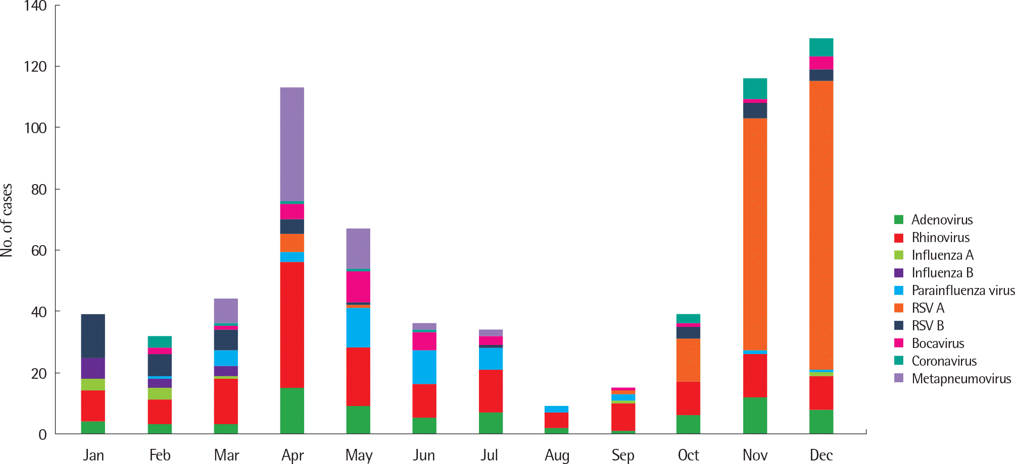
Table 1.
Demographics of study subjects (n=828)
| Variable | Value |
|---|---|
| Age (mo) | 17.0 (7.0–28.0) |
| Boys | 476 (57.5) |
| Birth weight (g) | 3,200 (2,900–3,480) |
| Gestational age (wk) | 39.0 (38.0–40.0) |
| Low birth weight∗ | 64 (7.7) |
| Preterm birth† | 76 (9.2) |
| Duration of hospitalization (day) | 6.0 (4.0–7.0) |
| Duration of fever (day) | 4.0 (2.0–6.0) |
| Lower respiratory tract infection | |
| Pneumonia:bronchiolitis:bronchitis | 617 (74.5):180 (21.7):31 (3.7) |
| Laboratory findings | |
| WBC (×103/µL) | 10.3 (7.7–13.8) |
| Eosinophils (%) | 0.6 (0.1–1.7) |
| ESR (mm/hr) | 16.0 (9.0–26.0) |
| CRP (mg/L) | 9.0 (2.4–23.8) |
| AST (IU/L) | 41.0 (35.0–49.0) |
| ALT (IU/L) | 18.0 (14.0–25.0) |
| Virus detection (n=628) | 519 (82.6) |
| Viral coinfection (n=628) | 135 (21.5) |
| Use of antibiotics | 103 (12.5) |
| Use of systemic steroid | 49 (5.9) |
| O2 supplement | 46 (5.6) |
Table 2.
Clinical characteristics according to the respiratory disease
| Variable | Pneumonia (n=617) | Bronchiolitis (n=180) | Bronchitis (n=31) | P-value |
|---|---|---|---|---|
| Age (mo) | 21.0 (4.0–7.0)∗ | 5.0 (3.0–11.8)† | 19.0 (10.0–36.0) | <0.001 |
| Low birth weight | 37 (6.0) | 24 (13.3) | 3 (9.7) | 0.006 |
| Preterm | 48 (7.8) | 25 (13.9) | 3 (9.7) | 0.044 |
| Duration of hospitalization (day) | 5.0 (4.0–7.0)† | 6.0 (4.0–7.0)† | 7.0 (5.0–8.0)‡ | 0.028 |
| Duration of fever (day) | 5.0 (3.0–6.0)∗,† | 1.0 (0.0–4.0) | 2.0 (1.0–5.0)‡ | <0.001 |
| Laboratory findings | ||||
| WBC (×103/µL) | 10.1 (7.6–13.5)∗ | 10.6 (8.7–14.2) | 11.0 (6.9–15.1) | 0.033 |
| Eosinophils (%) | 0.5 (0.1–1.5)∗ | 1.3 (0.2–2.5) | 0.7 (0.2–2.0) | <0.001 |
| ESR (mm/hr) | 17.0 (10.0–27.0)∗,† | 14.0 (7.0–28.0) | 13.0 (5.0–20.0)‡ | 0.019 |
| CRP (mg/L) | 10.8 (3.2–27.8)∗ | 4.4 (0.6–11.3)† | 12.9 (1.8–30.5) | <0.001 |
| AST (IU/L) | 41.0 (35.0–48.8)† | 42.0 (36.0–53.0)† | 37.0 (31.0–43.0)‡ | 0.015 |
| ALT (IU/L) | 16.0 (13.0–22.0)∗ | 24.0 (18.0–36.0)† | 17.0 (13.0–24.0) | <0.001 |
| No. of viruses PCR (+) | n=493 | n=164 | n=16 | 0.102 |
| Adenovirus | 59 (12.0) | 14 (8.5) | 2 (12.5) | 0.579 |
| Rhinovirus | 120 (24.3) | 39 (23.8) | 9 (56.3) | 0.170 |
| Influenza virus A, B | 22 (4.5) | 1 (0.6) | 1 (6.3) | 0.081 |
| Parainfluenza virus | 34 (6.9) | 11 (6.7) | 1 (6.3) | 0.917 |
| RSV A, B | 152 (30.8) | 86 (52.4) | 2 (12.5) | <0.001 |
| Bocavirus | 30 (6.1) | 3 (1.8) | 1 (6.3) | 0.121 |
| Coronavirus | 19 (3.9) | 5 (3.0) | 0 (0) | 0.620 |
| Metapneumovirus | 57 (11.6) | 5 (3.0) | 0 (0) | 0.002 |
| M. pneumoniae IgM (+) | 28 (4.5) | 2 (1.1) | 0 (0) | 0.052 |
| Use of antibiotics | 83 (13.5) | 10 (5.6) | 10 (32.3) | <0.001 |
| Use of systemic steroid | 18 (2.9) | 28 (15.6) | 3 (9.7) | <0.001 |
| O2 supplement | 17 (2.8) | 29 (16.1) | 0 (0) | <0.001 |
Table 3.
Comparison of clinical characteristics between normal birth weight children and low birth weight children
| Variable | Normal birth weight children (n=764) | Low birth weight children (n=64) | P-value |
|---|---|---|---|
| Age (mo) | 17.0 (8.0–28.0) | 20.0 (8.5–30.8) | 0.333 |
| Gestational age (wk) | 39.0 (38.0–40.0) | 33.0 (30.0–36.0) | <0.001 |
| Birth weight (g) | 3,250 (3,000–3,500) | 2,045 (1,565–2,300) | <0.001 |
| Duration of hospitalization | 6.0 (4.0–7.0) | 5.5 (4.0–7.0) | 0.981 |
| (day) | |||
| Duration of fever (day) | 4.0 (2.0–6.0) | 3.5 (0.5–5.0) | 0.094 |
| LRTI | 0.005 | ||
| Pneumonia | 580 (75.9) | 37 (57.8) | |
| Bronchiolitis | 156 (20.4) | 24 (37.5) | |
| Bronchitis | 28 (3.7) | 3 (4.7) | |
| Laboratory findings | |||
| WBC (×103/μL) | 10.1 (7.7–13.5) | 11.9 (8.9–16.2) | 0.008 |
| Eosinophils (%) | 0.6 (0.1–1.7) | 0.8 (0.1–2.0) | 0.698 |
| ESR (mm3/hr) | 16.0 (9.0–26.0) | 15.5 (10.0–24.8) | 0.496 |
| CRP (mg/L) | 8.3 (1.7–23.8) | 9.2 (2.4–23.9) | 0.705 |
| AST (IU/L) | 41.0 (35.0–49.0) | 41.5 (36.0–47.0) | 0.761 |
| ALT (IU/L) | 18.0 (14.0–25.0) | 20.0 (15.0–26.0) | 0.144 |
| No. of viruses PCR (+) | n=621 | n=52 | 0.657 |
| Adenovirus | 71 (11.4) | 4 (7.7) | 0.368 |
| Rhinovirus | 155 (25.0) | 13 (25.0) | 0.895 |
| Influenza virus A, B | 22 (3.5) | 2 (3.8) | 0.945∗ |
| Parainfluenza virus | 43 (6.9) | 3 (5.8) | 1.000∗ |
| RSV A, B | 220 (35.4) | 20 (38.5) | 0.794 |
| Bocavirus | 30 (4.8) | 4 (7.7) | 0.338∗ |
| Coronavirus | 22 (3.5) | 2 (3.8) | 1.000∗ |
| Metapneumovirus | 58 (9.3) | 4 (7.7) | 0.807∗ |
| M. pneumoniae IgM (+) | 26 (3.4) | 4 (6.3) | 0.281 |
| Use of antibiotics | 97 (12.7) | 6 (9.4) | 0.431 |
| Use of systemic steroid | 36 (4.7) | 13 (20.3) | <0.001∗ |
| O2 supplement | 35 (4.6) | 11 (17.2) | <0.001∗ |
Values are presented as median (interquartile range) or number (%). LRTI, lower respiratory tract infection; WBC, white blood cell; ESR, erythrocyte sedimentation rate; CRP, c-reactive protein; AST, aspartate transaminase; ALT, alanine transaminase; PCR, polymerase chain reaction; RSV, respiratory syncytial virus; M. pneumonia, Mycoplasma pneumonia.
Table 4.
Comparison of clinical characteristics according to the age group
 XML Download