Abstract
Purpose
To report a case of abducens nerve palsy and optic perineuritis caused by fungal sphenoidal sinusitis.
Case summary
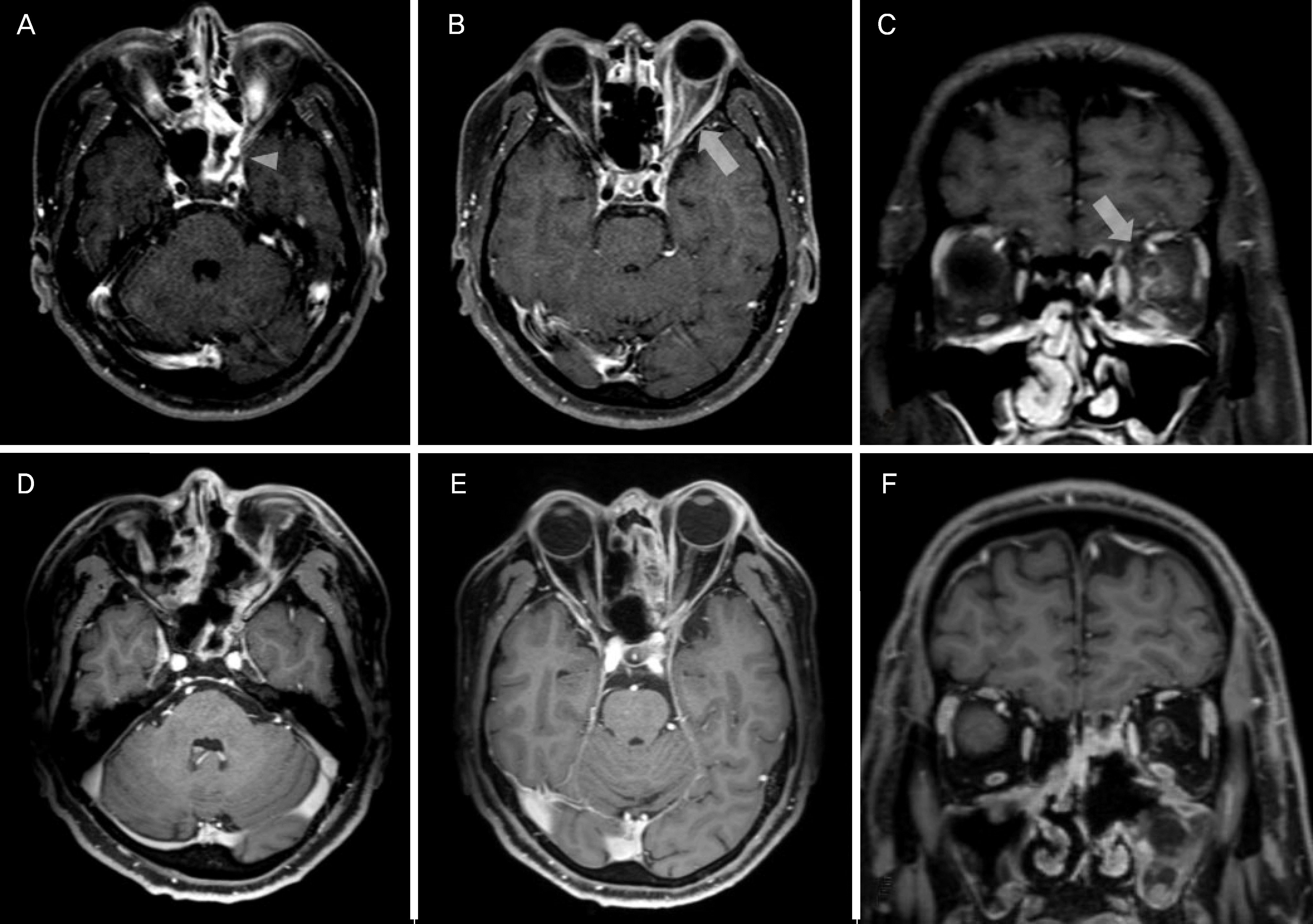
A 48-year-old male visited emergency department for retrobulbar pain, decreased vision, and horizontal diplopia for 3 days. He reported that previous medical history was non-specific, however, blood glucose level was 328 mg/dL (70–110). He had experienced severe headache for 7 days. The best corrected visual acuity was 20/20 at right eye and 20/25 at left eye. The pupil of left eye did not have relative afferent pupillary defect. Left mild proptosis was noted. The extraocular examination showed 30 prism diopters left esotropia at primary gaze and –4 abduction limitation of left eye. The left eye showed mild optic disc swelling and inferior field defect by field test. Brain magnetic resonance imaging showed enhancement of sphenoidal sinus, ethmoidal sinus, and around optic nerve at left eye. Three days after antibiotics treatment, the vision of left eye deteriorated to 20/40 and periorbital pain developed. The drainage and biopsy of sphenoidal sinus were performed. The histopathologic examination showed hyphae consistent with aspergillosis. The ocular symptoms were improved with anti-fungal treatment. Follow-up magnetic resonance imaging performed 1 month after treatment showed improvement of lesion at left orbit. Five months after surgery, the visual acuity of left eye was improved to 20/25. The patient showed orthotropia at primary gaze without limitation.
References
1. Witterick IJ, Vescan AD. Complications of rhinosinusitis. Kennedy DW, Hwang PH, editors. Rhinology. 1st ed.New York: Thieme Medical Publishers;2012. chap. 21.
2. Miller NR, Subramanian PS, Patel VR. Walsh and Hoyt's Clinical Neuro-Ophthalmology: the Essentials. 3rd ed.Philadelphia: Wolters Kluwer;2016. p. 341–71.
3. Chen L, Jiang L, Yang B, Subramanian PS. Clinical features of visual disturbances secondary to isolated sphenoid sinus inflammatory diseases. BMC Ophthalmol. 2017; 17:237.

4. Kline LB, Foroozan R. Neuro-Ophthalmology Review Manual. 7th ed.Thorofare: SLACK;2013. p. 83–92.
5. Jang JH, Kim YC, Chang SD, et al. A case of optic neuritis in acute sphenoid sinusitis. J Korean Ophthalmol Soc. 2007; 48:1742–6.

6. Kim DH, Kim HK, Nam JK, Park JG. A case of isolated aspergillus sphenoid sinusitis. J Korean Neurol Assoc. 2005; 23:402–4.
7. Lee HR, Kim HJ, Seong SY, Chang JH. A case of orbital apex syndrome related to sphenoid fungal sinusitis. Korean J Otorhinolaryngol-Head Neck Surg. 2010; 53:644–7.

8. Biousse V, Newman NJ. Neuro-ophthalmology Illustrated. 2nd ed.New York: Thieme;2016. p. 321–465.
9. Pane A, Miller NR, Burdon M. The neuro-ophthalmology survival guide. 2nd ed.Beijing: Elsevier;2017. p. 170–239.
10. Tamhankar MA, Biousse V, Ying GS, et al. Isolated third, fourth, and sixth cranial nerve palsies from presumed microvascular versus other causes: a prospective study. Ophthalmology. 2013; 120:2264–9.
11. Purvin V, Kawasaki A, Jacobson DM. Optic perineuritis: clinical and radiographic features. Arch Ophthalmol. 2001; 119:1299–306.
Figure 1.
Findings of ocular examination at initial visit. (A) Image of the patient in nine diagnostic position of gaze demonstrating esotropia at primary gaze and abduction limitation of the left eye. (B) Fundus examination shows mild swelling of optic disc at the left eye. (C) The visual field test reveals inferior visual field defect at the left eye (Humphrey Field Analyzer, Carl Zeiss Meditec, Inc., Dublin, CA, USA).

Figure 2.
Findings of brain magnetic resonance (MR) imaging during follow-ups. (A-C) The initial magnetic resonance (MR) imaging. (A) Axial T1-weighted gadolinium-enhanced MR image showing an enhancement of the circumferential mucosa in the sphenoid sinus and the ethmoidal sinus (arrowhead). (B, C) Axial and coronal T1-weighted gadolinium-enhanced MR images demonstrate an enhancement of the left optic nerve sheath (arrow). (D-F) The follow-up MR imaging at 1 month after the anti-fungal treatment. The patient underwent an endoscopic endonasal surgery for sinusitis. (E, F) Axial and coronal T1-weighted gadoli-nium-enhanced MR images showing a normalization of the left optic nerve.


 PDF
PDF ePub
ePub Citation
 Print
Print


 XML Download