

 PDF
PDF ePub
ePub Citation
 Print
Print


Abstract
Case summary
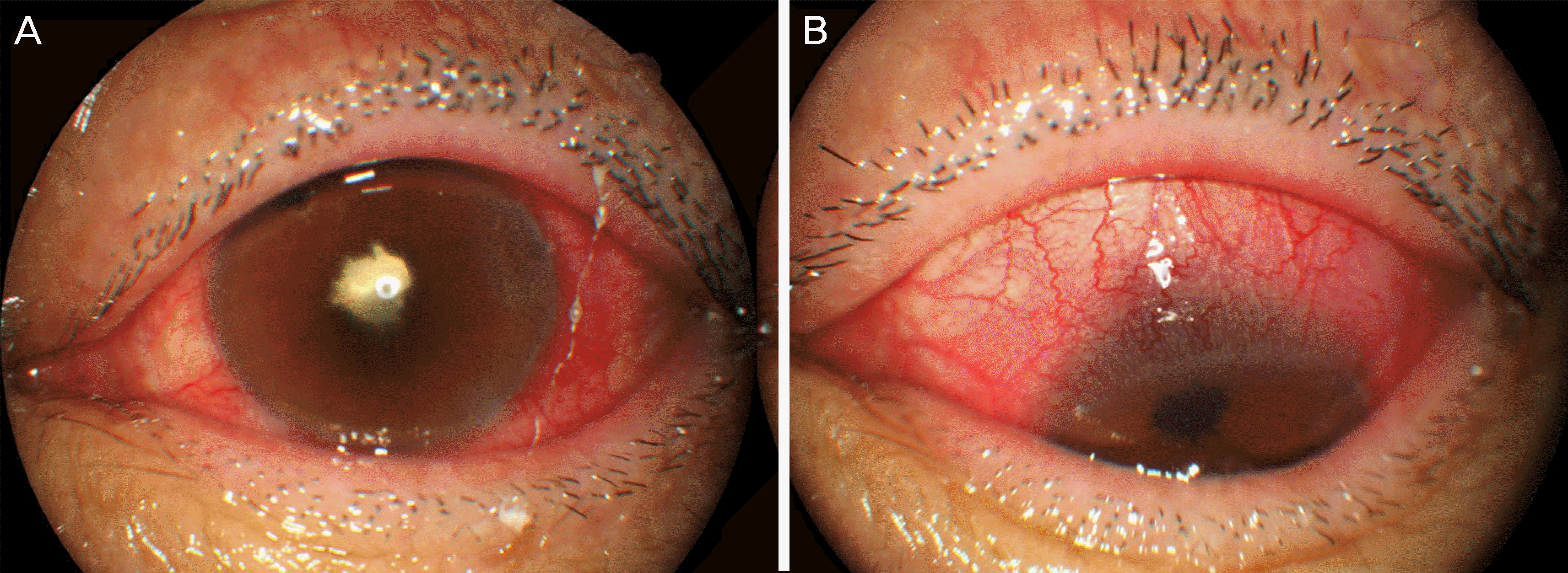
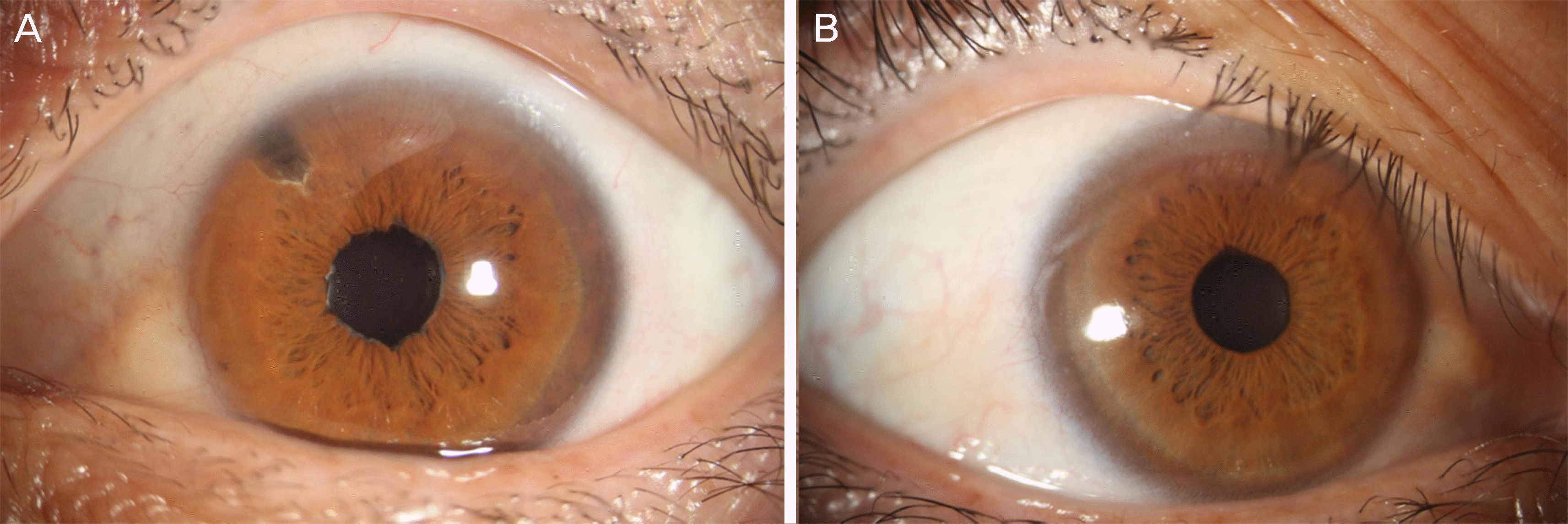
A 42-year-old male presented with decreased visual acuity in the left eye. Castleman disease had been diagnosed 21 months before and treated with systemic steroids and combined chemotherapy. Best-corrected visual acuity (BCVA) of the left eye was 0.02 and the intraocular pressure was 42 mmHg. Scleral edema and corneal edema were noted using a slit lamp examination. The anterior chamber cell was 2+ according to Standardization of Uveitis Nomenclature criteria. The fundus was invisible due to the anterior segment lesion. After one month, scleritis developed in the right eye and the patient complained of ocular pain. Topical steroids and non-steroidal anti-inflammatory drugs were prescribed. Due to recurrent scleritis and anterior uveitis, cataract extraction and laser iridectomy were performed on the left eye, and systemic steroids and the antimetabolite methotrexate were started. After 9 years of follow-up, all medications were stopped and there was no recurrence of inflammation, with a BCVA of 1.0 in both eyes.
Go to : 
References
1. Castleman B, Iverson L, Menendez VP. Localized mediastinal lymphnode hyperplasia resembling thymoma. Cancer. 1956; 9:822–30.

2. Herrada J, Cabanillas F, Rice L, et al. The clinical behavior of abdominal and multicentric Castleman disease. Ann Intern Med. 1998; 128:657–62.
3. Frizzera G, Peterson BA, Bayrd ED, Goldman A. A systemic abdominal disorder with morphologic features of Castleman's disease: clinical findings and clinicopathologic correlations in 15 patients. J Clin Oncol. 1985; 3:1202–16.
4. Chang Y, Cesarman E, Pessin MS, et al. Identification of herpesvi-rus-like DNA sequences in AIDS-associated Kaposi's sarcoma. Science. 1994; 266:1865–9.
5. Ishiyama T, Koike M, Nakamura S, et al. Interleukin-6 receptor abdominal in the peripheral B cells of patients with multicentric Castleman's disease. Ann Hematol. 1996; 73:179–82.
6. Pavlidis NA, Skopouli FN, Bai MC, Bourantas CL. A successfully treated case of multicentric angiofollicular hyperplasia with oral abdominal (Castleman's disease). Med Pediatr Oncol. 1990; 18:333–5.
7. Liberato NL, Bollati P, Chiofalo F, et al. Autoimmune hemolytic abdominal in multicentric Castleman's disease. Haematologica. 1996; 81:40–3.
8. Jorge R, Scott IU, Oliveira RC, et al. Ocular findings in a patient with Castleman's disease before and after treatment with immunosuppression and plasmapheresis. Ophthalmic Surg Lasers Imaging. 2010; 41:doi:. DOI: 10.3928/15428877-20100929-10.
9. Venizelos I, Papathomas TG, Papathanasiou M, et al. Orbital abdominal in Castleman disease. Surv Ophthalmol. 2010; 55:247–55.
10. Nishimoto N, Sasai M, Shima Y, et al. Improvement in Castleman's disease by humanized anti-interleukin-6 receptor antibody therapy. Blood. 2000; 95:56–61.
11. Inatani M, Kashii S, Nosaka K, Arima N. Orbital pseudotumor as an initial manifestation of multicentric Castleman's disease. Jpn J Ophthalmol. 2005; 49:505–8.
12. Severson GS, Harrington DS, Weisenburger DD, et al. Castleman's disease of the leptomeninges. Report of three cases. J Neurosurg. 1988; 69:283–6.
13. Tavoni A, Vitali C, Baglioni P, et al. Multicentric Castleman's abdominal in a patient with primary Sjogren's syndrome. Rheumatol Int. 1993; 12:251–3.
14. Koppens JM, Pon JA, Allen J, Sloan BH. Castleman's disease of the lacrimal gland. Clin Exp Ophthalmol. 2004; 32:108–10.
15. Park SH, Song SJ. Castleman's disease presenting with uveal abdominal syndrome. Korean J Ophthalmol. 2010; 24:182–5.
16. Kim U, Hwang JM. Optic neuropathy associated with Castleman disease. Korean J Ophthalmol. 2010; 24:256–9.
17. Liu DT, Shields JA, Li C, et al. Hypertensive choroidopathy in Castleman's disease. Graefes Arch Clin Exp Ophthalmol. 2011; 249:1901–3.
18. Oshitari T, Kajita F, Tobe A, et al. Refractory uveitis in patient with castleman disease successfully treated with tocilizumab. Case Rep Ophthalmol Med. 2012; 2012:968180.
19. Kozak I, Reid EG. Retinal vein occlusion during flare of abdominal Castleman's disease. Clin Ophthalmol. 2013; 7:1647–9.
20. Katz MS, Park S, Gritz DC. Posterior segment involvement in Castleman disease. Retin Cases Brief Rep. 2014; 8:209–11.
21. Lin P, Bhullar SS, Tessler HH, Goldstein DA. Immunologic abdominal as potential predictors of systemic autoimmune disease in abdominals with idiopathic scleritis. Am J Ophthalmol. 2008; 145:463–71.
22. Jabs DA, Mudun A, Dunn JP, Marsh MJ. Episcleritis and scleritis: clinical features and treatment results. Am J Ophthalmol. 2000; 130:469–76.
23. Murphy CC, Ayliffe WH, Booth A, et al. Tumor necrosis factor alpha blockade with infliximab for refractory uveitis and scleritis. Ophthalmology. 2004; 111:352–6.
Go to :
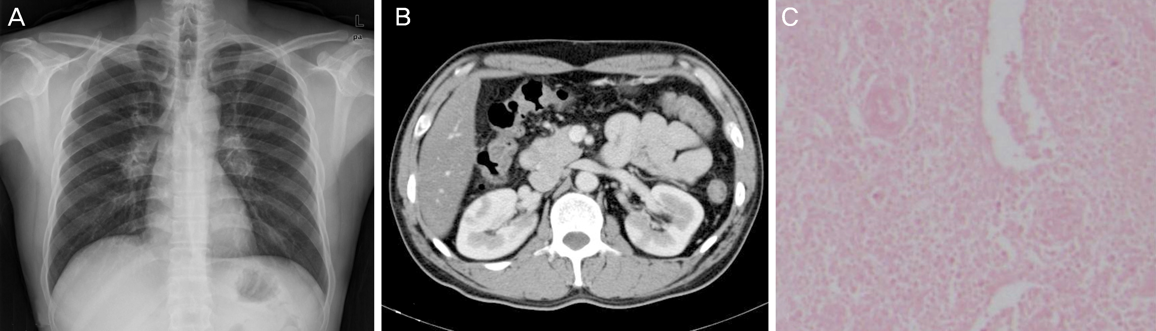 | Figure 1.Clinical and pathological findings of the patient. (A) Bihilar lymphadenopahy was noted in chest X-ray. (B) Computed tomography abdomen revealed multiple low density mass in both kidneys. (C) Histopathologic finding of inguinal lymph node revealed atypically proliferative follicular hyperplasia and increased interfollicular vascularity. Hyalinized germinal center was surrounded by lymphocytes, producing the “onion-skin appearance” (hematoxylin and eosin [H&E] stain, ×100). |
 XML Download