

 PDF
PDF ePub
ePub Citation
 Print
Print


Abstract
Purpose
To determine influences of intraoperative foveal traction during membrane peeling in idiopathic epiretinal membrane (ERM) surgery.
Methods
This retrospective observational study included 46 eyes of 46 patients with idiopathic ERM who underwent pars plana vitrectomy with ERM and internal limiting membrane peeling from February 2015 to September 2015. The presence of intraoperative foveal traction during membrane peeling was reviewed using video records. The main outcome measures were best-corrected visual acuity (BCVA), central foveal thickness (CFT), foveal contour, and photoreceptor inner segment/outer segment junction disruption using optical coherence tomography at baseline and at 1, 3, 6, and 12 months after surgery.
Results
Group 1 (ERM with intraoperative foveal traction) included 22 eyes, and group 2 (ERM without intraoperative foveal traction) included 24 eyes. Preoperatively, convex pattern ERM was observed more often in group 1. Group 1 had a significantly thicker CFT and a lower BCVA compared to group 2 at baseline and during the first 6 months, but the final postoperative BCVA and CFT were not significantly different between the groups at 12 months. Among 22 eyes, 12 eyes (54.5%) were restored to flat or concave ERM patterns at an average of 5.4 months after surgery in group 1, and 18 out of 24 eyes (75%) recovered at 2.4 months (p < 0.01) in group 2.
Figures and Tables
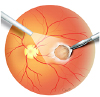 | Figure 1Schema of the retinal membrane peeling. A drawing shows foveal traction during membrane peeling.
|
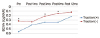 | Figure 2Mean longitudinal changes in best corrected visual acuity (BCVA). Traction group had a lower BCVA compared to non-traction group at baseline and during the first 6 months, but final postoperative BCVA were not significantly different between the groups at 12 months. mo = month(s). *p < 0.05, student's T-test.
|
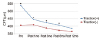 | Figure 3Mean longitudinal changes in central foveal thickness (CFT). Traction group had a thicker CFT compared to non-traction group at baseline and during the first 6 months, but final postoperative CFT were not significantly different between the groups at 12 months. mo = month(s). *p < 0.05, student's T-test.
|
Table 1
Preoperative data
Values are presented as mean ± SD or n (%) unless otherwise indicated.
ELM = external limiting membrane; IS/OS = inner segment/outer segment junction; CFT = central foveal thickness; VMT = vitreomacular traction; MPH = macular pseudohole; BCVA = best corrected visual acuity.
*Compared by Pearson's chi-squared test; †Compared by student's T-test; ‡Compared by logistic regression analysis; §Compared by Fisher's exact test.
![]()


Notes
References
1. Margherio RR, Cox MS Jr, Trese MT, et al. Removal of epimacular membranes. Ophthalmology. 1985; 92:1075–1083.

2. Michels RG. Vitrectomy for macular pucker. Ophthalmology. 1984; 91:1384–1388.
3. Wise GN. Clinical features of idiopathic preretinal macular fibrosis. Schoenberg Lecture. Am J Ophthalmol. 1975; 79:349–357.
4. Poliner LS, Olk RJ, Grand MG, et al. Surgical management of premacular fibroplasia. Arch Ophthalmol. 1988; 106:761–764.
5. Machemer R. The surgical removal of epiretinal macular membranes( macular puckers). Klin Monbl Augenheilkd. 1978; 173:36–42.
6. McDonald HR, Verre WP, Aaberg TM. Surgical management of idiopathic epiretinal membranes. Ophthalmology. 1986; 93:978–983.
7. Margherio RR, Cox MS Jr, Trese MT, et al. Removal of epimacular membranes. Ophthalmology. 1985; 92:1075–1083.
8. Hillenkamp J, Saikia P, Gora F, et al. Macular function and morphology after peeling of idiopathic epiretinal membrane with and without the assistance of indocyanine green. Br J Ophthalmol. 2005; 89:437–443.
9. Arndt C, Rebollo O, Séguinet S, et al. Quantification of metamorphopsia in patients with epiretinal membranes before and after surgery. Graefes Arch Clin Exp Ophthalmol. 2007; 245:1123–1129.
10. Wong JG, Sachdev N, Beaumont PE, Chang AA. Visual outcomes following vitrectomy and peeling of epiretinal membrane. Clin Experiment Ophthalmol. 2005; 33:373–378.
11. Park SJ, Lee JH. Clinical feature of macular preretinal membrane and visual changes after vitrectomy. J Korean Ophthalmol Soc. 1994; 35:824–829.
12. Rice TA, De Bustros S, Michels RG, et al. Prognostic factors in vitrectomy for epiretinal membranes of the macula. Ophthalmology. 1986; 93:602–610.
13. Kim SW, Park JH, Ahn DS, Yoon HS. Evaluation of each retinal layer thickness according to preoperative OCT patterns after idiopathic ERM removal. J Korean Ophthalmol Soc. 2014; 55:1843–1852.
14. Seo SJ, Lee SJ, Park JM. Surgical outcome according to morphology in epiretinal membrane based on optical coherence tomography (OCT). J Korean Ophthalmol Soc. 2013; 54:736–744.
15. Massin P, Allouch C, Hanouchine B, et al. Optical coherence tomography of idiopathic macular epiretinal membranes before and after surgery. Am J Ophthalmol. 2000; 130:732–739.
16. Kim JH, Kang SW, Kong MG, Ha HS. Assessment of retinal layers and visual rehabilitation after epiretinal membrane removal. Graefes Arch Clin Exp Ophthalmol. 2013; 251:1055–1064.
17. McCarty DJ, Mukesh BN, Chikani V, et al. Prevalence and associations of epiretinal membranes in the visual impairment project. Am J Ophthalmol. 2005; 140:288–294.
18. Kawasaki R, Wang JJ, Mitchell P, et al. Racial difference in the prevalence of epiretinal membrane between caucasians and asains. Br J Ophthalmol. 2008; 92:1320–1324.
19. Kwok AK, Lai TY, Li WW, et al. Indocyanine green-assisted internal limiting membraned removal in epiretinal membrane surgery: a clinical and histologic study. Am J Ophthalmol. 2004; 138:194–199.
20. Sorcinelli R. Surgical management of epiretinal membrane with indocyanine-green-assisted peeling. Ophthalmologica. 2003; 217:107–110.
21. Park DW, Dugel PU, Garda J, et al. Macular pucker removal with and without internal limiting membrane peeling: pilot study. Ophthalmology. 2003; 110:62–64.
22. Sivalingam A, Eagle RC Jr, Duker JS, et al. Visual prognoses correlated with the presence of interna-limiting membrane in histopathologic specimens obtained from epiretinal membrane surgery. Ophthalmology. 1990; 97:1549–1552.
23. Kinoshita T, Kovacs KD, Wagley S, Arroyo JG. Morphologic differences in epiretinal membranes on ocular coherence tomography as a predictive factor for surgical outcome. Retina. 2011; 31:1692–1698.
24. Joe SG, Lee KS, Lee JY, et al. Inner retinal layer thickness is the major determinant of visual acuity in patients with idiopathic epiretinal membrane. Acta Ophthalmol. 2013; 91:e242–e243.
25. Watanabe K, Tsunoda K, Mizuno Y, et al. Outer retinal morphology and visual function in patients with idiopathic epiretinal membrane. JAMA Ophthalmol. 2013; 131:172–177.
26. Suh MH, Seo JM, Park KH, Yu HG. Associations between macular findings by optical coherence tomography and visual outcomes after epiretinal membrane removal. Am J Ophthalmol. 2009; 147:473–480.
27. Inoue M, Arakawa A, Yamane S, Kadonosono K. Long-term outcome of preoperative disrupted inner/outer segment junctions assessed using spectral-domain optical coherence tomography in patients with idiopathic epiretinal membrane. Ophthalmologica. 2012; 228:222–228.
28. Shiono A, Kogo J, Klose G, et al. Photoreceptor outer segment length: a prognostic factor for idiopathic epiretinal membrane surgery. Ophthalmology. 2013; 120:788–794.
29. Falkner-Radler CI, Glittenberg C, Hagen S, et al. Spectral-domain optical coherence tomography for monitoring epiretinal membrane surgery. Ophthalmology. 2010; 117:798–805.
 XML Download