

 PDF
PDF ePub
ePub Citation
 Print
Print


Abstract
Purpose
The purpose of this study is to evaluate the recurrence rate and treatment effect of conservative treatment and surgical treatment with diamond burr in reccurent corneal erosion.
Methods
Between January 2010 and October 2017, 67 patients who were diagnosed with repeated corneal erosion, 55 patients underwent conservative treatment and 12 patients underwent surgical treatment with a diamond burr were evaluated for the duration of previous treatment, recurrence frequency, symptom severity, visual acuity, and the effect of treatment depending on recurrence. Surgical treatment was performed when corneal epithelial loosening occurred in more than 10% of the cases and the patient required surgical treatment. Conservative treatment was continued in patients who did not want surgical treatment.
Results
There were no differences in the mean age, sex, recurrence frequency, symptom severity, and duration of treatment. All 67 patients were unilateral, and 55 patients underwent conservative treatment. Of these, 23 patients (41.8%) showed improvement, and 32 patients relapsed within 6 months. Ten eyes (83.3%) of 12 eyes treated with a diamond burr showed improvement and two eyes recurred within 6 months. The recurrence rate was lower in the diamond burr group than in the conservative group (p = 0.011). In comparison with diamond burr and conservative treatment, the visual acuity of the diamond burr group improved statistically significantly compared to the group with conservative treatment (p = 0.002).
References
1. Ramamurthi S, Rahman MQ, Dutton GN, Ramaesh K. Pathogenesis, clinical features and management of recurrent corneal erosions. Eye (Lond). 2006; 20:635–44.

2. Ko BY, Lee GW. Clinical results of phototherapeutic keratectomy for refractory recurrent corneal erosion. J Korean Ophthalmol Soc. 2011; 52:392–400.
3. Reidy JJ, Paulus MP, Gona S. Recurrent erosions of the cornea: abdominal and treatment. Cornea. 2000; 19:767–71.
4. Lee SH, Kim TI, Chung SH, et al. A case of combined bacterial abdominal with recurrent corneal erosion. J Korean Ophthalmol Soc. 2007; 48:449–54.
5. Han MS, Lee JH, Lee SJ. Therapeutic effect of topical autologous serum in recurrent punctate corneal erosion. J Korean Ophthalmol Soc. 2004; 45:1639–44.
6. Suh Y, Kim MS. The longterm evaluation of recurrent corneal erosion. J Korean Ophthalmol Soc. 2002; 43:1570–6.
7. Shin DY, Chung SH. Efficacy of anterior stromal puncture using 5% NaCl eye drops for prolonged time in recurrent corneal erosion syndrome. J Korean Ophthalmol Soc. 2017; 58:503–8.
8. Watson S, Lee H. Interventions for recurrent corneal erosion: a Cochrane Systematic review. Eye (Lond). 2013; 27:1330–1.
9. del Castillo JM, de la Casa JM, Sardiña RC, et al. Treatment of abdominal corneal erosions using autologous serum. Cornea. 2002; 21:781–3.
10. Ziakas NG, Boboridis KG, Terzidou C, et al. abdominal follow up of autologous serum treatment for recurrent corneal erosions. Clin Experiment Ophthalmol. 2010; 38:683–7.
11. Benitez-Del-Castillo JM, Rodríguez-Bayo S, Fontan-Rivas E, et al. Treatment of recurrent corneal erosion with substance P-derived peptide and insulin-like growth factor I. Arch Ophthalmol. 2005; 123:1445–7.
12. Dursun D, Kim MC, Solomon A, Pflugfelder SC. Treatment of abdominal recurrent corneal erosions with inhibitors of matrix met-alloproteinase-9, doxycycline and corticosteroids. Am J Ophthalmol. 2001; 132:8–13.
13. Wang L, Tsang H, Coroneo M. Treatment of recurrent corneal abdominal syndrome using the combination of oral doxycycline and abdominal corticosteroid. Clin Experiment Ophthalmol. 2008; 36:8–12.
14. Singh RP, Raj D, Pherwani A, et al. Alcohol delamination of the corneal epithelium for recalcitrant recurrent corneal erosion abdominal: a prospective study of efficacy and safety. Br J Ophthalmol. 2007; 91:908–11.
15. Ryan G, Lee GA, Maccheron L. Epithelial debridement with abdominal burr superficial keratectomy for the treatment of recurrent corneal erosion. Clin Exp Ophthalmol. 2013; 41:621–2.
16. Avni Zauberman N, Artornsombudh P, Elbaz U, et al. Anterior stromal puncture for the treatment of recurrent corneal erosion abdominal: patient clinical features and outcomes. Am J Ophthalmol. 2014; 157:273–9.e1.
17. Tsai TY, Tsai TH, Hu FR, Hou YC. Recurrent corneal erosions treated with anterior stromal puncture by neodymium: yttrium-alu-minum-garnet laser. Ophthalmology. 2009; 116:1296–300.
18. Kim SY, Ko BY. Evaluation of anterior stromal puncture using Nd: YAG laser for refractory recurrent corneal erosion. J Korean Ophthalmol Soc. 2015; 56:331–8.
19. Choi M, Jung JW, Seo KY, et al. Comparison of Nd:YAG laser abdominal conservative management in the treatment of recurrent corneal erosion. J Korean Ophthalmol Soc. 2015; 56:687–93.
20. Hsu JK, Rubinfeld RS, Barry P, Jester JV. Anterior stromal puncture. Immunohistochemical studies in human corneas. Arch Ophthalmol. 1993; 111:1057–63.
21. Lee SW, Choi TH. Anterior stromal puncture with 26-gauge needle for recurrent corneal erosion: a report of five cases. J Korean Ophthalmol Soc. 2003; 44:511–6.
22. Galbavy EJ, Mobilia EF, Kenyon KR. Recurrent corneal erosions. Int Ophthalmol Clin. 1984; 24:107–31.
23. Trobe JD, Laibson PR. Dystrophic changes in the anterior cornea. Arch Ophthalmol. 1972; 87:378–82.
24. Buxton JN, Constad WH. Superficial epithelial keratectomy in the abdominal of epithelial basement membrane dystrophy. Ann Ophthalmol. 1987; 19:92–6.
25. Trokel SL, Srinivasan R, Braren B. Excimer laser surgery of the cornea. Am J Ophthalmol. 1983; 96:710–5.
26. Park JW, Kim JH. Phototherapeutic keratectomy for granular abdominal dystrophy. J Korean Ophthalmol Soc. 2003; 44:2465–72.
27. Das S, Seitz B. Recurrent corneal erosion syndrome. Surv Ophthalmol. 2008; 53:3–15.
28. Soong HK, Farjo Q, Meyer RF, Sugar A. Diamond burr superficial keratectomy for recurrent corneal erosions. Br J Ophthalmol. 2002; 86:296–8.
29. Gipson IK, Spurr-Michaud SJ, Tisdale AS. Anchoring fibrils forma complex network in human and rabbit cornea. Invest OphthalmolVis Sci. 1987; 28:212–20.
30. Judge D, Payant J, Frase S, Wood TO. Anterior stromal micro abdominal electron microscopic changes in the rabbit cornea. Cornea. 1990; 9:152–60.
31. Bae KH, Ahn M, Cho NC, You IC. Clinical presentation and abdominal outcomes of recurrent corneal erosion. J Korean Ophthalmol Soc. 2016; 57:555–61.
32. Thomas OW. Recurrent erosion. Tr Am Ophth Soc. 1984; 82:850–98.
33. Sridhar MS, Rapuano CJ, Cosar CB, et al. Phototherapeutic abdominal versus diamond burr polishing of bowman's membrane in the treatment of recurrent corneal erosions associated with anterior basement membrane dystrophy. Ophthalmology. 2002; 109:674–9.
Figure 2.
Combined ocular disease in RCE. Combined ocular disease at the time of diagnosis of recurrent corneal erosion. RCE = recurrent corneal erosion; DES = dry eye syndrome; MGD = meibomian gland disease.

Figure 3.
Causative factors. Causative factors in 67 eyes of 67 patients with recurrent corneal erosion. RCE = recurrent corneal erosion.
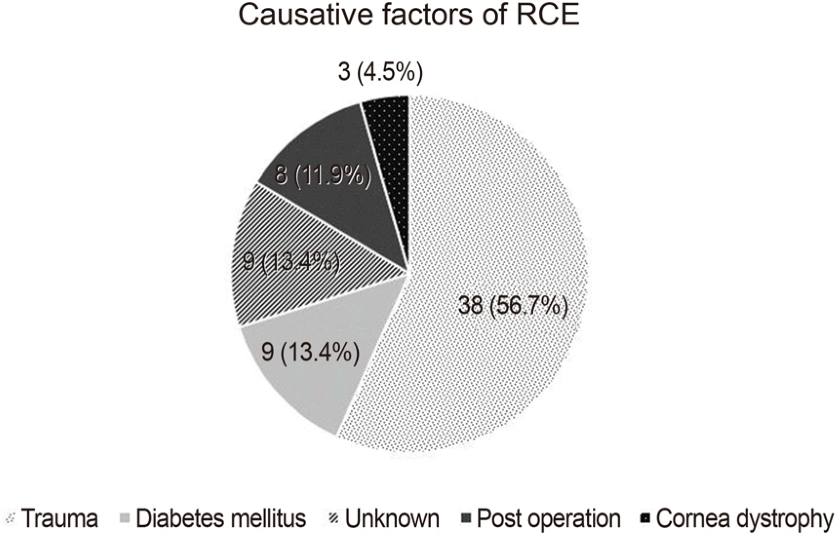
Figure 4.
Comparison of surgical and conservative treatment. A comparison of the recurrences between the surgical treatment with diamond burr group and conservative treatment group.
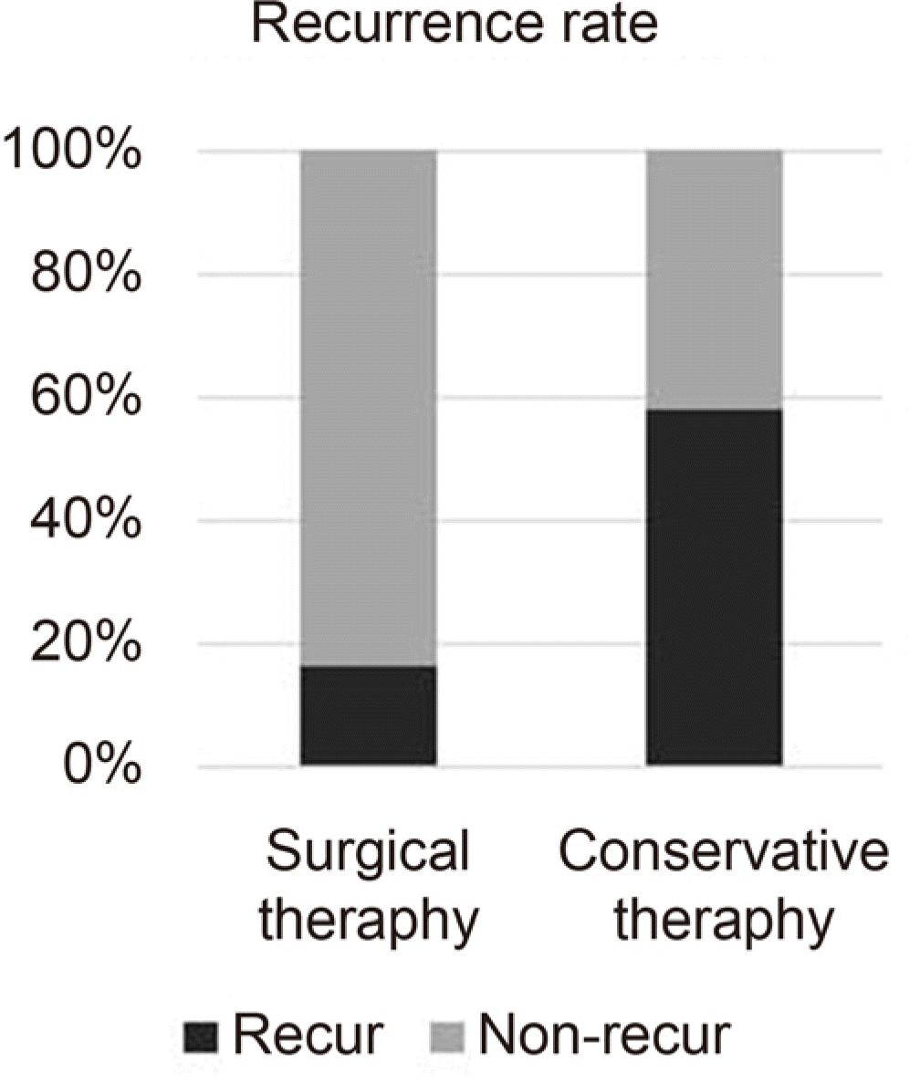
Table 1.
Demographic data of recurrent corneal erosion patients about age, previous recurrences, symptom severity, and duration of previous treatment
|
Surgical therapy |
Conservative therapy |
t | p-value* | |||
|---|---|---|---|---|---|---|
| Mean | S.D. | Mean | S.D. | |||
| Age | 42.67 | 10.990 | 46.60 | 14.836 | −0.866 | 0.390 |
| Previous recurrences Symptom severity36–37(stage1–4) | 2.42 1.75 | 1.730 0.754 | 3.58 1.73 | 5.597 0.706 | −0.710 0.100 | 0.480 0.921 |
| Previous treatment period (mo.) | 10.17 | 4.549 | 13.91 | 21.391 | −0.600 | 0.551 |
Table 2.
The best corrected visual acuity before treatment and after 6 months of treatment with diamond burr and conservative treatment group
|
Surgical therapy |
Conservative therapy |
t | p-value† | |||
|---|---|---|---|---|---|---|
| Mean | S.D. | Mean | S.D. | |||
| At the time of diagnosis (V/A) | 0.71 | 0.204 | 0.55 | 0.346 | 1.542 | 0.128 |
| After 6 months (V/A) | 0.95 | 0.090 | 0.81 | 0.259 | 3.250 | 0.002* |
 XML Download