

 PDF
PDF Citation
 Print
Print


INTRODUCTION
Regenerative endodontic procedures have become a viable therapeutic approach to save or extend the life of necrotic, immature teeth, as well as to preserve the alveolar bone and maintain optimal function in the long term [12]. Currently, the common clinical protocol for regenerative endodontic therapies includes disinfection of the root canal with an antibiotic paste for several weeks, followed by evoked apical bleeding to form a blood clot containing stem cells from the apical papilla (SCAP) and growth factors released from platelets [345]. Coupled with the release of growth factors sequestered in root dentin [67], this blood clot is thought to act as a scaffold to promote cellular growth and differentiation, followed by generation or ingrowth of tissue in the root canal space [8].
In contrast to a growing body of published regenerative endodontic cases in immature necrotic teeth, only one recent report [9] has attempted a modified regenerative endodontic procedure for mature necrotic permanent teeth since the initial reports by Östby [10] and Nygaard-Ostby and Hjortdal [11]. This limitation might be due to concerns in achieving adequate bleeding and subsequent angiogenesis through a narrow apical pathway [12], as well as the age-related decline in the amount and function of stem/progenitor cells that could be delivered into the empty canal space [913]. However, the presence of a wide-open apex and abundance of stem/progenitor cells residing in the periapical region do not necessarily warrant successful and predictable treatment outcomes, as evidenced by different tissue responses in immature permanent teeth [14].
The ultimate goals in the regenerative endodontic treatment of immature necrotic teeth are continuation of root development and regeneration of functional pulp tissue [15]. These goals partially apply to that of mature permanent teeth, since the latter already possess normal root dimensions (i.e., width and length) and a physiologic apical closure. Thus, implementation of current biologically-based treatment procedures might be a logical step toward achieving vital tissue formation in the root canal space of necrotic mature teeth. The present report describes the procedures and the outcome of a regenerative endodontic treatment approach in 2 previously-traumatized, necrotic mature incisors with apical periodontitis.
CASE REPORT
A healthy, 21-year-old female was referred to the Department of Endodontics for evaluation of teeth #21 and #22. Reportedly, the incisors had been traumatized in an accident at the age of 14, but the patient and parents did not seek dental treatment owing to the absence of tooth fracture or pain. Over the years, the teeth remained asymptomatic without discoloration, and more recently, presented with intermittent pain. Clinically, both tooth crowns were intact, had physiologic mobility and probing depths (< 3 mm), and showed little sensitivity to percussion. There were no signs of swelling or sinus tracts. Both teeth were non-responsive to a cold test (Endo-Ice, Coltene/Whaledent, Altstätten, Switzerland) and electric pulp test (Parkell Inc., Farmingdale, NY, USA), while the contralateral teeth responded positively to both tests. Radiographic examination revealed well-developed roots with closed apices and a large periradicular radiolucency involving both incisors (Figure 1). On the basis of clinical and radiographic findings, a diagnosis of pulp necrosis and symptomatic apical periodontitis was made. A biologically based endodontic treatment plan was considered, and possible treatment outcomes and applicability of conventional endodontic treatment in case of failure were thoroughly discussed with the patient. Following the patient's approval and written consent, the treatment was initiated at the same visit.
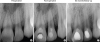
Figure 1
Preoperative (A), post-operative (B), and 60-month recall (C) radiographs of the patient. The post-operative and follow-up radiographs were corrected by using the TurboReg plug-in as a preparation for quantitative assessment of changes in root dimensions by using Image-J.
The teeth were anesthetized using 2% lidocaine with 1:100,000 epinephrine and isolated with rubber dam. Following endodontic access, a hemorrhagic, purulent exudate was observed. The canals were gently irrigated with approximately 20 mL of 5.25% NaOCl without instrumentation, and thereafter with 10 mL of saline. After drying the root canals with sterile paper points, a triple antibiotic paste (TAP) containing ciprofloxacin, metronidazole, and clindamycin (each 100 mg) was freshly prepared by mixing with sterile distilled water to a thin, creamy paste. The antibiotic paste was placed into the root canals using a lentulo spiral, and the access cavities were sealed with sterile cotton pellet and glass ionomer cement (Ketac Molar, 3M ESPE, Seefeld, Germany). The patient was recalled 4 weeks later.
Both teeth were asymptomatic at the second appointment. The teeth were anesthetized with 2% mepivacaine (Citanest, AstraZeneca, London, UK) without a vasoconstrictor followed by rubber dam isolation, and the antibiotic paste in the canal was removed by copious irrigation with 5.25% NaOCl [1617], and thereafter with 10 mL sterile distilled water. The root canals received a final rinse of 17% ethylenediaminetetraacetic acid for 1 minute, and the excess was removed with sterile paper points. Apical bleeding was induced by gentle instrumentation with sterile size 10 K-files 1–2 mm beyond the apices. After formation of a blood clot approximately 3 mm below the cement-enamel junction, ProRoot MTA (Dentsply Tulsa Dental, Tulsa, OK, USA) was mixed with sterile distilled water and carefully placed above the blood clot. Finally, a wet cotton pellet was placed over the MTA, and the access cavity was temporarily restored with conventional glass ionomer cement (Kavitan Plus, SpofaDental, Jičín, Czech Republic). The teeth were restored with acid-etch resin composite (3M ESPE Dental Products, St. Paul, MN, USA) 1 week later.
At 1-month follow-up, the teeth were asymptomatic and showed decreased periradicular radiolucency (Figure 1B). The patient was recalled every six months for clinical and radiographic evaluations. Complete resolution of the radiolucency, and regeneration of the periradicular tissues were evident at the 60-month follow-up radiograph (Figure 1C). Clinically, both teeth were asymptomatic with no sensitivity to percussion and palpation. As with the initial visit, the teeth were non-responsive to the cold test and the electric pulp test. To examine possible changes in root and root space dimensions, the radiographs were subjected to image analysis using Image-J software (V.1.44p, National Institutes of Health, Bethesda, MD, USA) as described previously [1819]. Using the preoperative radiograph as the source image, the recall and final radiographs were digitally aligned with the TurboReg plug-in (Biomedical Imaging Group, Swiss Federal Institute of Technology, Lausanne, Switzerland), followed by linear measurements in Image-J [1819]. The results of the measurements showed that the dimensions of the root space had remained unchanged during the 60-month follow-up period.
DISCUSSION
Revascularization is essential in both wound healing and the development of vital tissue in the root canal space [42021]. It has been reported that the size of the apical foramen could be a critical factor to promote revascularization, with a radiographic apical width of 1 mm or greater offering greater chance for cell migration and proliferation [12]. It should be noted; however, this conclusion was based on long-term follow-up of autotransplanted teeth without evoked apical bleeding [12], which may be a critical step in the revascularization phase of regenerative endodontic treatment in mature necrotic teeth [9]. A recent study using an animal model showed that an apical width of as small as 0.32 mm could be sufficient to permit ingrowth of vital tissue in the canal space [22]. With extreme caution in extrapolating results of animal experimentation to the clinical situation, it is logical to assume that the apical diameters of mature central and lateral incisors, which are greater than 0.32 mm (approximately 0.425 mm, 0.369 mm, respectively) [23], may offer a suitable dimension for promotion of angiogenesis, and that passing the files beyond the apex of necrotic mature teeth might be a practical method to further enlarge the apical diameter as well as to evoke apical bleeding, which both can enhance revascularization [924].
Although the exact nature of tissue formed in the canal of revascularized teeth is yet unknown, results of recent case reports involving histologic analysis of revascularized teeth [252627], and the resemblance of their findings with those obtained in the animal model [28] strongly suggest that the tissue within the pulp space is not functional pulp tissue, but rather a vascular, nonspecific vital tissue [29], comprised of both connective and hard tissue components [2526272829]. Based on these observations, the generic term ‘revitalization’ has been suggested to better describe the technique and the outcome of currently-employed regenerative endodontic procedures [2930]. Compared with necrotic immature teeth, the revitalization procedure of mature teeth may encounter additional challenges, owing to the scarcity or absence of SCAP, which are thought to play an important role for pulp/dentin regeneration in immature teeth [31]. According to Paryani and Kim [9], periodontal ligament stem cells, bone marrow mesenchymal stem cells, and surviving dental pulp stem cells residing around the root apex of mature necrotic teeth may initiate vital tissue growth when migration of these cells into the root canal are facilitated by evoked apical bleeding using gentle overinstrumentation. Their assumptions may be justified to some extent by the successful clinical and radiographic outcome of their case series involving closed-apex teeth of young patients (11 and 14 years old) [9]. Further, the observation of positive response to cold and electric pulp tests in one of their cases may suggest that some vital innervated tissue still existed within the root canal, and became produced by reestablishment of vascular support over the time [15]. Unlike those patients, the present case utilized the revitalization procedure in the closed-apex teeth of an older individual (21 years old). Since the teeth had been traumatized 7 years ago, it is highly unlikely that any vital tissue still existed in the root canal. Thus, if tissue ingrowth did take place in the canal space, it was probably the result of the migration of cells capable of revitalization through evoked apical bleeding and the maintenance of the blood clot, which provided sufficient scaffold for replacement of vital tissue over the long term [29]. A recent animal study on closed-apex teeth with apical periodontitis [32] showed that vital tissue formation can take place with a promising success rate (approximately 72%) in the presence of an induced blood clot. In addition, the outcome of this regenerative approach is not inferior to that obtained using an induced blood clot combined with platelet-rich plasma, bone marrow, or both.
Disinfection of the root canal space is a critical step in the regenerative endodontic treatment of necrotic permanent teeth. Although TAP may eliminate all possible bacteria from apical periodontitis, an undesirable side effect of the minocycline (a semi-synthetic tetracycline) was tooth discoloration. More recently, clindamycin has been substituted with minocycline because its spectrum of activity without causing discoloration [33]. Therefore, in the present case, a TAP consisting ciprofloxacin, metronidazole, and clindamycin was used, since combination antibiotics can effectively disinfect uninstrumented root canals [33]. It has been shown that high concentrations of TAP have a detrimental effect on stem cell survival [34], necessitating the use of diluted concentrations of antibiotics. Here, the antibiotic paste was applied as a very thin mixture, but it is still questionable whether the concentration of antibiotics in this consistency may prevent their possible adverse effects, particularly in mature permanent teeth of older patients where the smaller number and type of stem/progenitor cells are available. It was, however, evident that the TAP was effective in initiating the healing of the periradicular infection.
An ideal endodontic treatment is incomplete without prevention of reinfection by creating a bacteria-tight coronal seal. In regenerative endodontic procedures, this is commonly accomplished by placement of MTA as a pulp space barrier, followed by adhesive resin-based restorations. In the present case, the absence of pulpal and periapical complications at 60 months may partially be attributed to the successful coronal seal, although the longevity of this effect is yet to be demonstrated. A recent histologic analysis of an extracted revitalized premolar has shown that the ability of MTA in resisting bacterial penetration is questionable when the final adhesive restoration fails [27].
Although this case report shows a successful outcome of the revitalization protocol in mature permanent teeth, in the absence of histologic evidence, many issues remain to be clarified, with the possibility of an empty pulp space being the most important one. A revitalized tooth may remain asymptomatic with an empty canal space as long as a good coronal seal is maintained [2935]. Coupled with the fact that development of root apex is possible in the absence of regenerated pulp tissue [2935], the clinical and radiographic success of many revitalization cases may not be easily attributed to regeneration of a functional pulp or pulp-like tissue. Thus, until more effective regenerative procedures become employed in the routine, the outcome of current revitalization protocols should be interpreted with caution.
CONCLUSIONS
Based on clinical and radiographic outcomes of the present case, it can be concluded that the revitalization protocol involving root canal disinfection and evoked bleeding by gentle instrumentation beyond the apex provided an acceptable alternative to conventional root canal therapy over the 60-month follow-up period.
 XML Download