Journal List > Clin Exp Vaccine Res > v.7(2) > 1099676

Kim, Park, Seo, Hong, and Youn: Development of dual reporter imaging system for Francisella tularensis to monitor the spatio-temporal pathogenesis and vaccine efficacy

Abstract

Purpose

Study on the pathogen and the pathogen-related disease require the information at both cellular and organism level. However, lack of appropriate high-quality antibodies and the difference between the experimental animal models make it difficult to analyze in vivo mechanism of pathogen-related diseases. For more reliable research on the infection and immune-response of pathogen-related diseases, accurate analysis is essential to provide spatiotemporal information of pathogens and immune activity to avoid false-positive or mis-interpretations. In this regards, we have developed a method for tracking Francisella tularensis in the animal model without using the specific antibodies for the F. tularensis.

Materials and Methods

A dual reporter plasmid using GFP-Lux with putative bacterioferritin promoter (pBfr) was constructed and transformed to F. tularensis live vaccine strain to generate F. tularensis LVS (FtLVS)-GFP-Lux for both fluorescence and bioluminescence imaging. For vaccination to F. tularensis infection, FtLVS and lipopolysaccharide (LPS) from FtLVS were used.

Results

We visualized the bacterial replication of F. tularensis in the cells using fluorescence and bioluminescence imaging, and traced the spatio-temporal process of F. tularensis pathogenesis in mice. Vaccination with LPS purified from FtLVS greatly reduced the bacterial replication of FtLVS in animal model, and the effect of vaccination was also successfully monitored with in vivo imaging.

Conclusion

We successfully established dual reporter labeled F. tularensis for cellular and whole body imaging. Our simple and integrated imaging analysis system would provide useful information for in vivo analysis of F. tularensis infection as well as in vitro experiments, which have not been fully explained yet with various technical problems.

Introduction

Biological phenomenon is the product of tremendous network consist with various number of complex pathway. To explain this phenomenon as a whole system, accurate analyses of spatio-temporal information of in vivo animal model is required. Real-time imaging of specific molecules in living cells or organisms provides more reliable and accurate information. Although the various mechanisms of life have been developed, the role of molecular imaging is increasingly emphasized in exploring this vital response, as the network between them is still questionable.
Infectious disease is one of the demanding fields for molecular imaging analysis. Pathogenesis is a complex process, which is closely interplayed with various molecules of virulent factors and the immune systems, and varies with time point in each cell/or organ. However, these features can sometimes lead to misinterpretations related with limited spatio-temporal information in vivo. Spatio-temporal distribution of pathogens at the cellular or organism level provides valuable information to account for the detailed mechanisms of infection.
Discovery and development various fluorescent proteins opened a new era in cell biology by enabling researchers to visualize cellular molecules in living systems using optical imaging. By molecular cloning of fusing a fluorophore moiety with a wide variety of target proteins, fluorescence protein works as a successful reporter. Since fluorescence proteins require external excitation light source, luciferases can be used as alternatives of fluorescence proteins in limited light source because luciferases only need their substrate proteins to produce light. Various bacterial luciferases (Lux) from marine and terrestrial bacteria have been reported and cloned [1]. All Lux operons have a common gene organization of LuxCDABE, whereas significant differences exist in their sequences and properties with other Lux genes (I, R, F, G, and H). LuxAB encodes luciferase and LuxCDE encodes the fatty acid reductase complex responsible for synthesizing fatty aldehydes for the luminescence reaction. The Lux genes can be transferred into various different organisms to generate new luminescent species. Fusion of the Lux genes has also allowed the expression of luciferase under a single promoter in eukaryotic systems as well as bacteria. Unlike firefly luciferase, Lux system has its substrate and it can generate bioluminescence without adding exogenous substrate such as D-luciferin for firefly luciferase.
Previous studies of pathogenesis and immune-response were generally observed with fluorescence and bioluminescence imaging separately [2345]. For in vivo tracking of pathogen in live animal, bioluminescence imaging is most frequently used. For visualization of involved molecule with high-resolution, in vitro fluorescent staining is the first choice. This method sometimes has difficulties when appropriate antibody for analysis does not exist [67]. In many cases of infectious disease study, especially for high-risk pathogen or newly identified bacteria, immuno-staining methods with antibody cannot be applied in tracing study because labeling living bacteria with antibody may can retain their own physiology in the living organism. For this reason, it is difficult to study in vivo mechanism of several pathogens in live animals compare to well-characterized disease such as cancer [8910].
Francisella tularensis is the pathogen of high-risk infectious disease, tularemia. As extremely low dose (~10 colony-forming unit) of F. tularensis can lead to severe symptoms with rapid proceeding, vaccine are required for prevention of tularemia [111213]. Is spite of various effort for development, there is no official vaccine for tularemia approved by Food and Drug Administration [11121314]. Development of more efficient and trustable vaccine should be based on the molecular mechanism of pathogenesis and immune response, and molecular imaging methods can reveal the novel information on study of tularemia.
Constructions of either fluorescence or bioluminescence reporter plasmid for F. tularensis were reported [2345] but those reporters produce relatively lower number of photons. For in vivo imaging, reporter plasmid should generate higher number of transcripts to produce higher number of photon and established plasmids are not suitable for in vivo imaging. In this regards, we decided to develop a new reporter construct for visualizing F. tularensis pathogenesis. We used conjugated GFP and Lux genes under the control of bacterioferritin promoter to produce strong reporter expression for real-time dual imaging.

Materials and Methods

Animal experiments

Pathogen free BALB/c mice, 4 weeks old, were purchased and maintained in animal-bio safety level-2 facility. All experiments were accomplished under the provision of Institutional Animal Care and Use Committee of Korea National Institute of Health (KNIH).

Generation of F. tularensis LVS (FtLVS)-GFP-Lux

pKK214-GFP plasmid, isolated from FtLVS-GFP bacteria, FtLVS-GFP which is kindly gifted from Umea University, was used as a backbone vector for pKK214-GFP-Lux construct. For increasing sensitivity of reporter signals, putative bacterioferritin promoter in FtLVS was amplified, and substituted with GroEL promoter of pKK214-GFP plasmid using PacI/PstI. Lux operon was amplified from pXen 13 plasmid vector (Caliper Life Science, Hopkinton, MA, USA) and inserted to the EcoRI site, next to the M2-GFP gene in pKK214-GFP plasmid.
F. tularensis LVS (FtLVS, ATCC 29684), gifted from Division of Zoonoses, KNIH, was transformed with pKK214-GFP-Lux plasmid and FtLVS-GFP-Lux was isolated by using 2 µg/mL of tetracycline. All experiments using FtLVS and its derivatives were performed in bio safety level-2 facility accordance with bio-safe guideline of KNIH.

Lipopolysaccharide purification from F. tularensis LVS

Lipopolysaccharide (LPS) from FtLVS was purified using LPS extraction kit (Intron Biotechnology, Seongnam, Korea), followed to instructions from the manufacturer. Briefly, 5 mL of cultured FtLVS bacteria were harvest by centrifugation. Pellet of harvested bacteria was re-suspended with 1 mL of lysis buffer and vortexed vigorously. Then, 200 µL of chloroform (Sigma, St. Louis, MO, USA) were added, vortexed vigorously and incubated at room temperature for 5 minutes. Mixture was centrifuged for 10 minutes, and supernatant was transferred to new tube. Supernatant was added with 800 µL of purification buffer and incubated at −20℃ for 10 minutes. Mixture was then centrifuged to get the LPS pellet. Pellet was washed with 70% EtOH, and dissolved with distilled water by boiling for 2 minutes.

LPS vaccination and FtLVS-GFP-Lux infection

Mice were vaccinated with 20, 100, and 500 ng of LPS purified from FtLVS by intra-peritoneal injection. For control group, phosphate buffered saline (PBS) was injected as a control for LPS vaccination. Three weeks after vaccination, vaccinated/control mice were infected with 102–106 colony forming unit (CFU) of FtLVS-GFP-Lux and subjected to following analysis.

Measurement of fluorescence and luminescent signals

FtLVS-GFP-Lux were cultured in Luria-Bertani broth supplemented with 10% IsoVitaleX enrichment (Becton Dickinsons and Company, Sparks, MD, USA), harvested and serially diluted with PBS. Each sample was loaded in 96-well plate and subjected to measurement. Fluorescent signal was measured using SpectraMax M2 (Molecular Devices, San Jose, CA, USA) with 485 nm of excitation and 535 nm of emission filter. Luminescent signal was measured using Fluoroskan Agent FL with luminescence filter (Thermo Fisher Scientific, Miami, FL, USA).

Flow cytometry analysis

Each organ enucleated from mice were mashed for extraction of single cells. Each population of cells was re-suspended with PBS and examined using Cytomics FC500 (Beckman Coulter Inc., Brea, CA, USA) for measuring of green fluorescent signal and analyzed with CXP Software (Beckman Coulter Inc.).

Time lapse microscopy

Spleens enucleated from mice were mashed for harvesting of splenocytes. Isolated splenocytes were washed with PBS and incubated with red blood cell lysis buffer (Sigma) for removal of red blood cells. Then cells were re-suspended with RPMI without phenol-red, supplemented with 10% fetal bovine serum and subjected to time lapse microscopic analysis using BioStation IM (Nikon Corp., Tokyo, Japan). Splenocytes were cultured in 35 mm clear cover glass-bottom petri-dishes (SPL Life Sciences, Pocheon, Korea) at 37℃ with 5% CO2 concentration during image acquisition. Images were recorded at every 15 minutes for 72 hours with bright-field and fluorescent filter (excitation, 485 nm; emission, 510 nm). Snapshots matched with each time point were constructed as movie using BioStation IM software (Nikon Corp.).

Bioluminescence imaging

In vivo bioluminescence imaging was acquired and analyzed using In-Vivo Xtreme and accompanied analyzing software (Bruker Corp., Bremen, Germany). Analysis and acquisition were performed according to instructions from the manufacturer's manual. Before imaging, mice were anesthetized using arvertin (10% of 2,2,2 Tribromoethanol, Sigma). Luminescent signals from the mice were acquired for 20 seconds with a binning of 4×4 and 1.1 of fStop value. Bioluminescent signals were recorded as photons/sec/sr/cm2 and represented as pseudo-color image matched with signal intensity (red, least intensity; violet, most intensity; black, saturated).

Results

Construction of a plasmid with dual reporter and generation of reporter expressing F. tularensis

Plasmids containing fluorescence and bioluminescence reporter genes (pKK214-GFP-Lux) with constitutive bacterioferritin (Bfr) promoter was established for transforming FtLVS (Fig. 1A). Dual reporter expressing FtLVS was successfully generated (FtLVS-GFP-Lux) (Fig. 1B). To increase the imaging sensitivity, we introduced Bfr promoter to pKK214 vector. We compared the activity between pGloEL promoter and putative Bfr promoter in pKK214s for dual reporter expression. Both fluorescence and bioluminescence signals from transformed cells with plasmids under the control of Bfr promoter were higher than that of the cells with pGloEL, the original promoter of pKK214-GFP (Fig. 1C). Therefore, we used FtLVS-GFP-Lux with Bfr promoter to investigate spatiotemporal tracing of F. tularensis in the mouse model.
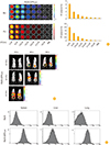
Next we analyzed the fluorescence and bioluminescence intensity from FtLVS-GFP-Lux to quantify the number of F. tularensis. Both fluorescence and bioluminescence signals from FtLVS-GFP-Lux increased with the number of F. tularensis represented by CFU (Fig. 2A). In addition, signals from FtLVS-GFP-Lux infected mouse also increased with the number of F. tularensis at the same time point after infection (Fig. 2B). We also monitored distribution of F. tularensis in the organ of infected mouse. Though GFP signals from the injected FtLVS-GFP-Lux (intravenously, 102 CFU/mouse) were not observed in the whole body of a mouse at 24 hours after infection (Fig. 2A), GFP signals were observed in the thymus, lung, liver, spleen and intestine at 24 hours after infection when we open the body of an infected mouse. For flow cytometry, we isolated cells from each F. tularensis (FtLVS or FtLVS-GFP-Lux) infected organs. When we compared the GFP signals from FtLVS or FtLVS-GFP-Lux infected organs, we could observe strong GFP signals from FtLVS-GFP-Lux infected organs such as liver, lung and spleen (Fig. 2C).
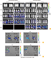
For spatio-temporal molecular imaging F. tularensis, we monitored FtLVS-GFP-Lux (102 CFU/mouse) infected mouse with various infection route such as intra-peritoneal, and intra-nasal injection (Fig. 3A). We analyzed bioluminescence signals from infected mice for a week (168 hours) with noninvasive imaging. Though bio-distribution of FtLVS-GFP-Lux in the infected mice with all injection route were similar, proliferation of FtLVS-GFP-Lux in the intra-nasal injected mice were slower than that in the intra-peritoneal injected mice. In nasally injected mice, lux signals were observed in the lung first, and the signals spread throughout body later. In peritoneally injected mice, lux signals observed in the injection site and then spread to spleen, liver, and lung.
When we compared fluorescence and bioluminescence imaging in the organ of infected mouse, bioluminescence signals were more sensitive than fluorescence signals (Fig. 3B). For fluorescence imaging, external excitation light source is required and the tissue penetration depth of each light source is different. However, our FtLVS-GFP-Lux contains LuxCDE as well as LuxAB (luciferase) and does not require the external light source. This may relate with lower sensitivity of GFP fluorescence compared to Lux bioluminescence.
Since we use live vaccine strain, we tested the vaccination effect of FtLVS by monitoring FtLVS-GFP-Lux in the infected mice. FtLVS (1×10³ CFU) were vaccinated to mice, challenged with FtLVS-GFP-LUX at 3 weeks after vaccination, and monitored the proliferation of FtLVS-GFP-LUX by fluorescence and bioluminescence. We isolated the organs from infected mice with or without vaccination, and monitored imaging signals (Fig. 3C). At 96 hours after challenge of FtLVS-GFP-LUX, vaccinated mouse clearly showed less fluorescence and bioluminescence signals in the target organ indicating less proliferation of FtLVS-GFP-LUX.

FtLVS-GFP-Lux imaging after LPS vaccination

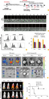
For evaluation of our imaging analysis as an efficient monitoring system of vaccine development, we tested the vaccine efficacy of LPS for F. tularensis infection. General protection efficacy of LPS from F. tularensis was reported [1112], but the detailed process of pathogenesis or vaccination was not fully investigated. With advantage of our dual-labeled F. tularensis monitoring system using FtLVS-GFP-Lux, we investigated the process of in vivo pathogenesis in mice with or without F. tularensis-LPS vaccination (Fig. 4A). Fluorescent signals from GFP from FtLVS-GFP-Lux were analyzed for time-lapse single cell imaging (Fig. 4B) and flow cytometry (Fig. 4C). Bioluminescent signals from Lux of FtLVS-GFP-Lux were analyzed for non-invasiveness pathogen tracing (Fig. 4E, F).
For visualizing replication pattern of F. tularensis, splenocytes from non-vaccinated and LPS (20 ng/mouse) vaccinated mice were subjected to time-lapse fluorescent microscopy. FtLVS-GFP-Lux was rapidly replicated within splenocyte of non-vaccinated control mouse throughout observation, while GFP-expressing FtLVS-GFP-Lux cannot be replicated and disappeared within splenocytes of LPS vaccinated mice (Fig. 4B). Then, we analyzed more detailed event of F. tularensis infection using fluorescent signals from FtLVS-GFP-Lux in cellular level by measuring the portion of infected cell in each target organ using flow cytometric analysis. At 72 hours after infection, GFP signals were observed in the cells from liver, spleen and lung of control mouse indicating FtLVS-GFP-Lux infection, while GFP signals were not observed in the cells from LPS vaccinated mice (Fig. 4C).
To decide the optimized vaccination amount of LPS, bacterial burden was also calculated by counting the infected cells in the target organ of mice with or without LPS vaccination. LPS vaccination clearly reduced F. tularensis infection and higher amount (500 ng) of LPS showed more effective vaccination results (Fig. 4D). We also monitored the proliferation of challenged FtLVS-GFP-Lux in the organ of infected mouse with different amount of LPS vaccination (Fig. 4E). LPS vaccinated mice showed lower bioluminescence signals indicating less FtLVS-GFP-Lux proliferation.
In the whole body imaging, FtLVS-GFP-Lux in the control non vaccinated mouse was multiplied at infection site (peritoneal cavity for intraperitoneal injection) first and propagated to other sites 24 hours after infection (Fig. 4F, upper panel). Liver is the first target site of propagation and, lung and spleen is also revealed as target organ of bacterial invasion. On the other hand, only limited signals of FtLVS-GFP-Lux from LPS vaccinated mouse were detected at infection site (Fig. 4F, lower panel). LPS vaccination also clearly increased survival rate of infected mouse (Fig. 4G).
Our results successfully demonstrated the efficacy of F. tularensis-LPS vaccination to the F. tularensis infection by using various imaging methods at the cellular, organ, and whole body level. These indicate that our FtLVS-GFP-Lux system can be very useful for the investigation of F. tularensis pathogenesis with various analysis methods such as histology, flow-cytometry, in vitro and in vivo molecular imaging.

Discussion

To evaluate our bacterial reporter imaging as an efficient monitoring system for vaccine development, we developed a sensitive imaging reporter to visualize F. tularensis and tested the efficacy of LPS vaccine for F. tularensis infection. In previous studies, LPS purified from F. tularensis has meaningful efficacy as vaccine for F. tularensis LVS infection [1112]. Our results from cellular, organ, and whole body analysis clearly confirmed that vaccination with F. tularensis-LPS efficiently prevent infection by FtLVS. These results suggest that our imaging analysis system with dual-labeled bacteria is valuable for study on infectious disease. Bioluminescent imaging capacitates real-time tracing of bacteria for analysis of pathogenic progress in whole organism level, and fluorescent imaging is able to be applied for the analysis of mechanisms using real-time single cell imaging, infected cell sorting, histology and other in vitro, ex vivo imaging.
Application field of our imaging analysis system is not restricted only in the study of F. tularensis, but our system can be expanded to general pathogens, especially in the case bacteria specific antibodies are not available. Any kinds of pathogens can be visualized and traced by substitution of appropriate replication origin and promoter for target bacteria. Furthermore, for several pathogens, attenuated bacteria with good vaccine efficacy are available [1314], and our system can be used for spatio-temporal tracing of bacteria pathogenesis. In study of vaccination mechanism of attenuated bacteria or individual analysis of sub-species which cannot be distinguished by antibody, our system would be very useful. As all subspecies of F. tularensis share same activity of replication-origin and promoter, each subspecies would be labeled by substitution with luminescence/fluorescence of different wavelength.
Recently, several kinds of near infra-red (NIR) fluorescent protein was developed and applied to some biological experiment [1516]. NIR fluorescent protein has relatively longer excitation and emission wave-length so can reduce the auto-fluorescence and blockage of light which is critical limitation of conventional fluorophores in application to non-invasiveness imaging. For future application, NIR can be another option to overcome the limitation of GFP.
Our dual imaging analysis system provides platform for in vivo study without specific antibodies for bacterial infectious disease in cellular and organism level. Moreover, there is large possibility of application using our system to understand certain biological events which specifically activated in certain condition by changing with conditional promoter.

Figures and Tables

Fig. 1

Construction of a plasmid with dual reporter and generation of reporter expressing Francisella tularensis. (A) Design of a dual reporter construct. (B) Generation of reporter expressing FtLVS. (C) Comparison of reporter activity under the different promoter. (D) Plate images of FtLVS-GFP-Lux with putative Bfr promoter. RFI, relative fluorescence intensity; RLI, relative luciferase intensity; CFU, colony forming unit; BLI, bioluminescent imaging; FLI, fluorescent imaging; FtLVS, F. tularensis LVS; Bfr, bacterioferritin.

cevr-7-129-g001
Fig. 2

Bioluminescence and fluorescence imaging of FtLVS-GFP-Lux and infected mouse. (A) Dose-dependent fluorescence and bioluminescence signals from FtLVS-GFP-Lux. (B) Dose-dependent fluorescent signals from FtLVS-GFP-Lux infected mouse model. Higher CFU of FtLVS-GFP-Lux infected mouse showed detectable fluorescent signal for in vivo imaging. (C) Flow cytometic analysis of cells from the organs of FtLVS-GFP-Lux infected mouse. FtLVS, Francisella tularensis LVS; CFU, colony forming unit; BLI, bioluminescent imaging; FLI, fluorescent imaging.

cevr-7-129-g002
Fig. 3

Spatio-temporal imaging of FtLVS-GFP-Lux infected mouse. (A) Spatio-temporal bioluminescence imaging of FtLVS-GFP-Lux (102 CFU/mouse) infected mouse by injection route. (B) Fluorescent and luminescent signals from each organ from LVS-GFP-LUX infected mice. (C) Monitoring vaccination effects of FtLVS (1×10³ CFU) in each organ after challenging with FtLVS-GFP-LUX (3 weeks after vaccination, 1×10⁶ CFU). FtLVS, Francisella tularensis LVS; CFU, colony forming unit; FLI, fluorescent imaging; BLI, bioluminescent imaging.

cevr-7-129-g003
Fig. 4

FtLVS-GFP-Lux imaging after LPS vaccination. (A) Experimental plan to evaluate FtLVS-GFP-Lux imaging system. (B) Time-lapse imaging of splenocytes from non-vaccinated mouse and LPS vaccinated mouse. (C) Flow cytometry analysis of cells from organs of non-vaccinated mouse and LPS vaccinated mouse. (D) Bacterial burden on target organ after FtLVS-GFP-Lux challenge by LPS amount of vaccination. (E) Ex-vivo bioluminescence imaging of FtLVS-GFP-Lux infected mouse by LPS amount of vaccination. (F) Whole body bioluminescence imaging of FtLVS-GFP-Lux infected mouse with or without LPS vaccination. (G) Survival rate of infected mice with or without LPS vaccination. FtLVS, Francisella tularensis LVS; LPS, lipopolysaccharide; FACS, fluorescence-activated cell sorting; PBS, phosphate buffered saline; CFU, colony forming unit.

cevr-7-129-g004

Notes

No potential conflict of interest relevant to this article was reported.

References

1. Meighen EA. Molecular biology of bacterial bioluminescence. Microbiol Rev. 1991; 55:123–142.
crossref
2. Golovliov I, Baranov V, Krocova Z, Kovarova H, Sjostedt A. An attenuated strain of the facultative intracellular bacterium Francisella tularensis can escape the phagosome of monocytic cells. Infect Immun. 2003; 71:5940–5950.
crossref
3. Bina XR, Miller MA, Bina JE. Construction of a bioluminescence reporter plasmid for Francisella tularensis. Plasmid. 2010; 64:156–161.
crossref
4. Giorno R, Mallozzi M, Bozue J, et al. Localization and assembly of proteins comprising the outer structures of the Bacillus anthracis spore. Microbiology. 2009; 155:1133–1145.
crossref
5. Glomski IJ, Piris-Gimenez A, Huerre M, Mock M, Goossens PL. Primary involvement of pharynx and peyer's patch in inhalational and intestinal anthrax. PLoS Pathog. 2007; 3:e76.
crossref
6. Louie M, Read S, Simor AE, et al. Application of multiplex PCR for detection of non-O157 verocytotoxin-producing Escherichia coli in bloody stools: identification of serogroups O26 and O111. J Clin Microbiol. 1998; 36:3375–3377.
crossref
7. Rajwa B, Dundar MM, Akova F, et al. Discovering the unknown: detection of emerging pathogens using a label-free light-scattering system. Cytometry A. 2010; 77:1103–1112.
crossref
8. Shachaf CM, Kopelman AM, Arvanitis C, et al. MYC inactivation uncovers pluripotent differentiation and tumour dormancy in hepatocellular cancer. Nature. 2004; 431:1112–1117.
crossref
9. Burton JB, Johnson M, Sato M, et al. Adenovirus-mediated gene expression imaging to directly detect sentinel lymph node metastasis of prostate cancer. Nat Med. 2008; 14:882–888.
crossref
10. Nasu M, Carbone M, Gaudino G, et al. Ranpirnase interferes with NF-kappaB pathway and MMP9 activity, inhibiting malignant mesothelioma cell invasiveness and xenograft growth. Genes Cancer. 2011; 2:576–584.
crossref
11. Oyston PC, Sjostedt A, Titball RW. Tularaemia: bioterrorism defence renews interest in Francisella tularensis. Nat Rev Microbiol. 2004; 2:967–978.
crossref
12. Pechous RD, McCarthy TR, Zahrt TC. Working toward the future: insights into Francisella tularensis pathogenesis and vaccine development. Microbiol Mol Biol Rev. 2009; 73:684–711.
crossref
13. Hong KJ, Park PG, Seo SH, Rhie GE, Hwang KJ. Current status of vaccine development for tularemia preparedness. Clin Exp Vaccine Res. 2013; 2:34–39.
crossref
14. Green M, Choules G, Rogers D, Titball RW. Efficacy of the live attenuated Francisella tularensis vaccine (LVS) in a murine model of disease. Vaccine. 2005; 23:2680–2686.
crossref
15. Wang X, Zhang X, Zhou D, Yang R. Live-attenuated Yersinia pestis vaccines. Expert Rev Vaccines. 2013; 12:677–686.
16. Herwig L, Rice AJ, Bedbrook CN, et al. Directed evolution of a bright near-infrared fluorescent rhodopsin using a synthetic chromophore. Cell Chem Biol. 2017; 24:415–425.
crossref
TOOLS
ORCID iDs

Young-Hwa Kim
https://orcid.org/0000-0003-0517-9559

Pil-Gu Park
https://orcid.org/0000-0002-3024-3439

Sang-Hwan Seo
https://orcid.org/0000-0002-4518-8858

Kee-Jong Hong
https://orcid.org/0000-0003-2054-7014

Hyewon Youn
https://orcid.org/0000-0003-1006-2689

Similar articles