

 PDF
PDF ePub
ePub Citation
 Print
Print


INTRODUCTION
Cedar pollinosis (CP) is a specific seasonal allergic disease, especially in Japan, that afflicts up to 30% of people during February to April.12 Besides symptom-relieving medications, allergen-specific immunotherapy (AIT) is one of the most effective treatments for CP. Many years after administration of subcutaneous immunotherapy (SCIT) using standardized cedar pollen extract since the 1960s,3 the first sublingual immunotherapy (SLIT) was approved for use in 2014.4 There is much clinical and scientific evidence regarding the effectiveness and safety of SLIT for patients with CP;567 additionally, this therapy is easier to administer and safer to use than SCIT. In cases treated with SCIT, systemic injections were required and severe side effects including fatal anaphylaxis were feared. However, the mechanisms by which SLIT and SCIT exert their efficacies have not been fully elucidated.
Despite the usefulness of SLIT, it has been reported that up to 30% of treated patients do not respond to this therapy.89 We have also confirmed this fact in our recent clinical study;1011 nevertheless, the existence of patients who are high-responders (HRs) or non-responders (NRs) to SLIT may be helpful in understanding the mechanisms of SLIT. Thus, by detailed comparison between HRs and NRs, we have delineated an essential biological signaling pathway responsible for the efficacy of SLIT. Although the difference between the HR and NR groups were not obvious even in the transcriptomic analysis,11 using a multivariate analysis of microarray data on mRNA expression in CD4+ T cells and basophils, we have suggested the involvement of apoptosis-related pathways in the efficacy of SLIT in patients with CP.
MATERIALS AND METHODS
Clinical study of SLIT for the treatment of CP
The study design, recruitment of patients, administration of cedar pollen extracts and evaluation of clinical efficacies have been described elsewhere.10 Briefly, 202 patients with Japanese CP, who lived in Tokyo, Japan, were over 20 years of age, and were being treated at the Nippon Medical School Hospital, the Tokyo Metropolitan Hospitals and clinics in Tokyo were included in this study. All patients showed symptoms of allergic rhinitis such as sneezing, rhinorrhea and nasal congestion during the pollen season from February to April for at least 3 consecutive years. They were positive on skin-tests as well as for immunoglobulin E (IgE) against the cedar pollen allergen. The study was registered in the University Hospital Medical Information Network Clinical Trials Registry Database (UMIN000016532) and was conducted in accordance with the Declaration of Helsinki and Good Clinical Practice guidelines. All experimental procedures were approved by the ethical committee of Tokyo Metropolitan Institute of Medical Science (Approval No.17-10), Nippon Medical School Hospital (No.18-3), Tokyo Metropolitan Komagome Hospital (No.18-473), Tokyo Metropolitan Hiroo Hospital (No.18-14), Tokyo Metropolitan Fuchu Hospital (No.18-2), Tokyo Metropolitan Otsuka Hospital (No.2006-1), and Tokyo Metropolitan Health and Medical Treatment Corporation Ebara Hospital (No.18-1). Endo ENT Clinic and Hirooka Clinic were also participated in this study under the supervision of Nippon Medical School Hospital. All patients provided informed written consent prior to participation.
All subjects were individually administered an allergen from cedar pollen.10 Briefly, patients held the allergen under their tongues for 2 minutes and then spat it out. Starting with 1 drop consisting of 2 Japanese allergy units (JAU) per mL, the amount of allergen administered to the patients was gradually increased up to 20 drops of 2,000 JAU per mL as the maintenance dose at 5 weeks. The administration of the allergen started from July 2006 and further continued for 2 years.
Nasal symptoms of individual patients observed between February 1 and April 30 in 4 pollen seasons from 2006 to 2009 were investigated using the Japanese guideline for allergic rhinitis.112 Briefly, the numbers of sneezes and nose-blowing, and the extents of nasal congestion and eye itchiness were daily recorded by each patient, using an allergy diary according to the classification developed by Okuda.13 The quality of life (QOL) of the patients were determined 3 times, at the end of February, the middle of March, and the middle of April, respectively, for each year using a Japan Rhinoconjunctivitis Quality of Life Questionnaire No.1 (JRQLQ No1). At the end of each pollen season, the clinical efficacy of SLIT was evaluated based on the descriptions in the allergy diaries and the JRQLQ No1 as previously described.6
Cell preparation and microarray analysis
The preparation of CD4+ T cells and basophils, and the subsequent microarray analysis of mRNA expression have been described elsewhere.1011 Briefly, blood samples were obtained twice, namely, in June 2006 and 2008, respectively, just prior to the start of and following the end of allergen administration. CD4+ T cells and basophils were purified from peripheral blood mononuclear cells as CD4+ and CD123+CD11clow cell populations, respectively, using a FACSAria cell sorting system (BD Biosciences, San Diego, CA, USA).
Total RNA was extracted from the stored cells. Microarray analyses for the extracted RNA from HRs and NRs, before and after undergoing SLIT, were comparably performed using GeneChip Human Gene 1.0 ST Array (Agilent Technologies, Santa Clara, CA, USA) with GeneChip Scanner 3000 7G (Affymetrix Inc., Santa Clara, CA, USA) and Affymetrix GeneChip Command Console Software. Just before the microarray analysis, the quality of RNA was examined by Qubit RNA IQ Assay Kit (ThermoFisher Scientific, Waltham, MA, USA).
Multivariate pathway analysis
The microarray data from CD4+ T cells were processed using the orthogonal partial least squares (OPLS) analysis with SIMCA-P™ software (Umetrics, Umeå, Sweden). Highly reliable markers that may distinguish between the 2 groups were extracted by OPLS-discriminant analysis (OPLS-DA). The target group pairs were 1) HRs before and after undergoing SLIT, 2) NRs before and after undergoing SLIT, and 3) HRs and NRs before undergoing SLIT. The resulting candidate markers were processed for pathway analysis using the MetaCore™ software (version 6.24, build 67895; Thomson Reuters, Philadelphia, PA, USA). This analysis was repeated using the microarray data from basophils to compare HRs and NRs after undergoing SLIT.
RESULTS
The detailed results of this clinical study have been described elsewhere.10 Consistent with the results of previous investigations,1415 in our study approximately 30% of patients did not show any improvements in their symptoms even after undergoing the 2-year treatment with SLIT. The first 33 HR patients whose conditions were extremely improved and the last 34 NR patients whose conditions were either unchanged or exacerbated were selected. After the exclusion of the samples from cypress pollen-specific IgE-positive patients and those with damaged RNA or DNA, 25 samples each from HRs and NRs were processed for microarray analyses.
Using OPLS-DA with a confidence interval of 0.4 for the microarray data from CD4+ T cells, 1,145, 1,475 and 652 genes were extracted in the comparison between HRs before and after undergoing SLIT (Supplementary Fig. S1A), NRs before and after SLIT (Supplementary Fig. S1B), and HRs and NRs before undergoing SLIT (Supplementary Fig. S1C), respectively. The HRs and NRs after undergoing SLIT were not distinguishable by OPLS-DA.
These extracted genes were analyzed by pathway analysis. In the comparison between HRs before and after undergoing SLIT, an apoptosis pathway involving Caspase-10, Apo-2L (TNFSF10), Acinus, tumor necrosis factor (TNF)-related apoptosis-inducing ligand (TRAIL) receptor with a truncated death domain (TRUNDD, TNF receptor superfamily member 10D [TNFRSF10D]) and Aif was identified with a G-score of 1,680.23 (Table 1 and Figure A). However, no significant pathways with high G-scores were determined in the comparison between NRs before and after undergoing SLIT or between HRs and NRs before undergoing SLIT (data not shown).
Table 1
Top 10 list of GO process networking molecules differentially expressed in CD4+ T cells of HR patients between before and after SLIT
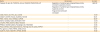
The predicted GO processes were only shown for the highest G-score networking molecules (MetaCore™ version 6.24, build 67895).
GO, gene ontology; HR, high-responders; SLIT, sublingual immunotherapy; TRUNDD, tumor necrosis factor-related apoptosis-inducing ligand receptor with a truncated death domain; TNFRSF10D, tumor necrosis factor receptor superfamily member 10D.
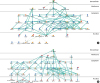
Figure
The pathway in CD4+ T cells and basophils that is expected to be related to the efficacy of SLIT. The candidate markers extracted from CD4+ T cells (A) as shown in Supplementary Fig. S1 that may distinguish HRs before and after undergoing SLIT and those from basophils (B) as shown in Supplementary Fig. S2 that may distinguish HRs and NRs after undergoing SLIT were processed for pathway analysis using the MetaCore™ software. The most reliable pathway, wherein the extracted markers are connected by a bold line, is shown.
SLIT, sublingual immunotherapy; HR, high-responders; NR, non-responders.
Using OPLS-DA with a confidence interval of 0.4 for the microarray data from basophils, 780 genes were extracted in the comparison between HRs and NRs after undergoing SLIT (Supplementary Fig. S2). Then, as in the case of microarray data from CD4+ T cells, an apoptosis induction pathway involving TNF-receptor (R)1, tBid, receptor for advanced glycation end products (RAGE), Bid and Caspase-3 was identified with a G-score of 133.08 (Table 2 and Figure B).
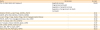
Table 2
Top 10 list of GO process networking molecules differentially expressed in basophils after SLIT between HR and NR patients
DISCUSSION
To delineate the mechanisms by which SLIT is effective for the treatment of CP, biomarker sets that are different between HRs and NRs both before and after undergoing therapy were investigated by multivariate analysis of microarray data on mRNA expression in CD4+ T cells and basophils.
CD4+ T cells play crucial roles in antigen recognition; therefore, they are implicated in the effects of AIT.1617181920 By comparing data on HRs before and after undergoing SLIT, an apoptosis pathway was identified. Consistent with this finding, the AIT-induced apoptosis in the T cells of atopic patients has previously been reported.212223 However, several other groups have demonstrated that Fas-induced apoptosis is unrelated to the effects of AIT.2425 We recently demonstrated that some taste receptors expressed on CD4+ T cells were related to the efficacy of SLIT. Although further functional analysis is required to confirm those possibilities including the contribution of taste receptors, our present findings support the hypothesis that the effect of SLIT is achieved, at least in part, by the induction of apoptosis in CD4+ T cells.
The involvement of basophils in the pathogenesis of allergic diseases has recently been discussed.2627 Like in CD4+ T cells, an apoptosis-inducing pathway has been identified in basophils too; this is an intriguing observation. The apoptosis of T cells is a well-known biological response, but the same has not been commonly recognized in basophils. However, Matsumoto et al.28 and Förster et al.29 recently reported the presence of a Fas-mediated apoptosis pathway in basophils. Since basophils play important roles in the pathogenesis of allergic diseases,30 the induction of apoptosis in basophils may be an additional mechanism for the efficacy of treatment with SLIT.
In conclusion, our present findings suggest the possibility that the effectiveness of SLIT in the treatment of CP is mainly caused by the induction of apoptosis in CD4+ T cells and basophils. Further investigations are needed to prove this hypothesis and to study whether apoptosis is induced in T cells and basophils by the same mechanisms.
 XML Download