Journal List > Tuberc Respir Dis > v.81(1) > 1099561

Overton, Varma, and Post: Comparison of Interferon-γ Release Assays and the Tuberculin Skin Test for Diagnosis of Tuberculosis in Human Immunodeficiency Virus: A Systematic Review

Abstract

Background

It remains uncertain if interferon-γ release assays (IGRAs) are superior to the tuberculin skin test (TST) for the diagnosis of active tuberculosis (TB) or latent tuberculosis infection (LTBI) in immunosuppressed populations including people with human immunodeficiency virus (HIV) infection. The purpose of this study was to systematically review the performance of IGRAs and the TST in people with HIV with active TB or LTBI in low and high prevalence TB countries.

Methods

We searched the MEDLINE database from 1966 through to January 2017 for studies that compared results of the TST with either the commercial QuantiFERON-TB Gold in Tube (QFTGT) assay or previous assay versions, the T-SPOT.TB assay or in-house IGRAs. Data were summarized by TB prevalence. Tests for concordance and differences in proportions were undertaken as appropriate. The variation in study methodology was appraised.

Results

Thirty-two studies including 4,856 HIV subjects met the search criteria. Fourteen studies compared the tests in subjects with LTBI in low TB prevalence settings. The QFTGT had a similar rate of reactivity to the TST, although the first-generation version of that assay was reactive more commonly. IGRAs were more frequently positive than the TST in HIV infected subjects with active TB. There was considerable study methodology and population heterogeneity, and generally low concordance between tests. Both the TST and IGRAs were affected by CD4 T-cell immunodeficiency.

Conclusion

Our review of comparative data does not provide robust evidence to support the assertion that the IGRAs are superior to the TST when used in HIV infected subjects to diagnose either active TB or LTBI.

Introduction

Infection with Mycobacterium tuberculosis (MTb) continues to be a major cause of morbidity and mortality throughout the world, with a disproportionate burden occurring in individuals infected with the human immunodeficiency virus (HIV). In 2015, the World Health Organization (WHO) estimated there were 10.4 million new cases of tuberculosis (TB) worldwide with 1.2 million of these in people co-infected with HIV resulting in 400,000 deaths1. HIV infected patients with latent MTb infection (LTBI) have a 5%–10% annual risk of developing active disease2. Unlike active TB, LTBI does not produce clinical symptoms and, although the host is infected, it does not result in cultivable organisms. There is no gold standard for the diagnosis of LTBI. Until recently, the diagnosis of LTBI has relied on the tuberculin skin test (TST), which is based on a delayed hypersensitivity response to MTb within the host. It has a number of limitations including cross reaction against the bacille Calmette-Guerin (BCG) vaccine strain and environmental mycobacteria, reported low sensitivity and anergy in HIV infected patients (resulting in false-negative results), operator variability and the requirement of two consultations3.
Interferon-γ release assays (IGRAs) are an attractive alternative to the TST. There are a number of commercial IGRAs that have been studied: the QuantiFERON-TB Gold in Tube (QFTGT) (Cellestis, Carnegie, Australia) and earlier versions including QuantiFERON-TB (QFT) and QuantiFERON-Gold (QFTG) assays, as well as the T-SPOT.TB (TS.TB) (Oxford Immunotec, Abingdon, UK) assay. The QFTGT measures the release of interferon (IFN)-γ by T cells, through an enzyme linked immunosorbent assay (ELISA), from a stimulated whole blood sample, in response to antigens including the region of difference 1 antigen, culture filtrate protein 10 (CFP-10), early secreted antigen target 6 (ESAT-6), and TB antigen 7.7 peptides. This assay has more antigens than the previous versions. More recently a fourth generation QuantiFERON TB Gold Plus has been released and is currently being evaluated4. In contrast to the QuantiFERON assays, the TS.TB utilizes an enzyme linked immunospot (ELISPOT) method, on a sample of peripheral blood mononuclear cells. In contrast to the TST, indeterminate results may be generated with IGRAs usually due to failure of production of IFN-γ after stimulation with a potent mitogen used as a positive control. Indeterminate results imply abnormal T-cell function or technical error5. The frequency of indeterminate IGRA results in HIV infected individuals may increase with increasing levels of immunodeficiency 6.
As there is no gold standard for the diagnosis of LTBI, the evaluation of IGRAs has been undertaken in subjects with active TB as a marker of true MTb infection. A number of studies and systematic reviews have assessed the performance of these assays in HIV negative subjects, which identified some of the advantages of the IGRA compared to the TST789. IGRA do not appear to react with exposure to certain non-tuberculous mycobacteria or BCG and test reactivity has been shown to correlate with either risk factors associated with acquisition of TB, or exposure to cases of active TB101112. However, as these assays depend on intact T-cell function it is plausible that such assays may perform differently in the immunocompromised host.
Our aim was to systematically review the performance of IGRAs and the TST in HIV infected subjects with active TB or LTBI in different TB prevalence settings.

Materials and Methods

This study was conducted and reported in concordance with PRISMA guidelines for systematic reviews13. Ethics approval was not required for completion of this systematic review.

1. Search strategy

A MEDLINE search for English language articles published between 1966 and January 2017 was undertaken. The search strategy used search terms including (TB infection OR TB disease) AND [(QuantiFERON OR ELISPOT) OR (interferon gamma release assays) OR (tuberculin skin test) OR (t-cell assay)] AND (HIV OR AIDS). Manufacturers of the commercial assays were contacted for any additional material of relevance.

2. Study selection

Three independent reviewers assessed article titles and abstracts for selection for full text review. Articles excluded were commentary, guidelines, policy, review, case studies, those with HIV negative or unknown status patients, immune reconstitution studies, in vitro studies, non-diagnostic, behavioural or modelling studies, serological, therapeutic and vaccine studies. After full text review only studies that presented data in a format allowing comparison between TST and the IGRAs were included. The population included HIV-1 or HIV-2 infected adults or children. Data from studies that recruited subjects of mixed HIV seropositivity were included only if the data pertaining to the HIV infected group could be extracted. TB referred to pulmonary or extra pulmonary disease due to MTb infection. The IGRAs included the commercial assays QFT, QFTG, QFTGT, and TS.TB. In-house ELISPOT (IHE) assays were included if they used antigens against ESAT-6 and CFP-10. Studies were included if the IGRAs were performed on samples from peripheral blood.

3. Data extraction

Once identified studies were separated into studies of active TB disease or LTBI, based on the case definitions employed by the authors, which included variable assessments of clinical, radiological (including computed tomography scan or plain X-ray), microbiological (including detection of acid fast bacilli on light microscopic examination and/or growth in culture media and/or polymerase chain reaction) and/or histological parameters as listed in the study methodology. Countries were determined to be a low incidence TB country if they reported less than 20 cases of TB per 100,000 people and/or were not one of the 63 high TB/HIV burden countries as per WHO definitions1. Study methodology and review of potential bias was extracted in a standardized format and included: recruitment method and exclusion criteria, inclusion of patients with a past history of active TB or LTBI, inpatient or outpatient setting of the study, age (classified into adult or child) and ethnicity of the study population. We assessed the laboratory methods detailed by the authors, including definitions of test positivity (for both TST and IGRA), indeterminate (IGRA only) and whether technicians were blinded to the TST result. HIV specific data including mean or median CD4 T-cell count and proportion of patients taking antiretroviral therapy were also extracted.

4. Statistical analyses

We calculated the proportion of reactive assays and 95% confidence intervals using paired data. To test the measurement of agreement between IGRA and TST Cohen's kappa statistic (k) and p-values reported in studies was used. Statistical calculators were sourced from VasserStats: Website for Statistical Computation (http://vassarstats.net/). Meta-analysis was not undertaken due to heterogeneity of study methodology (population, setting, type of IGRA, type of TST, and non-consecutive recruitment).

Results

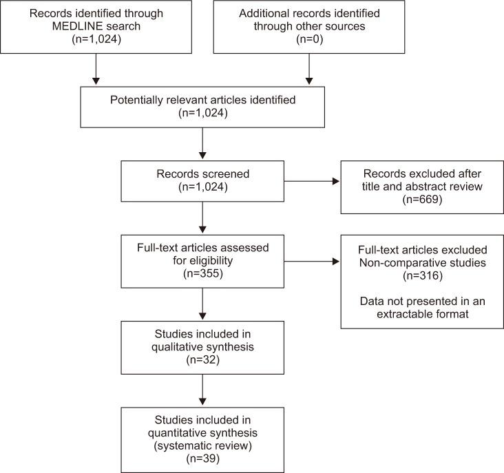
Our search identified a total of 1024 articles (Figure 1) of which 32 studies with a total of 4,856 HIV infected subjects were selected for final review.

1. Active TB disease

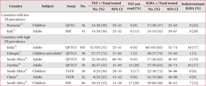
We identified nine studies in which IGRA and TST were performed in 431 patients with active TB (Table 1)141516171819202122. Only two studies1621 reported a greater proportion of positive results in IGRAs than the TST. There was inter- and intra-test variability in test positivity and proportion of indeterminate results. Concordance between the TST and QFTGT was reported in only one Indian study and was found to be low (k=0.27)16. There was considerable heterogeneity in the study design including the case definition of active TB disease and exclusion criteria including varying durations of anti-tuberculous therapy (ATT) at the time of testing (Table 2)141516171819202122.

2. LTBI

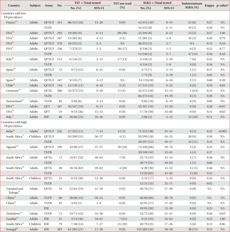
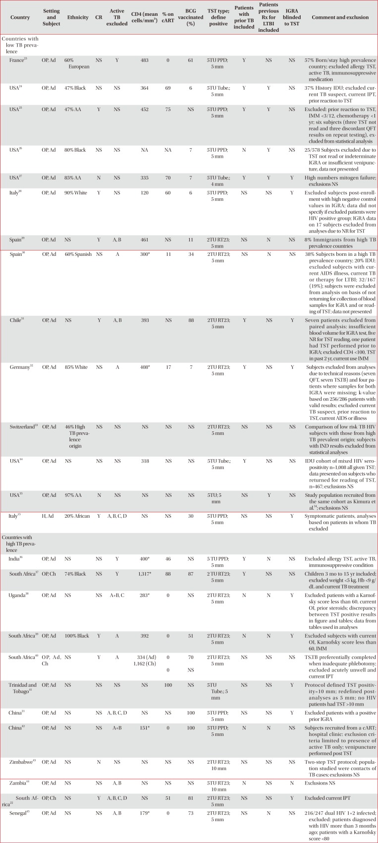
Fourteen studies from low TB prevalence countries were analysed comprising a total of 2,959 HIV adult subjects (Table 3)1521222324252627282930313233343536373839404142434445. There was significant heterogeneity in the studies (Table 4)1521222324252627282930313233343536373839404142434445. There were only three studies where significant differences between IGRA and TST were identified although the direction of difference was inconsistent. The TS.TB (but not the QFTG) had greater reactivity than the TST in one study from Germany32. In contrast the TST had greater reactivity than an IHE in an Italian study15. The TST also had greater reactivity than both the QFTGT and TS.TB in an anti-retroviral naïve French cohort23. The rate of positive reactions was higher in the first generation QFT compared to the TST based on two studies with a total sample size of 201 subjects, in which the study population were from the same cohort of mainly injecting drug users (IDU) in urban United States (Table 4)3435. These findings were not replicated with the more recent versions of the QuantiFERON assay or other IGRAs. Concordance between IGRAs and the TST ranged from extremely poor (k=0.02) to strong (k=0.69). Some authors did not present complete data on all test outcomes in subjects who were excluded from statistical analyses due to an indeterminate IGRA or “technical failure of the sample,” potentially introducing bias (Table 4)25283234. A significant proportion of subjects in some of these studies did not return to have the TST read2832.
Twelve studies (eight in adult subjects) comprising a total of 1,466 subjects (401 children) from high TB prevalence countries were analysed. There were generally higher rates of positive results in all assays (IGRAs and TST) when compared to studies that took place in lower TB prevalence settings. The results were variable with one study finding higher rates of positivity in the TST37 and five with the IGRA being positive more frequently than the TST2136384245.
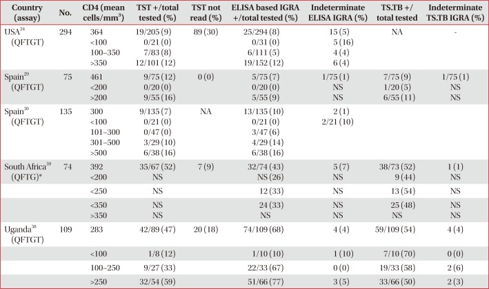
The rate of reactivity of both IGRAs and TST was affected by the level of HIV associated CD4 T-cell deficiency (Table 5)2429303839.

Discussion

The performance characteristics of the TST, including the previously discussed limitations, are well described in HIV populations and other risk groups. Direct comparative studies of new assays with the TST provide data to assist clinicians when considering which test to use in the diagnosis of LTBI in HIV positive patients. Our analysis highlighted three main issues. Subjects with active TB had higher rates of reactivity in IGRAs when compared to the TST, although neither test type had sufficient sensitivity to be useful diagnostically. Secondly, the TST had similar rates of reactivity as the IGRAs when used in subjects without evidence of active TB. There was variability in rates of test reactivity and concordance between the TST and the different generations of the ELISA and ELISPOT based assays when all three tests were used in the same studies. Lastly, the heterogeneity of study methodology and population precluded a definitive conclusion on test superiority.
In the absence of a gold standard test for LTBI, active TB is commonly used as a surrogate when assessing the performance characteristics of IGRAs. However, they have different immunological and clinical phenotypes and are not equivalent conditions. Although this review identified that the QFTGT performed better than the TST in symptomatic patients with clinical or confirmed TB, it would not be accurate enough to exclude all cases of active TB. There was relatively limited data on the use of the TS.TB within this setting.
Studies used different criteria to include and exclude cases of active TB infection. The clinical evaluation of HIV TB co-infected patients is challenging, particularly with more advanced states of immunodeficiency. The clinical presentation can be varied with higher rates of disseminated disease, lower rates of sputum smear positivity and atypical radiological appearances 4647. This heterogeneity may affect the findings presented here.
The spectrum of the kinetics of the IGRA response to TB infection has not been established. Studies in mainly HIV negative subjects have analysed IHE during ATT, which report intra- and inter-patient variability in the measured MTb immune response and of test reversion from positive to negative in the TS.TB484950. The effect of a prior TST on an individual who subsequently has a further TST can lead to the “boosting” phenomenon, whereby an increased reaction to the injected mycobacterial antigen is observed, attributed to the immunological recall of a previous hypersensitivity reaction51. However, it is unclear how a prior TST can affect the test performance of the IGRAs. The few studies that have investigated this issue, primarily within HIV seronegative subjects, suggest the possibility of boosting the IGRA response52. The spectrum of this effect has not been established. These test performance characteristics of the IGRAs may have impacted on the studies in our review, a number of which included subjects who had been commenced on ATT (with variable treatment duration), had a prior TST, treated past TB infection or isoniazid preventative therapy prior to the IGRA being undertaken.
The population groups studied were diverse. Some of the subjects in studies from low prevalence countries included subjects born in high TB incidence countries. A number of other features varied between studies including the physical performance status of subjects, the inclusion of mainly IDU populations, the proportion of individuals on combination antiretroviral therapy, the degree of immunosuppression and HIV-2 seropositivity. Additionally, there was substantial study design differences including some studies with non-consecutive recruitment, variable definition of TST reactivity and the type and amount of purified protein derivative used.
All assays appeared to be affected by the degree of CD4 T-cell depletion, with the possible exception of the TS.TB assay. The likelihood of a positive reaction in the TST or ELISA based IGRA at lower CD4 strata appears negligible. There is insufficient data to suggest that an ELISA based IGRA performs better than the TST in patients with HIV related immunodeficiency. The proportion of indeterminate IGRA in patients with LTBI was lower than that observed in the studies in patients with active TB. This may reflect the impact of severe immunosuppression and malnutrition occurring within this patient group.
Previous studies have assessed IGRAs within HIV subjects and have reported increased “sensitivity” in diagnosing LTBI in a high and low TB prevalence settings in comparison to the TST6445354. This increased “sensitivity” usually represents increased rates of assay reactivity rather than true sensitivity as there is no gold standard. However, most of these studies did not directly compare the results of concurrent TST and IGRA testing in individual subjects. Prior systematic reviews and meta-analysis of the test performance of the IGRA in HIV infected individuals have attempted to ascertain the sensitivity and specificity of the QFTGT and TS.TB in predicting active and LTBI55565758. However, studies were included that did not perform direct comparisons with the TST and studies using older generation commercial IGRA as well as IHE and TS.TB were excluded.
There are limitations of this review. We have presented data on the first generation commercial QFT test that is no longer commonly available. Most studies had small sample sizes and study heterogeneity precluded formal meta-analysis. Finally, the data included in our review were unable to answer which test performs better in the diagnosis of LTBI due to the lack of a gold standard. We reported on cross sectional differences in test reactivity without longitudinal outcome data. A number of the studies had high non-return rates for TST reading, affecting the outcome measured for TST. This represents the operational result, rather than the intrinsic utility of the assay, although the two visits required for the test is clearly an operational challenge.
Our review of comparative data does not provide robust evidence to support the assertion that the IGRAs are superior to the TST when used in HIV infected subjects without evidence of active TB. Further longitudinal outcome studies are required to establish the prognostic value of a positive IGRA. In the interim, clinicians should be cognizant of the limitations of the data and variable test performance when considering which test to use to diagnose LTBI in this population.

Notes

Conflicts of Interest: No potential conflict of interest relevant to this article was reported.

References

1. World Health Organization. Global tuberculosis control: WHO report 2011. Geneva: World Health Organization;2011.
2. Targeted tuberculin testing and treatment of latent tuberculosis infection. This official statement of the American Thoracic Society was adopted by the ATS Board of Directors, July 1999. This is a Joint Statement of the American Thoracic Society (ATS) and the Centers for Disease Control and Prevention (CDC). This statement was endorsed by the Council of the Infectious Diseases Society of America. (IDSA), September 1999, and the sections of this statement. Am J Respir Crit Care Med. 2000; 161(4 Pt 2):S221–S247. PMID: 10764341.
3. Wang L, Turner MO, Elwood RK, Schulzer M, FitzGerald JM. A meta-analysis of the effect of Bacille Calmette Guerin vaccination on tuberculin skin test measurements. Thorax. 2002; 57:804–809. PMID: 12200526.
crossref
4. Telisinghe L, Amofa-Sekyi M, Maluzi K, Kaluba-Milimo D, Cheeba-Lengwe M, Chiwele K, et al. The sensitivity of the QuantiFERON(R)-TB Gold Plus assay in Zambian adults with active tuberculosis. Int J Tuberc Lung Dis. 2017; 21:690–696. PMID: 28482964.
5. Richeldi L. An update on the diagnosis of tuberculosis infection. Am J Respir Crit Care Med. 2006; 174:736–742. PMID: 16799073.
crossref
6. Brock I, Ruhwald M, Lundgren B, Westh H, Mathiesen LR, Ravn P. Latent tuberculosis in HIV positive, diagnosed by the M. tuberculosis specific interferon-gamma test. Respir Res. 2006; 7:56. PMID: 16579856.
crossref
7. Dheda K, van Zyl Smit R, Badri M, Pai M. T-cell interferon-gamma release assays for the rapid immunodiagnosis of tuberculosis: clinical utility in high-burden vs. low-burden settings. Curr Opin Pulm Med. 2009; 15:188–200. PMID: 19387262.
8. Pai M, Dheda K, Cunningham J, Scano F, O'Brien R. T-cell assays for the diagnosis of latent tuberculosis infection: moving the research agenda forward. Lancet Infect Dis. 2007; 7:428–438. PMID: 17521596.
crossref
9. Mori T. Usefulness of interferon-gamma release assays for diagnosing TB infection and problems with these assays. J Infect Chemother. 2009; 15:143–155. PMID: 19554399.
crossref
10. Andersen P, Munk ME, Pollock JM, Doherty TM. Specific immune-based diagnosis of tuberculosis. Lancet. 2000; 356:1099–1104. PMID: 11009160.
crossref
11. Menzies D, Pai M, Comstock G. Meta-analysis: new tests for the diagnosis of latent tuberculosis infection: areas of uncertainty and recommendations for research. Ann Intern Med. 2007; 146:340–354. PMID: 17339619.
crossref
12. Diel R, Loddenkemper R, Meywald-Walter K, Niemann S, Nienhaus A. Predictive value of a whole blood IFN-gamma assay for the development of active tuberculosis disease after recent infection with Mycobacterium tuberculosis. Am J Respir Crit Care Med. 2008; 177:1164–1170. PMID: 18276940.
13. Moher D, Liberati A, Tetzlaff J, Altman DG. PRISMA Group. Preferred reporting items for systematic reviews and meta-analyses: the PRISMA statement. PLoS Med. 2009; 6:e1000097. PMID: 19621072.
crossref
14. Stavri H, Ene L, Popa GL, Duiculescu D, Murgoci G, Marica C, et al. Comparison of tuberculin skin test with a whole-blood interferon gamma assay and ELISA, in HIV positive children and adolescents with TB. Roum Arch Microbiol Immunol. 2009; 68:14–19. PMID: 19507622.
15. Vincenti D, Carrara S, Butera O, Bizzoni F, Casetti R, Girardi E, et al. Response to region of difference 1 (RD1) epitopes in human immunodeficiency virus (HIV)-infected individuals enrolled with suspected active tuberculosis: a pilot study. Clin Exp Immunol. 2007; 150:91–98. PMID: 17680823.
crossref
16. Syed Ahamed Kabeer B, Sikhamani R, Swaminathan S, Perumal V, Paramasivam P, Raja A. Role of interferon gamma release assay in active TB diagnosis among HIV infected individuals. PLoS One. 2009; 4:e5718. PMID: 19479058.
crossref
17. Idh J, Abate E, Westman A, Elias D, Janols H, Gelaw A, et al. Kinetics of the QuantiFERON-TB Gold In-Tube test during treatment of patients with sputum smear-positive tuberculosis in relation to initial TST result and severity of disease. Scand J Infect Dis. 2010; 42:650–657. PMID: 20465490.
crossref
18. Tsiouris SJ, Coetzee D, Toro PL, Austin J, Stein Z, El-Sadr W. Sensitivity analysis and potential uses of a novel gamma interferon release assay for diagnosis of tuberculosis. J Clin Microbiol. 2006; 44:2844–2850. PMID: 16891501.
crossref
19. Raby E, Moyo M, Devendra A, Banda J, De Haas P, Ayles H, et al. The effects of HIV on the sensitivity of a whole blood IFN-gamma release assay in Zambian adults with active tuberculosis. PLoS One. 2008; 3:e2489. PMID: 18560573.
20. Liebeschuetz S, Bamber S, Ewer K, Deeks J, Pathan AA, Lalvani A. Diagnosis of tuberculosis in South African children with a T-cell-based assay: a prospective cohort study. Lancet. 2004; 364:2196–2203. PMID: 15610806.
21. Jiang W, Shao L, Zhang Y, Zhang S, Meng C, Xu Y, et al. High-sensitive and rapid detection of Mycobacterium tuberculosis infection by IFN-gamma release assay among HIV-infected individuals in BCG-vaccinated area. BMC Immunol. 2009; 10:31. PMID: 19476627.
crossref
22. Davies MA, Connell T, Johannisen C, Wood K, Pienaar S, Wilkinson KA, et al. Detection of tuberculosis in HIV-infected children using an enzyme-linked immunospot assay. AIDS. 2009; 23:961–969. PMID: 19287300.
crossref
23. Bourgarit A, Baron G, Breton G, Tattevin P, Katlama C, Allavena C, et al. Latent tuberculosis infection screening and 2-year outcome in antiretroviral-naive HIV-infected patients in a low-prevalence country. Ann Am Thorac Soc. 2015; 12:1138–1145. PMID: 26213798.
crossref
24. Luetkemeyer AF, Charlebois ED, Flores LL, Bangsberg DR, Deeks SG, Martin JN, et al. Comparison of an interferon-gamma release assay with tuberculin skin testing in HIV-infected individuals. Am J Respir Crit Care Med. 2007; 175:737–742. PMID: 17218620.
25. Jones S, de Gijsel D, Wallach FR, Gurtman AC, Shi Q, Sacks H. Utility of QuantiFERON-TB Gold in-tube testing for latent TB infection in HIV-infected individuals. Int J Tuberc Lung Dis. 2007; 11:1190–1195. PMID: 17958980.
26. Weinfurter P, Blumberg HM, Goldbaum G, Royce R, Pang J, Tapia J, et al. Predictors of discordant tuberculin skin test and QuantiFERON(R)-TB Gold In-Tube results in various high-risk groups. Int J Tuberc Lung Dis. 2011; 15:1056–1061. PMID: 21740668.
27. Talati NJ, Seybold U, Humphrey B, Aina A, Tapia J, Weinfurter P, et al. Poor concordance between interferon-gamma release assays and tuberculin skin tests in diagnosis of latent tuberculosis infection among HIV-infected individuals. BMC Infect Dis. 2009; 9:15. PMID: 19208218.
crossref
28. Richeldi L, Losi M, D'Amico R, Luppi M, Ferrari A, Mussini C, et al. Performance of tests for latent tuberculosis in different groups of immunocompromised patients. Chest. 2009; 136:198–204. PMID: 19318676.
crossref
29. Latorre I, Martinez-Lacasa X, Font R, Lacoma A, Puig J, Tural C, et al. IFN-gamma response on T-cell based assays in HIV-infected patients for detection of tuberculosis infection. BMC Infect Dis. 2010; 10:348. PMID: 21143955.
crossref
30. Santin M, Casas S, Saumoy M, Andreu A, Moure R, Alcaide F, et al. Detection of latent tuberculosis by the tuberculin skin test and a whole-blood interferon-gamma release assay, and the development of active tuberculosis in HIV-seropositive persons. Diagn Microbiol Infect Dis. 2011; 69:59–65. PMID: 21146715.
31. Balcells ME, Perez CM, Chanqueo L, Lasso M, Villanueva M, Espinoza M, et al. A comparative study of two different methods for the detection of latent tuberculosis in HIV-positive individuals in Chile. Int J Infect Dis. 2008; 12:645–652. PMID: 18534887.
crossref
32. Stephan C, Wolf T, Goetsch U, Bellinger O, Nisius G, Oremek G, et al. Comparing QuantiFERON-tuberculosis gold, T-SPOT tuberculosis and tuberculin skin test in HIV-infected individuals from a low prevalence tuberculosis country. AIDS. 2008; 22:2471–2479. PMID: 19005270.
crossref
33. Hoffmann M, Reichmuth M, Fantelli K, Schoch OD, Fierz W, Furrer H, et al. Conventional tuberculin skin testing versus T-cell-based assays in the diagnosis of latent tuberculosis infection in HIV-positive patients. AIDS. 2007; 21:390–392. PMID: 17255757.
crossref
34. Kimura M, Converse PJ, Astemborski J, Rothel JS, Vlahov D, Comstock GW, et al. Comparison between a whole blood interferon-gamma release assay and tuberculin skin testing for the detection of tuberculosis infection among patients at risk for tuberculosis exposure. J Infect Dis. 1999; 179:1297–1300. PMID: 10191241.
35. Converse PJ, Jones SL, Astemborski J, Vlahov D, Graham NM. Comparison of a tuberculin interferon-gamma assay with the tuberculin skin test in high-risk adults: effect of human immunodeficiency virus infection. J Infect Dis. 1997; 176:144–150. PMID: 9207360.
36. Mathad JS, Bhosale R, Balasubramanian U, Kanade S, Mave V, Suryavanshi N, et al. Quantitative IFN-gamma and IL-2 response associated with latent tuberculosis test discordance in HIV-infected pregnant women. Am J Respir Crit Care Med. 2016; 193:1421–1428. PMID: 26765255.
37. Mandalakas AM, Kirchner HL, Walzl G, Gie RP, Schaaf HS, Cotton MF, et al. Optimizing the detection of recent tuberculosis infection in children in a high tuberculosis-HIV burden setting. Am J Respir Crit Care Med. 2015; 191:820–830. PMID: 25622087.
crossref
38. Leidl L, Mayanja-Kizza H, Sotgiu G, Baseke J, Ernst M, Hirsch C, et al. Relationship of immunodiagnostic assays for tuberculosis and numbers of circulating CD4+ T-cells in HIV infection. Eur Respir J. 2010; 35:619–626. PMID: 19608590.
crossref
39. Rangaka MX, Wilkinson KA, Seldon R, Van Cutsem G, Meintjes GA, Morroni C, et al. Effect of HIV-1 infection on T-Cell-based and skin test detection of tuberculosis infection. Am J Respir Crit Care Med. 2007; 175:514–520. PMID: 17158278.
crossref
40. Mandalakas AM, Hesseling AC, Chegou NN, Kirchner HL, Zhu X, Marais BJ, et al. High level of discordant IGRA results in HIV-infected adults and children. Int J Tuberc Lung Dis. 2008; 12:417–423. PMID: 18371268.
41. Baboolal S, Ramoutar D, Akpaka PE. Comparison of the QuantiFERON(R)-TB Gold assay and tuberculin skin test to detect latent tuberculosis infection among target groups in Trinidad & Tobago. Rev Panam Salud Publica. 2010; 28:36–42. PMID: 20857019.
crossref
42. Zhang M, Wang H, Liao M, Chen X, Graner M, Zhu X, et al. Diagnosis of latent tuberculosis infection in bacille Calmette-Guerin vaccinated subjects in China by interferon-gamma ELISpot assay. Int J Tuberc Lung Dis. 2010; 14:1556–1563. PMID: 21144240.
43. Mutsvangwa J, Millington KA, Chaka K, Mavhudzi T, Cheung YB, Mason PR, et al. Identifying recent Mycobacterium tuberculosis transmission in the setting of high HIV and TB burden. Thorax. 2010; 65:315–320. PMID: 20388756.
44. Chapman AL, Munkanta M, Wilkinson KA, Pathan AA, Ewer K, Ayles H, et al. Rapid detection of active and latent tuberculosis infection in HIV-positive individuals by enumeration of Mycobacterium tuberculosis-specific T cells. AIDS. 2002; 16:2285–2293. PMID: 12441800.
45. Karam F, Mbow F, Fletcher H, Senghor CS, Coulibaly KD, LeFevre AM, et al. Sensitivity of IFN-gamma release assay to detect latent tuberculosis infection is retained in HIV-infected patients but dependent on HIV/AIDS progression. PLoS One. 2008; 3:e1441. PMID: 18197251.
46. Huang L, Crothers K. HIV-associated opportunistic pneumonias. Respirology. 2009; 14:474–485. PMID: 19645867.
crossref
47. Lee MP, Chan JW, Ng KK, Li PC. Clinical manifestations of tuberculosis in HIV-infected patients. Respirology. 2000; 5:423–426. PMID: 11192558.
crossref
48. Hill PC, Jackson-Sillah DJ, Fox A, Brookes RH, de Jong BC, Lugos MD, et al. Incidence of tuberculosis and the predictive value of ELISPOT and Mantoux tests in Gambian case contacts. PLoS One. 2008; 3:e1379. PMID: 18167540.
crossref
49. Bosshard V, Roux-Lombard P, Perneger T, Metzger M, Vivien R, Rochat T, et al. Do results of the T-SPOT.TB interferon-gamma release assay change after treatment of tuberculosis? Respir Med. 2009; 103:30–34. PMID: 18977647.
50. Ribeiro S, Dooley K, Hackman J, Loredo C, Efron A, Chaisson RE, et al. T-SPOT.TB responses during treatment of pulmonary tuberculosis. BMC Infect Dis. 2009; 9:23. PMID: 19250549.
crossref
51. Cobelens F, Borgdorff M. Boosting, BCG, and time of reading in tuberculin skin testing. Lancet. 2001; 358:2014.
52. van Zyl-Smit RN, Zwerling A, Dheda K, Pai M. Within-subject variability of interferon-g assay results for tuberculosis and boosting effect of tuberculin skin testing: a systematic review. PLoS One. 2009; 4:e8517. PMID: 20041113.
crossref
53. Cattamanchi A, Ssewenyana I, Davis JL, Huang L, Worodria W, den Boon S, et al. Role of interferon-gamma release assays in the diagnosis of pulmonary tuberculosis in patients with advanced HIV infection. BMC Infect Dis. 2010; 10:75. PMID: 20302657.
crossref
54. Dheda K, Chang JS, Breen RA, Haddock JA, Lipman MC, Kim LU, et al. Expression of a novel cytokine, IL-4delta2, in HIV and HIV-tuberculosis co-infection. AIDS. 2005; 19:1601–1606. PMID: 16184029.
crossref
55. Chen J, Zhang R, Wang J, Liu L, Zheng Y, Shen Y, et al. Interferon-gamma release assays for the diagnosis of active tuberculosis in HIV-infected patients: a systematic review and metaanalysis. PLoS One. 2011; 6:e26827. PMID: 22069472.
crossref
56. Cattamanchi A, Smith R, Steingart KR, Metcalfe JZ, Date A, Coleman C, et al. Interferon-gamma release assays for the diagnosis of latent tuberculosis infection in HIV-infected individuals: a systematic review and meta-analysis. J Acquir Immune Defic Syndr. 2011; 56:230–238. PMID: 21239993.
crossref
57. Santin M, Munoz L, Rigau D. Interferon-gamma release assays for the diagnosis of tuberculosis and tuberculosis infection in HIV-infected adults: a systematic review and meta-analysis. PLoS One. 2012; 7:e32482. PMID: 22403663.
58. Ayubi E, Doosti-Irani A, Sanjari Moghaddam A, Sani M, Nazarzadeh M, Mostafavi E. The clinical usefulness of tuberculin skin test versus interferon-gamma release assays for diagnosis of latent tuberculosis in HIV patients: a meta-analysis. PLoS One. 2016; 11:e0161983. PMID: 27622293.
crossref
Figure 1

Selection and assessment of articles for inclusion in review.

trd-81-59-g001
Table 1

Performance of TST and various IGRAs in HIV infected individuals with evidence of active TB
trd-81-59-i001
Country Subject Assay No. TST +/Total tested TST not read (%) IGRA +/Total tested Indeterminate IGRA (%)
No. (%) 95% CI No. (%) 95% CI
Countries with low TB prevalence
 Romania14 Children QFTG 36 14/36 (39) 25–55 0 (0) 17/36 (47) 32–63 9 (25)
 Italy15 Adults IHE 45 14/39 (36) 23–52 6 (13) 24/45 (53) 39–67 9 (20)
Countries with high TB prevalence
 India16 Adults QFTGT 105 33/105 (31) 23–41 0 (0) 68/105 (65) 55–74 18 (17)
 Ethiopia17 Children and adults* QFTGT 38 27/37 (73) 57–85 1 (3) 26/37 (70) 54–82 1 (3)
 South Africa18 Adults QFTGT 26 22/26 (85) 66–94 0 (0) 17/26 (65) 46–81 5 (19)
 Zambia19 Adults QFTGT 59 26/47 (55) 41–69 12 (20) 37/59 (63) 50–74 10 (17)
 South Africa20 Children TS.TB 30 9/25 (36) 20–55 5 (17) 22/30 (73) 56–86 0 (0)
 China21 Adults TS.TB 32 8/32 (25) 13–42 0 (0) 21/32 (66) 48–80 0 (0)
 South Africa22 Children IHE 60 10/43 (23) 13–38 17 (28) 29/60 (48) 36–61 7 (12)

Detailed description of methodological differences between studies in Table 2.

*Study population aged 15–60 years old.

TST: tuberculin skin test; IGRA: interferon-γ release assay; HIV: human immunodeficiency virus; TB: tuberculosis; CI: confidence interval; QFTG: QuantiFERON Gold; IHE: in house ELISPOT assay; QFTGT: QuantiFERON Gold in Tube; TS.TB: T-SPOT.TB.

Table 2

Methodological differences between studies comparing TST and various IGRAs in subjects with evidence of active TB disease

trd-81-59-i002
Country Setting and Subject CR TB site CD4 (mean cells/mm3) % on cART BCG vaccinated (%) TST type; induration positive Patients with prior TB ATT duration (wk) IGRA blinded to TST Comment
Countries with low TB prevalence
 Romania14 H, Ch NS P, EP NS NS 100 2TU IC65; 5 mm NS Any or complete NS Children aged 12–18 years; exclusions NS
 Italy15 H, Ad NS P, EP 179 48 51 5TU PPD; 5 mm NS N Y Exclusions NS
Countries with high TB prevalence
 India16 H, Ad NS P, EP 116 0 NS 2TU RT23; 5 mm N <2 NS Included non-microbiologically confirmed TB in case definition. Ten percent dual HIV 1+2 infection; excluded ESRF, IMM, prior TST, cART, silicosis.
 Ethiopia17 OP, Ad, Ch Y P 219 0 24 2TU RT23; 10 mm N 0 NS Included non-microbiologically confirmed TB in case definition. Excluded subjects <15 years or >60 years old; hospitalized patients, pregnancy, concurrent other disease.
 South Africa18 OP, Ad NS P, EP NS NS NS 2TU RT23; 10 mm Y <1 NS Self-reported HIV status
 Zambia19 OP, Ad Y P 212 13 NS 2TU RT23; 5 mm Y <4 NS TST reading between 48–164 hr; excluded ATT >4 wk
 South Africa20 H, OP, Ch Y P, EP NS 0 NS 2TU RT23; any induration N <4 Y Included non-microbiologically confirmed TB in case definition. Children aged up to 14 yr. Ten percent ELISPOT assays not analyzed (bacterial contamination).
 China21 NS, Ad NS P NS 62 100 5TU PPD; 5 mm NS Any Y Data in text and flow chart not consistent; text data analyzed; exclusions NS.
 South Africa22 H, OP, Ch NS P, EP NS 25 39 2TU RT23; 5 mm NS <1 Y Included non-microbiologically confirmed TB in case definition. Exclusions NS.

TST: tuberculin skin test; IGRA: interferon-γ release assay; TB: tuberculosis; CR: consecutive recruitment; cART: combination antiretroviral therapy; BCG: bacille Calmette-Guerin; ATT: anti-tuberculous therapy; H: hospital; Ch: children; NS: not stated in text; P: pulmonary TB; EP: extra pulmonary TB; TU IC65: Romanian purified protein derivative; Ad: adults; N: no; Y: yes; PPD: purified protein derivative; TU RT23: international tuberculin units of purified protein derivative Staten Serum Institute, Copenhagen, Denmark; HIV: human immunodeficiency virus; ESRF: end stage renal failure; IMM: immunosuppressant including steroids; OP: outpatient.

Table 3

Performance of TST and various IGRAs in HIV infected individuals without evidence of active TB infection
trd-81-59-i003
Country Subject Assay No. TST +/Total tested TST not read (%) IGRA +/Total tested Indeterminate IGRA (%) Kappa p-value*
No. (%) 95% CI No. (%) 95% CI
Countries with low TB prevalence
 France23 Adults QFTGT 415 66/415 (16) 13–20 0 (0) 43/415 (10) 8–14 23 (6) 0.27 NA
TS.TB 34/415 (8) 6–11 10 (2) 0.30 NA
 USA24 Adults QFTGT 294 19/205 (9) 6–14 89 (30) 25/294 (8) 6–12 15 (5) 0.37 1.00
 USA25 Adults QFTGT 207 13/201 (6) 4–11 3 (1) 11/201 (5) 3–9 10 (5) 0.38 0.79
 USA26 Adults QFTGT 578 18/533 (3) 2–5 NA 26/553 (5) 3–7 NA 0.15 0.18
 USA27 Adults QFTGT 336 7/278 (2) 1–5 58 (17) 9/336 (3) 1–5 6 (2) 0.23 0.77
TS.TB 14/336 (4) 2–7 47 (14) 0.16 0.18
 Italy28 Adults QFTGT 133 6/116 (5) 2–11 17 (13) 5/116 (4) 2–10 7 (6) 0.52 NA
TS.TB 4/116 (3) 1–8 0 (0) 0.16 NA
 Spain29 Adults QFTGT 75 9/75 (12) 6–21 0 (0) 5/75 (7) 3–15 1 (1) 0.37 NA
TS.TB 7/75 (9) 5–18 1 (1) 0.02 NA
 Spain30 Adults QFTGT 167 9/135 (7) 4–12 NA 13/135(10) 6–16 2 (1) 0.60 0.28
 Chile31 Adults QFTGT 116 12/110 (11) 6–18 5 (4) 17/115 (15) 9–22 0 (0) 0.59 0.05
 Germany32 Adults QFTG 286 33/275 (12) 9–16 11 (4) 52/275 (19) 15–24 1 (0.4) 0.33 NA
TS.TB 66/275 (24) 19–29 8 (3) 0.20 NA
 Switzerland33 Adults TS.TB 85 5/85 (6) 2–13 0 (0) 9/85 (10) 6–19 8 (9) 0.69 NA
 USA34 Adults QFT 167 16/167 (10) 6–15 0 (0) 32/167 (19) 14–26 0 (0) 0.28 <0.01
 USA35 Adults QFT 34 9/34 (26) 15–43 0 (0) 17/34 (50) 34–66 0 (0) 0.41 0.02
 Italy15 Adults IHE 66 16/66 (24) 16–36 0 (0) 3/66 (4) 2–12 12 (18) 0.11 NA
Countries with high TB prevalence
 India36 Adults QFTGT 252 27/252 (11) 7–15 12 (5) 71/252 (28) 23–34 8 (3) 0.25 <0.005
 South Africa37 Children QFTGT 93/299 (31) 26–37 4 (1) 59/299 (20) 16–25 28 (9) 0.50 NA
TS.TB 39/297 (13) 10–17 34 (11) 0.45 NA
 Uganda38 Adults QFTGT 109 42/89 (47) 37–57 20 (18) 74/109 (68;) 59–76 4 (4) 0.34 NA
TS.TB 59/109 (54) 45–63 4 (4) 0.37
 South Africa39 Adults QFTG 74 35/67 (52) 40–64 7 (9) 32/74 (43) 32–54 5 (7) 0.58 NA
TS.TB 38/74 (51) 40–62 1 (1) 0.60
 South Africa40 Adults QFTG 20 10/16 (62) 39–82 4 (20) 6/20 (30) 14–52 3 (15) 0.46 NA
TS.TB 13/20 (65) 43–82 2 (10) 0.43
 South Africa40 Children QFTG 23 6/23 (26) 12–46 0 (0) 2/12 (17) 5–45 0 (0) 0.44 NA
TS.TB 12/23 (52) 33–71 0 (0) −0.02
 Trinidad and Tobago41 Adults QFTG 70 12/64 (19) 11–30 0 (0) 26/70 (37) 27–49 6 (8) NA NA
 China21 Adults TS.TB 68 28/68 (41) 30–53 0 (0) 46/68 (68) 59–78 0 (0) NA NA
 China42 Adults TS.TB 93 3/93 (3) 1–9 0 (0) 16/93 (17) 11–26 0 (0) NA NA
IHE 19/93 (20) 13–30 0 (0) 0.23 NA
 Zimbabwe43 Adults TS.TB 73 33/73 (45) 34–56 0 (0) 22/73 (30) 21–41 0 (0) 0.40 0.03
 Zambia44 Adults IHE 21 5/14 (36) 16–61 7 (33) 9/21 (43) 24–63 0 (0) 0.25 1.00
 South Africa22 Children IHE 79 7/48 (14) 7–27 31 (39) 20/79 (25) 17–36 5 (6) 0.12 0.06
 Senegal45 Adults IHE 285 61/285 (21) 17–26 0 (0) 125/285 (44) 38–50 38 (13) 0.23 NA

Detailed description of methodological differences between studies in Table 4.

*McNemars test. Statistically significant. Pregnant women.

TST: tuberculin skin test; IGRA: interferon-γ release assay; HIV: human immunodeficiency virus; TB: tuberculosis; QFTGT: QuantiFERON Gold in Tube; TS.TB: T-SPOT.TB; NA: not available; QFTG: QuantiFERON Gold; QFT: QuantiFERON; IHE: in house ELISPOT assay.

Table 4

Methodological and population differences between studies comparing TST and various IGRAs in subjects without evidence of active TB infection
trd-81-59-i004
Country Setting and Subject Ethnicity CR Active TB excluded CD4 (mean cells/mm3) % on cART BCG vaccinated (%) TST type; define positive Patients with prior TB included Patients previous Rx for LTBI included IGRA blinded to TST Comment and exclusion
Countries with low TB prevalence
 France23 OP, Ad 60% European NS Y 483 0 61 5TU PPD; 5 mm Y NS NS 57% Born/stay high prevalence country; excluded allergy TST, active TB, immunosuppressive medication
 USA24 OP, Ad 47% Black NS NS 364 69 6 5TU Tube.; 5 mm Y Y NS 37% History IDU; excluded current TB suspect, current IPT, prior reaction to TST
 USA25 OP, Ad 47% AA Y NS 452 75 NS 5TU PPD; 5 mm Y Y NS Excluded: prior reaction to TST, IMM <3/12, chemotherapy <1 yr; six subjects (three TST not read and three discordant QFT results on repeat testing), excluded from statistical analysis
 USA26 OP, Ad 80% Black NS NS NA NA 7 5TU PPD; 5 mm N Y NS 25/578 Subjects excluded due to TST not read or indeterminate IGRA or insufficient venipuncture, data not presented
 USA27 OP, Ad 85% AA N NS 335 70 7 5TU Tube.; 4 mm Y Y Y High numbers mitogen failure; exclusions NS
 Italy28 OP, Ad 90% White Y NS 120 60 6 5TU PPD; 5 mm NS NS Y Excluded subjects post-enrollment with high negative control values in IGRA; data did not specify if excluded patients were HIV positive group; IGRA data on 17 subjects excluded from analyses due to NR for TST
 Spain29 OP, Ad NS Y A, B 461 NS 11 2TU RT23; 5 mm NS N NS 8% Immigrants from high TB prevalence countries
 Spain30 OP, Ad 60% Spanish NS A 300* 11 34 2TU RT23; 5 mm NS N NS 38% Subjects born in a high TB prevalence country; 20% IDU; excluded subjects with current AIDS illness, current TB or therapy for LTBI; 32/167 (19%); subjects were excluded from analysis on basis of not returning for collection of blood samples for IGRA and or reading of TST; data not presented
 Chile31 OP, Ad NS Y A, B 393 NS 88 2TU RT23; 5 mm Y NS Y Seven patients excluded from paired analysis: insufficient blood volume for IGRA test, five NR for TST reading, one patient had TST performed prior to IGRA; excluded CD4 <100, TST in past 2 yr, current use IMM
 Germany32 OP, Ad 85% White NS A 408* 17 7 2TU RT23; 5 mm Y NS Y Subjects excluded from analyses due to technical reasons (seven QFT, seven TS.TB) and four patients where samples for both IGRA were missing; k-value based on 256/286 patients with valid results; excluded current TB suspect, prior reaction to TST, current AIDS or illness
 Switzerland33 OP, Ad 46% High TB prevalence origin NS NS NS NS NS 2TU RT23; 5 mm NS NS NS Comparison of low risk TB HIV subjects with those from high TB prevalent origin; subjects with IND results excluded from statistical analyses
 USA34 OP, Ad NS NS NS 318 NS NS 5TU Tube.; 5 mm Y NS NS IDU cohort of mixed HIV seropositivity n=1,008 all given TST; data presented on subjects who returned for reading of TST, n=467; exclusions NS
 USA35 OP, Ad 97% AA N NS NS NS NS 5TU; 5 mm NS Y NS Study population recruited from the same cohort as Kimura et al.34; exclusions NS
 Italy15 H, Ad 20% African Y A, B, C, D NS NS 30 5TU PPD; 5 mm NS NS Y Symptomatic patients, analyses based on patients in whom TB excluded
Countries with high TB prevalence
 India36 OP, Ad NS NS Y 400* 46 NS 5 TU PPD; 5 mm Y N NS Excluded allergy TST, active TB, immunosuppressive condition
 South Africa37 OP, Ch 74% Black NS Y 1,317* 88 87 2 TU RT23; 5 mm Y NS NS Children 3 mo to 15 yr included; excluded weight <5 kg, Hb <9 g/dL and current TB treatment
 Uganda38 OP, Ad NS NS A+B, C 283* 0 NS 2TU RT23; 5 mm N N Y Excluded: patients with a Karnofsky score less than 60, current OI, prior steroids; discrepancy between TST positive results in figure and tables; data from tables used in analyses
 South Africa39 OP, Ad 100% Black Y A 392 0 51 2TU RT23; 5 mm N N Y Excluded subjects with current OI, Karnofsky score less than 60, IMM
 South Africa40 O P, A d, Ch NS Y A 334 (Ad) 0 70 2TU RT23; 5 mm NS NS NS TS.TB preferentially completed when inadequate phlebotomy; excluded acutely unwell and current IPT
1,162 (Ch) 0 NS
 Trinidad and Tobago41 OP, Ad NS NS NS NS 100 NS 5TU Tube.; 5 mm NS NS Y Protocol defined TST positivity=10 mm ; redefined post-analyses as 5 mm ; no HIV patients had TST >10 mm
 China21 OP, Ad NS NS A, B, C, D NS NS 100 5TU PPD; 5 mm NS NS Y Excluded patients with a positive prior IGRA
 China42 OP, Ad NS NS A+B 151* 0 100 5TU PPD; 5 mm N N NS Subjects recruited from a cART; hospital clinic: exclusion criteria limited to presence of active TB only; venipuncture performed post TST
 Zimbabwe43 OP, Ad NS N NS NS NS NS 2TU RT23; 10 mm NS NS NS Two-step TST protocol; population studied were contacts of TB cases; exclusions NS
 Zambia44 OP, Ad NS NS A, B NS NS NS 5TU RT23; 10 mm N NS N Exclusions NS
 South Africa22 OP, Ch NS Y A, B, C, D NS 51 81 2TU RT23; 5 mm NS NS Y Excluded current IPT
 Senegal45 OP, Ad NS NS A, B 179* 0 73 2TU RT23; 5 mm NS N NS 216/247 dual HIV 1+2 infected; excluded: patients diagnosed with HIV more than 3 months ago; patients with a Karnofsky core <80

*Median.

TST: tuberculin skin test; Rx: radiotherapy; IGRA: interferon-γ release assay; TB: tuberculosis; CR: consecutive recruitment; cART: combination antiretroviral therapy; BCG: bacille Calmette-Guerin; LTBI: latent tuberculosis infection; OP: outpatient; Ad: adults; NS: not stated in text; Y: yes; N: no; TU: international tuberculin units; PPD: purified protein derivative; Tube.: tubersol purified protein derivative; IDU: injecting drug use; IPT: isoniazid preventative therapy; AA: African American; IMM: immunosuppressant including steroids; QFT: QuantiFERON-TB; A: clinical evidence of TB; B: radiological features consistent with TB; TU RT23: international tuberculin units of purified protein derivative Staten Serum Institute, Copenhagen, Denmark; AIDS: acquired immune deficiency syndrome; TS.TB: T-SPOT.TB; HIV: human immunodeficiency virus; H: hospital; C: acid fast bacilli-positive smear; D: TB Culture positive; OI: opportunistic infection; Ch: children; Hb: hemoglobin.

Table 5

Performance of TST and IGRAs stratified by CD4 T-cell count in patients without active TB infection
trd-81-59-i005
Country (assay) No. CD4 (mean cells/mm3) TST +/total tested (%) TST not read (%) ELISA based IGRA +/total tested (%) Indeterminate ELISA IGRA (%) TS.TB +/ total tested Indeterminate TS.TB IGRA (%)
USA24 (QFTGT) 294 364 19/205 (9) 89 (30) 25/294 (8) 15 (5) NA -
<100 0/21 (0) 0/31 (0) 5 (16)
100–350 7/83 (8) 6/111 (5) 4 (4)
>350 12/101 (12) 19/152 (12) 6 (4)
Spain29 (QFTGT) 75 461 9/75 (12) 0 (0) 5/75 (7) 1/75 (1) 7/75 (9) 1/75 (1)
<200 0/20 (0) 0/20 (0) NS 1/20 (5) NS
>200 9/55 (16) 5/55 (9) NS 6/55 (11) NS
Spain30 (QFTGT) 135 300 9/135 (7) NA 13/135 (10) 2 (1)
<100 0/21 (0) 0/21 (0) 2/21 (10)
101–300 0/47 (0) 3/47 (6)
301–500 3/29 (10) 4/29 (14)
>500 6/38 (16) 6/38 (16)
South Africa39 (QFTG)* 74 392 35/67 (52) 7 (9) 32/74 (43) 5 (7) 38/73 (52) 1 (1)
<200 NS NS (26) NS 9 (44) NS
<250 NS 12 (33) NS 13 (54) NS
<350 NS 24 (33) NS 25 (48) NS
>350 NS NS NS NS NS
Uganda38 (QFTGT) 109 283 42/89 (47) 20 (18) 74/109 (68) 4 (4) 59/109 (54) 4 (4)
<100 1/8 (12) 1/10 (10) 1 (10) 7/10 (70) 0 (0)
100–250 9/27 (33) 22/33 (67) 0 (0) 19/33 (58) 2 (6)
>250 32/54 (59) 51/66 (77) 3 (5) 33/66 (50) 2 (3)

*Proportion (%) of positive IGRA individuals within CD4 count stratification, denominator not available.

TST: tuberculin skin test; IGRA: interferon-γ release assay; TB: tuberculosis; ELISA: enzyme linked immunosorbent assay; TS.TB: T-SPOT.TB; QFTGT: QuantiFERON Gold in Tube; NA: not available; NS: not stated in text; QFTG: QuantiFERON Gold.

TOOLS
Similar articles