

 PDF
PDF ePub
ePub Citation
 Print
Print


Introduction
Introduction of computed tomography (CT) scans for the diagnosis of lung cancer (LCA) have led to a remarkable increase in the detection of small nodules that are not visible in conventional radiographs1. Small nodules with so-called ground-glass opacity in CT scans (ground-glass nodules or GGNs) may be caused by various disorders, including inflammatory disease or fibrosis. In addition, focal GGNs are associated with early-stage lung adenocarcinomas2, and usually have better prognoses than solid-type lung adenocarcinomas34. Adenocarcinoma is steadily increasing in both women and men and is the most common type of LCA in Korea5.
GGN lung adenocarcinomas also grow very slowly (volume doubling time >700 days)6. Surgical resection is the mainstay modality for pathologic examination and curative treatment, due to the unfeasibility of percutaneous biopsy for smallest GGNs. However, the small tumor size alone does not represent a sufficient indication for curative surgery, with regard to the surgical treatment of peripheral LCA, because even adenocarcinomas smaller than 2 cm have considerable potential for lymph node metastasis7. Therefore, the 2017 National Comprehensive Cancer Network guidelines recommend positron emission tomography (PET)/CT scans (category 2A), not brain magnetic resonance imaging (MRI), for stage IA non-small cell lung cancer (NSCLC)8.
However, there is as yet no consensus for preoperative staging of GGNs. Therefore, we retrospectively evaluated the need for the routine use of PET/CT and brain MRI for the detection of mediastinal and distant metastases in patients with GGN lung adenocarcinoma with a diameter of 3 cm or less on chest CT scans.
Materials and Methods
1. Patients and recorded parameters
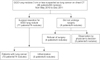
This study was a retrospective analysis of prospectively collected dataset at the Samsung Medical Center (a 1,960-bed university-affiliated, tertiary referral hospital in Seoul, South Korea) between May 2010 and December. In total, 72 patients with 74 pathology-proven adenocarcinomas, who had undergone surgical resection for curative intent, were included (Figure 1). In preoperative staging, all of the enrolled patients had GGNs of 3 cm or less in diameter on chest CT scans.
We reviewed the medical records (demographic data, history, operation reports, pathology reports, imaging studies, and clinical and pathologic TNM staging9 of all of the enrolled patients). The presence of recurrence or metastasis after surgical treatment, if any, was also recorded. Radiologically, the CT and PET/CT findings were also assessed in terms of GGN size, internal characteristics of the GGN, multiplicity, marginal characteristics, and maximum standardized uptake value (SUVmax). This study was approved by the Institutional Review Board of the Samsung Medical Center, which waived the requirement for informed consent from individual patients, given the retrospective nature of the study.
2. Imaging acquisition and analysis
Chest CT scans, PET/CT scans, and brain MRIs were performed using our standard protocols1011. GGNs were defined as a hazy increase in lung attenuation, which did not obscure the underlying vascular markings in the CT scans. The longest diameter of the tumor was measured. GGNs were classified into two subgroups in terms of their solidity: pure or part-solid GGNs12. A pure GGN was defined as a discrete pulmonary nodular abnormality with homogeneous attenuation that was not as high as that of the surrounding soft-tissue structures (e.g., blood vessels) or the encompassed lung. A part-solid GGN was defined as a lesion containing both ground-glass and solid soft-tissue attenuation components. Regarding marginal characteristics, the presence of a lobulated or spiculated margin was documented. A lobulated margin was defined when a portion of the surface of a lesion showed a scalloped configuration. A spiculated margin was defined as fine linear strands extending radially beyond a lesion10. The location of a tumor was considered to be central when it was located in the inner third of the lung field of a CT scan, and peripheral when in the outer two-thirds7.
3. Follow-up strategies for GGNs and initial preoperative staging
If a GGN displayed an internal solid portion or presented a diameter greater than 10 mm, video-assisted thoracoscopic surgery was initially considered. If a GGN was less than 10 mm in diameter and did not possess a solid portion, it was observed for at least 3 months to determine if it would resolve on its own. If the lesion remained stable, the CT examination was repeated after 6 months and then annually thereafter for a pure GGN, with follow-up examinations repeated every 3 months for the first year and every 6 months thereafter for a part-solid GGN. An increase or decrease in the size of a GGN was defined as a change of ≥2 mm from the size in the initial CT scan6. Surgical resections were planned for growing GGNs, those that showed new development, and those in which the internal solid portion grew, during the follow-up examination.
If surgical resection was necessary, staging work-ups of the relevant patients were undertaken according to the American Joint Committee on Cancer13. Preoperative work-ups included documenting clinical history, and clinical examinations (including neurological examinations), blood tests, standard chest X-ray, chest CT scan (including upper abdomen and adrenal glands), brain MRI, PET/CT, spirometry, and flexible bronchoscopy, as necessary. The determination of the clinical TNM stage was based on the chest CT scan, PET/CT, and brain MRI findings9. No patients underwent percutaneous fine needle aspiration or transbronchial biopsy via bronchoscopy.
4. Surgical procedures and pathologic evaluations
Data on the type of surgery, maximum tumor dimensions of the surgical specimens, and pathologic staging, were evaluated. A lobectomy was performed with systematic hilar and mediastinal lymph node dissection in principle.
An experienced lung pathologist interpreted the histopathologic findings. When multiple lesions were resected in a patient, they were considered to be synchronous multiple primary lung cancers, and were staged separately when there was no regional or distant lymph node metastasis14. The histologic classification of adenocarcinoma followed the International Association for the Study of Lung Cancer/American Thoracic Society/European Respiratory Society classification15. Each patient was assigned a final TNM stage based on the clinical, surgical, and pathologic data, together with an additional diagnostic work-up to evaluate the abnormal findings on PET/CT, and follow-up information according to the revised International System for Staging Lung Cancer9. All of the patients who underwent surgical resection were followed from the day of surgery, using a previously described method16.
5. Statistical analysis
Because the majority of data did not follow a normal distribution, the results are presented as the medians and interquartile ranges (IQRs) for continuous variables, and as numbers and percentages for categorical variables. The stage accuracy assessed by chest CT alone and combination modalities using chest CT, PET/CT, and brain MRI were calculated by standard definitions. Concordance over the final stage between the stage as assessed by CT alone, and that assessed by the combined modalities was compared to a kappa value. All of the tests were two-sided, and a p-value less than 0.05 was considered statistically significant. Data were analyzed using IBM SPSS Statistics version 19.0 (IBM Corp., Armonk, NY, USA).
Results
1. Clinical characteristics of the study patients
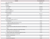
The demographic and clinical characteristics of the enrolled patients are listed in Table 1. The median age of the enrolled patients was 59 years, and 39 patients (54.2%) were male. Forty-two patients (58.3%) had never smoked. GGN was detected in chest CT scans of six patients who underwent chest CT for persistent respiratory symptoms, in 61 (84.7%) who underwent routine health screening, and in five (6.9%) who had no pulmonary symptoms but who underwent a chest CT examination as part of the follow-up of a previous malignancy.
A PET/CT scan and brain MRI were performed for all of the enrolled patients. Any suspicious metastatic lesions were not detected in brain MRIs. However, an incidental abnormality in PET/CT scans was observed in 11 patients, including the thyroid (n=7, but only three [4.2%] in the focal uptake), adrenal gland (n=2), colon (n=1), and mediastinum (n=1). Among these, only two patients had a thyroid malignancy. The absence of metastasis or malignancy was proven by adrenal CT (n=2) and negative biopsy results (n=8) (Table 1).
2. Radiographic characteristics and preoperative stage based on clinical imaging of each nodule
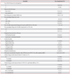
Of the enrolled 72 patients with 74 pathologically confirmed malignant GGNs, 70 had a solitary GGN and two had two nodules. The median GGN diameter was 18 mm on chest CT scans. The GGN margin was lobulated or spiculated in 25 (33.8%). The numbers of pure and part-solid GGNs were 35 (47.3%) and 39 (52.7%), respectively. Most GGNs (86.5%) were located in a peripheral location, with the most common sites for the lesions being the right upper and right lower lobes. For 13 nodules, the PET/CT scan was interpreted as an unidentifiable fluoro-2-deoxy-D-glucose uptake. In addition, we were not able to calculate the SUVmax in six patients with six nodules, because they underwent the PET/CT scan at another hospital. Therefore, the median SUVmax for the remaining 68 GGNs was 1.65 (IQR, 1.03–2.55) (Table 2).
In two patients with two GGNs, the GGN characteristics in images differed between the two GGNs, and there was no regional or distant lymph node metastasis on radiological imaging. Therefore, these GGNs were considered to be synchronous primary lung cancers and were staged separately. On the basis of the chest CT alone, all of the GGNs were categorized as stage IA. However, the preoperative evaluation of nodal and distant metastasis by PET/CT provided a false-positive N2 in one case and adrenal metastasis in two cases. Therefore, the preoperative stage was classified as stage IA in 71 nodules (95.9%), stage IIIA in one (1.4%), and stage IV in two (2.7%), with the addition of PET/CT scan and brain MRI (Table 2).
3. Surgical procedure and pathologic stage
The median CT follow-up period was 125 days (range, 28–625 days) in 55 patients (76.4%). The CT follow-up findings revealed that the GNN grew in size in 37 patients, with a progression of the internal solid portion in 20, and was stable in four patients.
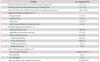
All of the enrolled patients underwent integrated PET/CT and brain MRI within a median of 14 days before resection. Pathologic specimens were obtained by lobectomy in 60 patients (83.8%), by wedge resection in 11 (15.3%), and by segmentectomy in one patient (1.4%). Most of the GGNs were histologically diagnosed as invasive adenocarcinoma (n=53, 71.6%) (Table 3). None of the tumors displayed lymph node involvement or lymphatic invasion, and only one tumor showed visceral pleural involvement. No procedurally related mortality or morbidity was identified.
The final stage of the 74 GGNs, after surgery and additional diagnostic work-up, was stage IA in 70 patients (94.6%), stage IB in three (4.1%), and stage IIA in one (1.4%) (Table 3). Only one patient with a pathologic stage IIA was treated with adjuvant chemotherapy. Neither local recurrence nor a distant metastasis occurred in any of the 72 patients during the follow-up period (median, 710 days; IQR, 508–818 days).
4. Radiology and pathology correlation
This study included 35 pure and 39 part-solid GGNs. The median longitudinal diameter of solid component in patients with part-solid GGN was 7 mm (IQR, 5–14) (Table 2). Solid component measuring >5 mm was found in 29 of 39 part-solid GGNs (74.4%). Final pathological diagnosis of 35 pure GGNs included nine adenocarcinoma in situ, four minimally invasive adenocarcinoma, and 22 invasive adenocarcinomas (invasive component >0.5 cm in great dimension). Final pathological diagnosis of 39 part-solid GGNs included one adenocarcinoma in situ, seven minimally invasive adenocarcinoma, and 31 invasive adenocarcinomas
5. Comparisons of the concordance between CT alone and the combined modalities in the final stage
In the final stage, as determined by surgery and additional diagnostic work-up for the incidentally abnormal findings on PET/CT scan, four cases were upstaged to IB (three patients were changed from T1b to T2a) and IIA (one patient with visceral pleural involvement) following surgery, compared to the preoperative staging assessed by both chest CT alone and the combined staging modalities including chest CT, PET/CT, and brain MRI. However, three cases were downstaged to IA from stage IIIA (one with a false-positive mediastinal lesion confirmed by surgery) and from stage IV (two cases with a benign adrenal adenoma confirmed by an adrenal CT scan), as assessed by the combined modalities (Table 4). Therefore, the accuracy in stage determination was higher for chest CT alone than for the combined modality of PET/CT and brain MRI, and similar findings were also seen in the T (extent of the primary tumor) status and N (regional lymph node) descriptor (Table 4). In addition, these combined modalities required unwarranted evaluation for incidental findings in several cases, compared to staging with chest CT alone (Tables 1, 4). Furthermore, the concordance between chest CT alone and the combined modalities of PET/CT and brain MRI did not statistically differ (kappa value, 0.712).
Discussion
We performed a comparative study of the accuracy in staging between chest CT alone and a combination of modalities including chest CT, PET/CT, and brain MRI, in patients with GGN lung adenocarcinomas. No mediastinal or distant metastasis was observed in these patients. A false-positive interpretation of mediastinal (n=1) and adrenal (n=2) lesions, and two thyroid malignancies, were incidentally found with a PET/CT scan.
Our results for mediastinal metastasis are similar to a previous study17 but are lower than the results from two other studies1819, whose values ranged from 2.3% to 2.9%. Similar to our results, distant metastasis was not reported in any of the studies, although preoperative staging work-ups with PET/CT scan were performed in only one study (only 34% of the enrolled patients)19, and brain MRI was not used in any study. In contrasts, we performed both PET/CT and brain MRI in all of our enrolled patients, and could determine the clinical roles of PET/CT and brain MRI in staging GGN lung adenocarcinomas, compared to previous studies171819. Our study showed that there were no statistically significant differences for the determination of overall stage in patients with ground-glass nodular lung adenocarcinomas, between chest CT alone and a combination of modalities including chest CT, PET/CT, and brain MRI. Therefore, the patients with GGN lung adenocarcinomas with stage IA on chest CT scan seems to be suitable for omitting PET/CT scans and brain MRIs to avoid increasing cost caused by the unwarranted additional tests for false-positive interpretation by PET/CT or brain MRI, considering low prevalence of mediastinal nodal and distant metastases3420.
In our study, an incidental uptake on PET/CT was detected in 10 cases, including the thyroid (n=7, 9.7% but only 3 [4.2%] in focal uptake), adrenal gland (n=2, 2.8%), and colon (n=1, 1.4%). Among them, only two cases were confirmed to be thyroid cancer. Because PET/CT is widely used in staging and the metastasis work-up of patients with LCA, the routine use of PET/CT significantly increases the probability of detecting unexpected diseases unrelated to the primary neoplasm. Similar to our study, incidental focal uptake in the thyroid gland was observed in 4.3% of patients who had undergone initial NSCLC staging, and 66% of these were confirmed as thyroid cancer21. In other studies, incidental colonic uptake with a clinical significance was observed in 1.1%–3.4% of patients, and approximately 35%–56% of these represented significant pathology unrelated to LCA2223. Because more than 50% of the incidental thyroid and colonic uptake lesions on PET/CT during the initial NSCLC staging had a diagnosis with clinical significance, additional evaluations should be considered, although metastasis from LCA to the thyroid or colon is rare (less than 1%)2425, and it in general, neither influences treatment decisions nor alters the prognosis. However, adrenal metastases from LCA are not uncommon. About 4% of patients with otherwise operable NSCLC have a unilateral adrenal mass, and up to 40% of these may be malignant and present as a solitary site of metastasis26. Because the detection of adrenal metastasis from operable NSCLC substantially changes the stage, adrenal biopsy or other secondary imaging is recommended to accurately characterize adrenal masses27. In this study, two patients had adrenal uptake on PET/CT, but additional evaluation using an adrenal CT scan suggested the possibility of adrenal adenoma.
To fully appreciate our results, the limitations of this study must be acknowledged. The major limitation was the potential for selection bias. This was the retrospective study of prospectively collected dataset performed on a small number of patients from a single center. Thus, these factors may limit the generalizability of our findings to other institutions or patient populations. The conclusion of this study should be interpreted conservatively. Furthermore, the duration of our follow-up CT was heterogeneous and our study was conducted at a university-affiliated tertiary referral hospital with a comprehensive cancer center that had a large number of cancer patients referred from other hospitals. Therefore, the initial presumptive diagnosis of LCA might have easily been made and any minimal change in the GGN was rapidly detected. Moreover, the surgical approach was also more easily decided. Finally, we included both pure and part-sold GGNs in this study. However, part-solid GGNs may have more aggressive clinical courses than pure GGNs. Therefore, a large-scale prospective study for patients with part-solid GGNs is required to elucidate the role of preoperative PET/CT and brain MRI in the future.
In conclusion, PET/CT and brain MRI have no additional benefit for the staging of patients with GGN lung adenocarcinoma prior to surgery. Therefore, routine PET/CT scans and brain MRIs do not seem to be mandatory for staging GGNs, considering the indolent nature of GGNs and the need for unwarranted additional tests for false-positive interpretation with PET/CT and brain MRI.
 XML Download