Journal List > Ewha Med J > v.41(3) > 1099542

Kwak, Kim, Ban, Sohn, and Hong: Comparison of Clinical Manifestation and Laboratory Findings between Adenoviral Infection with or without Kawasaki Disease

Abstract

Objectives

Adenovirus infection, which has been known to mimic Kawasaki disease (KD), is one of the most frequent conditions observed during differential diagnosis when considering KD. Accordingly, it is essential to being able to differentiate between these two diseases. Therefore, we performed multiplex reverse transcriptase- polymerase chain reaction and tissue-Doppler echocardiography to distinguish between adenovirus patients and KD patients.

Methods

A total of 113 adenoviral infection patients (female 48, male 65) diagnosed from January 2010 to June 2016 were evaluated. We divided adenoviral infection patients into two groups: group 1, which consisted of individuals diagnosed with KD according to the KD American Heart Association criteria (n=62, KD with adenovirus infection); and group 2, which comprised individuals only diagnosed with adenovirus infection (n=51). Laboratory data were obtained from each patient including N-terminal pro-brain natriuretic peptide. Echocardiographic measurements were compared between two groups. In addition, reverse transcriptase-polymerase chain reaction was performed using nasopharyngeal secretions to diagnose adenoviral infection.

Results

Conjunctival injection, cervical lymphadenopathy, polymorphous skin rash, abnormalities of the lip or oral mucosa and abnormalities of extremities were significantly higher in group 1 than group 2. Moreover, group 1 had significantly higher C-reactive protein and alanine aminotransferase levels, as well as lower platelet counts and albumin levels than group 2. Coronary artery diameter was significantly greater in group 1 than group 2.

Conclusion

In patients with adenoviral infection with unexplained prolonged fever, echocardiography and C-reactive protein can be used to differentiate KD with adenoviral infection from adenoviral infection alone.

References

1. McCrindle BW, Rowley AH, Newburger JW, Burns JC, Bolger AF, Gewitz M, et al. Diagnosis, treatment, and long-term management of Kawasaki disease: a scientific statement for health professionals from the American Heart Association. Circulation. 2017; 135:e927–e999.
crossref
2. Newburger JW, Takahashi M, Gerber MA, Gewitz MH, Tani LY, Burns JC, et al. Diagnosis, treatment, and long-term management of Kawasaki disease: a statement for health professionals from the Committee on Rheumatic Fever, Endocarditis, and Kawasaki Disease, Council on Cardiovascular Disease in the Young, American Heart Association. Pediatrics. 2004; 114:1708–1733.
crossref
3. Newburger JW, Takahashi M, Burns JC, Beiser AS, Chung KJ, Duffy CE, et al. The treatment of Kawasaki syndrome with intravenous gamma globulin. N Engl J Med. 1986; 315:341–347.
crossref
4. Burns JC, Glode MP. Kawasaki syndrome. Lancet. 2004; 364:533–544.
crossref
5. Jordan-Villegas A, Chang ML, Ramilo O, Mejias A. Concomitant respiratory viral infections in children with Kawasaki disease. Pediatr Infect Dis J. 2010; 29:770–772.
crossref
6. Rowley AH, Baker SC, Shulman ST, Garcia FL, Fox LM, Kos IM, et al. RNA-containing cytoplasmic inclusion bodies in ciliated bronchial epithelium months to years after acute Kawasaki disease. PLoS One. 2008; 3:e1582.
crossref
7. Okano M, Thiele GM, Sakiyama Y, Matsumoto S, Purtilo DT. Adenovirus infection in patients with Kawasaki disease. J Med Virol. 1990; 32:53–57.
crossref
8. Esper F, Shapiro ED, Weibel C, Ferguson D, Landry ML, Kahn JS. Association between a novel human coronavirus and Kawasaki disease. J Infect Dis. 2005; 191:499–502.
crossref
9. Shike H, Shimizu C, Kanegaye JT, Foley JL, Schnurr DP, Wold LJ, et al. Adenovirus, adeno-associated virus and Kawasaki disease. Pediatr Infect Dis J. 2005; 24:1011–1014.
crossref
10. Chang LY, Lu CY, Shao PL, Lee PI, Lin MT, Fan TY, et al. Viral infections associated with Kawasaki disease. J Formos Med Assoc. 2014; 113:148–154.
crossref
11. Song E, Kajon AE, Wang H, Salamon D, Texter K, Ramilo O, et al. Clinical and virologic characteristics may aid distinction of acute adenovirus disease from Kawasaki disease with incidental adenovirus detection. J Pediatr. 2016; 170:325–330.
12. Ferone EA, Berezin EN, Durigon GS, Finelli C, Felicio MC, Storni JG, et al. Clinical and epidemiological aspects related to the detection of adenovirus or respiratory syncytial virus in infants hospitalized for acute lower respiratory tract infection. J Pediatr (Rio J). 2014; 90:42–49.
crossref
13. Rocholl C, Gerber K, Daly J, Pavia AT, Byington CL. Adenoviral infections in children: the impact of rapid diagnosis. Pediatrics. 2004; 113(1 Pt 1):e51–e56.
crossref
14. Fukuda S, Ito S, Fujiwara M, Abe J, Hanaoka N, Fujimoto T, et al. Simultaneous development of Kawasaki disease following acute human adenovirus infection in monozygotic twins: a case report. Pediatr Rheumatol Online J. 2017; 15:39.
crossref
15. Turnier JL, Anderson MS, Heizer HR, Jone PN, Glode MP, Dominguez SR. Concurrent respiratory viruses and Kawasaki disease. Pediatrics. 2015; 136:e609–e614.
crossref
16. Japan Kawasaki Disease Research Committee. Report of subcommittee on standardization of diagnostic criteria and reporting of coronary artery lesions in Kawasaki disease. Tokyo: Ministry of Health and Welfare; 1984.
17. Lee SB, Choi HS, Son S, Hong YM. Cardiac function in Kawasaki disease patients with respiratory symptoms. Korean Circ J. 2015; 45:317–324.
crossref
18. Jaggi P, Kajon AE, Mejias A, Ramilo O, Leber A. Human adenovirus infection in Kawasaki disease: a confounding bystander? Clin Infect Dis. 2013; 56:58–64.
crossref
19. Kim JH, Yu JJ, Lee J, Kim MN, Ko HK, Choi HS, et al. Detection rate and clinical impact of respiratory viruses in children with Kawasaki disease. Korean J Pediatr. 2012; 55:470–473.
crossref
20. Heim A, Ebnet C, Harste G, Pring-Akerblom P. Rapid and quantitative detection of human adenovirus DNA by real-time PCR. J Med Virol. 2003; 70:228–239.
crossref
21. Edwards KM, Thompson J, Paolini J, Wright PF. Adenovirus infections in young children. Pediatrics. 1985; 76:420–424.
crossref
22. Tabain I, Ljubin-Sternak S, Cepin-Bogovic J, Markovinovic L, Knezovic I, Mlinaric-Galinovic G. Adenovirus respiratory infections in hospitalized children: clinical findings in relation to species and serotypes. Pediatr Infect Dis J. 2012; 31:680–684.
23. Colvin JM, Muenzer JT, Jaffe DM, Smason A, Deych E, Shannon WD, et al. Detection of viruses in young children with fever without an apparent source. Pediatrics. 2012; 130:e1455–e1462.
crossref
24. Barone SR, Pontrelli LR, Krilov LR. The differentiation of classic Kawasaki disease, atypical Kawasaki disease, and acute adenoviral infection: use of clinical features and a rapid direct fluorescent antigen test. Arch Pediatr Adolesc Med. 2000; 154:453–456.
25. Kawasaki Y, Hosoya M, Katayose M, Suzuki H. Correlation between serum interleukin 6 and C-reactive protein concentrations in patients with adenoviral respiratory infection. Pediatr Infect Dis J. 2002; 21:370–374.
crossref

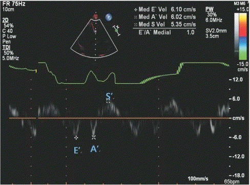
Fig. 1.
Myocardial velocity by tissue Doppler imaging in Kawasaki disease patient. E’, early diastolic myocardial velocity; A’, late diastolic myocardial velocity; S’, systolic myocardial velocity.
emj-2018-41-3-45f1.tif
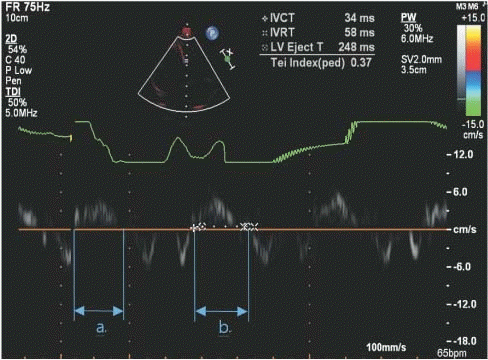
Fig. 2.
Tei index by tissue Doppler imaging in Kawasaki disease patient. Tei index=(a-b)/b=(IVCT+IVRT)/LVET. IVCT, isovolumetric contraction time; IVRT, isovolumetric relaxation time; LVET, left ventricle ejection time.
emj-2018-41-3-45f2.tif
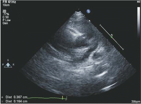
Fig. 3.
Right coronary artery dilatation by echocardiography in Kawasaki disease patient.
emj-2018-41-3-45f3.tif
Table 1.
Comparison of clinical characteristics between group 1 and group 2
Clinical characteristics Group 1 (n=62) Group 2 (n=51) P-value
Boys 37 (59.7) 28 (54.9) 0.609
Girls 25 (40.3) 23 (45.1) 0.703
Age (year) 4 (2–4) 3 (1–4) 0.045
Fever duration (>5 days) 38 (61.3) 26 (51.0) 0.271
Conjunctival injection 39 (62.9) 16 (31.4) 0.001
Cervical lymphadenopathy 20 (32.3) 2 (3.9) <0.001
Polymorphous skin rash 22 (35.5) 1 (2.0) <0.001
Abnormalities of lip or oral mucosa 19 (30.6) 2 (3.9) <0.001
Abnormalities of extremities 8 (12.9) 1 (2.0) 0.039

Values are presented as number (%) or number (range).

P value obtained from the Mann-Whitney test.

P value obtained from the Fisher’s exact test.

Group 1, KD with adenoviral infection; Group 2, adenoviral infection.

Table 2.
Comparison of laboratory data between two groups
Laboratory data Group 1 (n=62) Group 2 (n=51) P-value
Hb (g/dL) 11.4 (11.0–12.1) 11.8 (11.2–12.3) 0.116
WBC (/μL) 9,570 (7,417–12,677) 10,030 (7,500–12,440) 0.892
Neutrophil (%) 59.1 (49.9–65.7) 57.0 (46.5–67.0) 0.604
Platelet (x109/L) 230.0 (193.0–282.2) 257.0 (215.0–302.0) 0.044
ESR (mm/hr) 36.0 (26.0–49.5) 31.0 (18.5–41.0) 0.063
CRP (mg/dL) 5.65 (2.81–7.66) 2.52 (1.09–5.12) <0.001
AST (IU/L) 26.5 (17.5–32.3) 32.0 (28.0–35.0) <0.001
ALT (IU/L) 16.5 (13.0–29.3) 15.0 (12.0–18.0) 0.044
Total protein (g/dL) 6.5 (6.3–6.7) 6.5 (6.1–6.7) 0.858
Albumin (g/dL) 3.7 (3.6–3.9) 3.8 (3.7–4.0) 0.006
NT-pro BNP (pg/mL) 192.0 (82.5–316.5) 144.5 (144.0–220.0) 0.089

Values are expressed as the mean (range).

Group 1, KD with adenoviral infection; Group 2, adenoviral infection.

Hb, hemoglobin; WBC, white blood cell count; ESR, erythrocyte sedimentation rate; ALT, alanine aminotransferase; AST, aspartate aminotransferase; CRP, C-reactive protein; NT-proBNP, N-terminal pro-brain natriuretic peptide; RCA, right coronary artery.

Table 3.
Comparison of echocardiographic finding between two groups
Echo findings Group 1 (n=62) Group 2 (n=51) P value
RCA (mm) 2.30 (1.75–3.95) 1.70 (1.30–2.00) <0.001
LCA (mm) 1.80 (1.50–2.00) 1.60 (1.50–1.90) 0.067
EF (%) 69.45 (65.25–75.10) 71.55 (65.08–76.73) 0.392
FS (%) 38.15 (35.10–42.75) 39.60 (35.03–44.20) 0.423
TDI
 E’ (cm/sec) 1.80 (1.50–2.00) 9.73 (8.68–11.13) 0.020
 A’ (cm/sec) 69.45 (65.25–75.10) 4.56 (3.86–6.52) 0.805
 S’ (cm/sec) 38.15 (35.10–42.75) 5.47 (4.91–6.34) 0.066
Tei index 1.80 (1.50–2.00) 0.43 (0.38–0.47) 0.302
 IVCT (msec) 69.45 (65.25–75.10) 66.00 (54.00–79.00) 0.277
 IVRT (msec) 38.15 (35.10–42.75) 48.00 (41.00–58.00) 0.834
 LVET (msec) 1.80 (1.50–2.00) 277.00 (252.50–298.00) 0.696

Values are expressed as the mean (range).

Group 1, KD with adenoviral infection; Group 2, adenoviral infection.

RCA, right coronary artery; LCA, left coronary artery; EF, ejection fraction; FS, fractional shortening; TDI, tissue Doppler imaging; E', early diastolic myocardial velocity; A', late diastolic myocardial velocity; S', systolic myocardial velocity; IVCT, isovolumetric contraction time; IVRT, isovolumetric relaxation time; LVET, left ventricle ejection time.

TOOLS
Similar articles