

 PDF
PDF ePub
ePub Citation
 Print
Print


Persistent left superior vena cava (PLSVC) is a congenital thoracic venous malformation. In most cases, it drains into the right atrium via the coronary vein without causing any clinical manifestations. Very rarely, however, it may drain into the left atrium directly or via the unroofed coronary sinus, producing a right-to-left shunt, putting the patient at high risk of hemodynamic instability, syncope, embolism, or brain abscess.123 We report a case of a persistent left superior vena cava draining into the left atrium in a patient with left pontine infarction, which was not detected by transcranial Doppler ultrasonography (TCD) using agitated saline test with microbubbles from right antecubital vein , but was diagnosed on contrast transesophageal echocardiography (TEE) and computed tomography (CT).
CASE
A 77-year-old right-handed woman presented to the emergency room with symptoms of right-sided weakness and dysarthria that had occurred 5 hours before admission. Her vital signs on admission were: BP 110/70 mmHg, pulse 88 beats/min, and temperature 36.6℃. She was diagnosed with hypertension 10 years ago. Her social and family history was unremarkable. Neurological examination performed in the emergency room yielded the following findings: dysarthria, right-side central facial palsy, and grade 4 muscle strength in both, the right upper and lower extremities. The National Institutes of Health Stroke Scale (NIHSS) score on admission was 4.
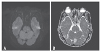
The brain magnetic resonance imaging performed on the day of admission showed high signal intensity in the left upper pons (Fig. 1), but magnetic resonance angiography did not reveal any remarkable segmental stenosis of the intracranial vessels. Thoracic and abdominal examination revealed no visible abnormalities, no palpable masses in the neck, and no abnormal sound in the carotid artery and abdominal aorta. Chest X-ray showed mild cardiomegaly and aortic arch dilation.
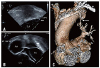
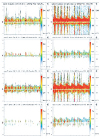
No intracranial stenosis or microthrombosis was observed on the TCD performed on the third day of admission to identify the cause of infarction, and no right-to-left shunt was detected in the test performed after injection of saline with microbubbles through the right antecubital vein. No arrhythmia was noted in the 24 hours ECG performed on the fourth day of stroke unit admission, and transthoracic echocardiography and TTE performed on the seventh day of admission revealed no abnormalities in the left ventricular ejection fraction and the ventricular, atrial, and coronary sinus sizes. There was no arterial septal defect or patent foramen ovale, which is associated with a thrombus or an intracardiac shunt. However, a large number of contrast bubbles were visible in the left atrium in the TEE bicaval view after injection of agitated saline with microbubbles through the left antecubital vein. No shunt associated with atrial septal defect was observed even under Valsalva maneuver during microbubble contrast study using agitated saline. However, it was observed that microbubbles drained through the left pulmonary vein (Fig. 2A, B). A chest CT scan showed that a persistent left superior vena cava was directly connected to the left atrium via the left superior pulmonary vein (Fig. 2C). During the TCD performed on the ninth day of admission, after injection of agitated saline with microbubbles through the left antecubital vein, a curtain of microembolic signals was observed (Fig. 3). Most of the neurologic symptoms except for dysarthria resolved and the patient was discharged on the ninth day with NIHSS score 2. Although surgery or coil embolization was planned to prevent the cerebral infarction due to paradoxical embolism cause by right-to-left shunt, it was decided to put her under medication with aspirin 100 mg and clopidogrel 75 mg, considering the patient's age and her own wishes.
DISCUSSION
This case is a patient of persistent left superior vena cava (PLSCV) which was not detected right to left shunt on TCD with saline agitated test from right antecubital vein. But RLS was detected on TEE and TCD with saline agitated test from left antecubital vein.
PLSVC is a thoracic venous malformation found in approximately 10% of patients with congenital heart disease. It is a rare disease with a prevalence of 0.3–0.5% in the general population.4 In over 90% of PLSVC cases, it drains into the right atrium through the coronary sinus and does not form a RLS or lead to hemodynamic problems. Affected patients are at higher risk of air embolism associated with intravenous injection as well as stroke or intracranial abscess associated with septic embolism or thromboembolism.5
TCD and TEE using agitated saline with microbubbles are efficient techniques for confirming the presence of RLS. In the presence of RLS, bubbles drain directly to the left atrium without passing through the lungs, and micro-embolic signals are observed in both left atrium and cerebral blood flow.6
The guideline about contrast saline agitation test gives no detailed information for the side of vein.7 But, usually, when performing TEE, the right antecubital vein is preferred by many hospitals and medical staff for convenient access because patients usually take the left lateral decubitus position. As demonstrated in the case presented, if PLSVC drains into the left atrium, it may not be discovered when agitated saline with microbubbles is injected through the right antecubital vein. Although the importance of injecting into the left arm during saline agitated studies has been discussed in the cardiac imaging literature.89 The issue has received little attention in the neurology literature and therefore may be less well recognized by neurologists who are evaluating patients which is suspected of cryptogenic stroke or presence of RLS. It is important to adhere to the correct protocol for infusion of saline with microbubbles through the left brachial vein and contrast bubbles, which is essential in accurately diagnosing rare cases such as PLSVC presented in this report.
 XML Download