초록
Studies of interictal epileptiform discharges are essential for improving the diagnosis, classifi-cation, and management of epilepsy. In this case series we sought to identify the clinical and neurophysiological significance of bifid spikes, whose pattern bears a strong resemblance to the cardiac M pattern. We hypothesize that, analogous to the cardiac M pattern, the cerebral M pattern is generated by a conduction defect associated with asynchronous spatiotemporal averaging of electrical signals in the cortex, resulting in the signals reaching the scalp with different latencies. Unlike the cardiac M pattern, the pathology underlying the cerebral M pattern is unknown, although congenital CNS anomalies may be a culprit.
Go to : 
REFERENCES
1.Goldensohn ES., Purpura DP. Intracellular potentials of cortical neurons during focal epileptogenic discharges. Science. 1963. 139:840–842.

2.Maulsby RI. Some guidelines for assessment of spikes and sharp waves in EEG tracings. Am J EEG Technol. 1971. 11:3–16.

3.Ebersole JS. Defining epileptogenic foci: past, present, future. J Clin Neurophysiol. 1997. 14:470–483.

4.Olejniczak P. Neurophysiological basis of EEG. J Clin Neurophyiol. 2006. 23:186–189.
5.Daube JR., Stead SM. Basics of neurophysiology. Daube JR, Rubin DI, editors. Textbook of clinical neurophysiology. 3rd ed.New York: Oxford;2009. p. 69–96.

6.Janati AB., Umair M., Alghassab N., Al-Shurtan KS. Positive sharp waves in the EEG of children and adults. Neurol Sci. 2014. 35:707–713.

7.Janati AB., Alghassab N., Umair M. Focal triphasic sharp waves and spikes in the electroencephalogram. Neurol Sci. 2015. 36:221–226.

8.Janati AB., AlGhasab NS., Alshammari RA., saad AlGhassab A., Al-Aslami Yossef Fahad. Sharp slow waves in the EEG. Neurodi-agn J. 2016. 56:83–94.

9.Hashiguchi K., Morioka T., Yoshida F., Miyagi Y., Nagata S., Sakata A, et al. Correlation between scalp-recorded electroencephalo-graphic and electrographic activities during ictal period. Seizure. 2007. 16:238–247.
10.Da Costa D., Brady WJ., Edhouse J. Bradycardias and atrioventricu-lar conduction block. BMJ. 2002. 2:535–538.
11.Shimomura J., Kubota Y., Tottori T., Matsuda K., Mihara T., Fujiwara T, et al. Surgery for catastrophic epilepsy with temporo-parieto-oc-cipital cortical dysplasia: clinical features and surgical outcome of three patients. Epilepsia. 2000. 41(S9):67–68.
12.Gambardella A., Palmini A., Anderman F., Dubeau F., Da Costa JC., Quesney LF, et al. Usefulness of focal rhythmic discharges on scalp EEG of patients with focal cortical dysplasia and intractable epilepsy. Electroencephalogr Clin Neurophysiol. 1996. 98:243–249.

13.Trevelyan AJ., Sussillo D., Watson BO., Yuste R. Modular propagation of epileptiform activity: evidence for an inhibitory veto in neocortex. J Neurosci. 2006. 26:12447–12455.

14.Walker J. The propagation of epileptiform events across the corpus callosum [Thesis]. Middletown (CT): Wesleyan University;2009 May. p. 58.
Go to : 
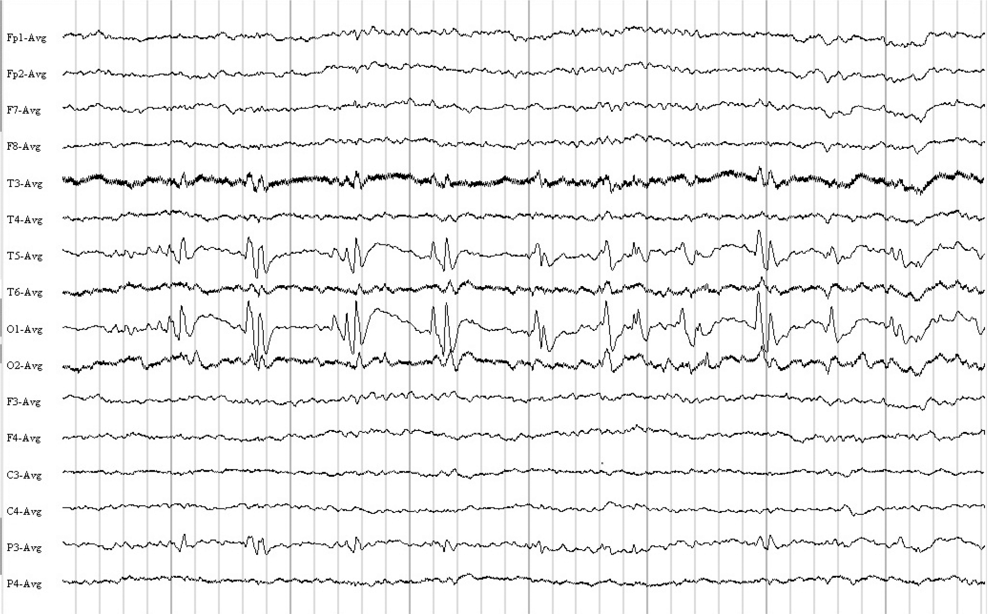 | Fig. 1.Nine-year-old female with partial motor (right-sided) seizures with secondary generalization. Patient asleep. The EEG shows quasi-pe-riodic "bifid spikes") in the left occipital-temporal area. EEG, electroencephalogram. |
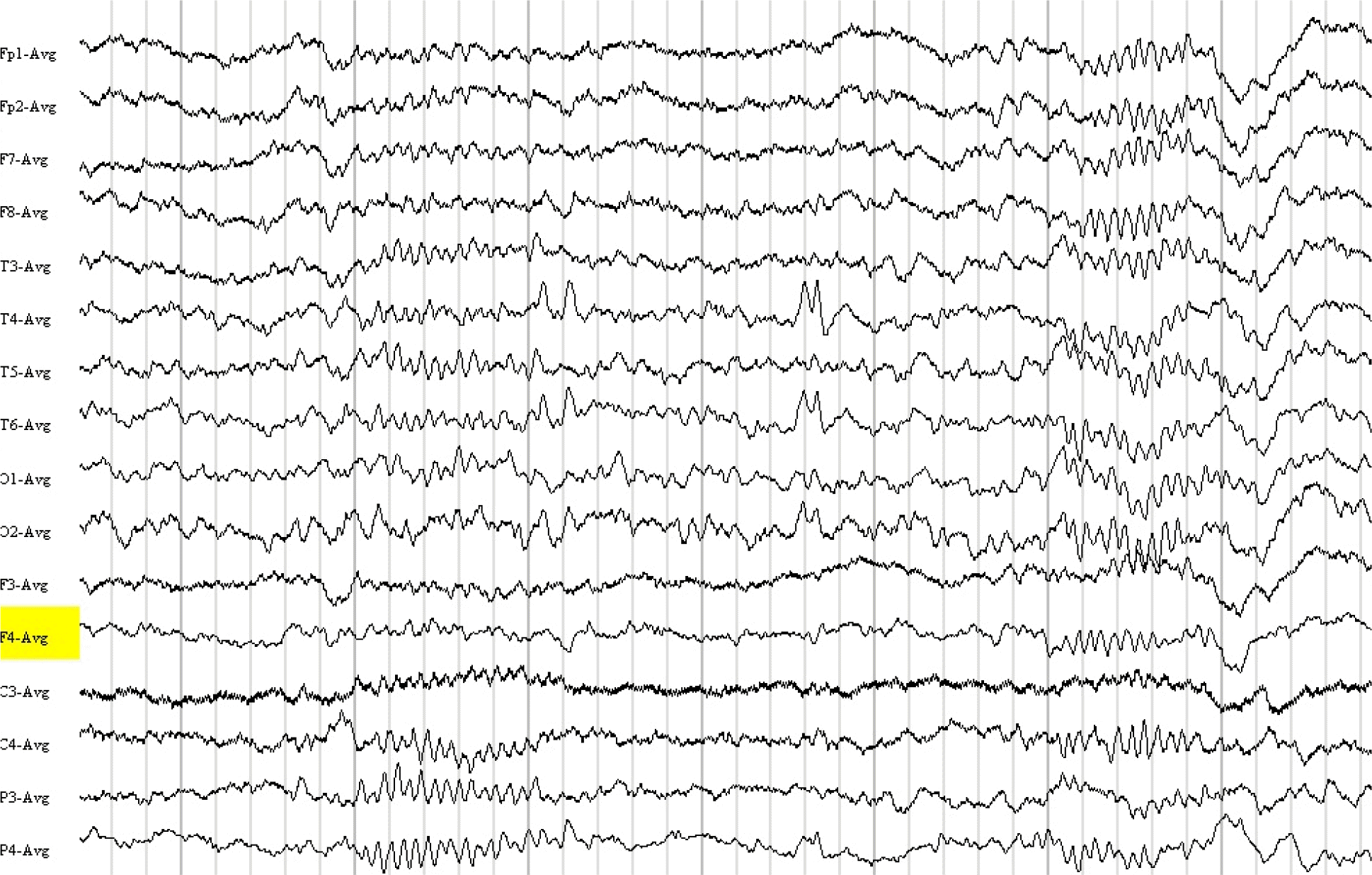 | Fig. 2.Thirteen-year-old male with generalized convulsive epileptic seizures preceded by fear. Patient asleep. The EEG sample shows "bifid spikes" in the right temporal-occipital region. EEG, electroencephalogram. |
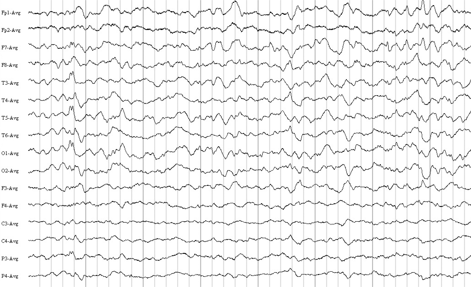 | Fig. 3.The patient is a 30-year-old female with mental retardation and generalized convulsive epileptic seizures. Patient asleep. The EEG shows "bifid spikes" in the left temporal-occipital region. Intermixed are focal, at times "notched" delta in the left temporal region. EEG, electroencephalogram. |
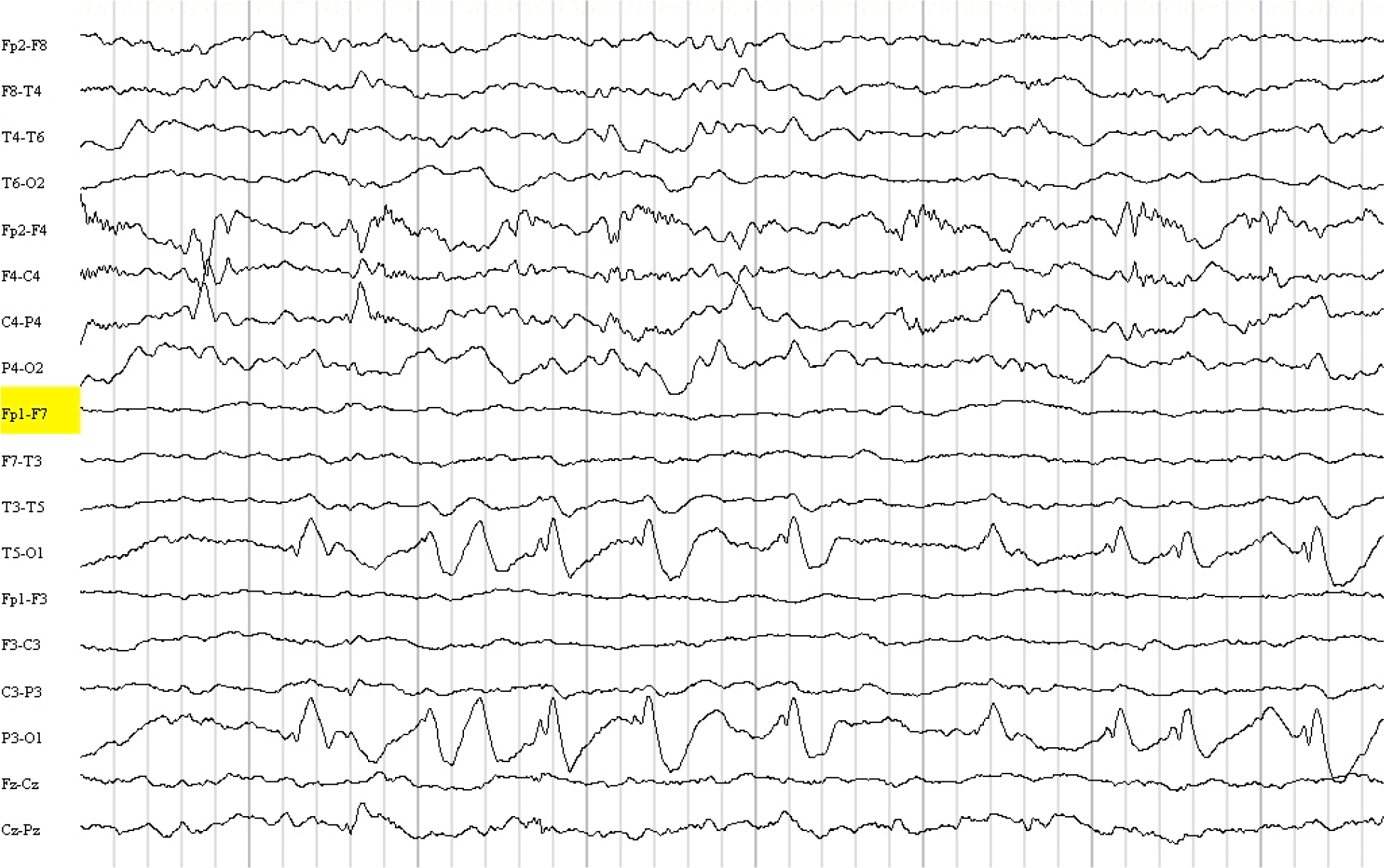 | Fig. 4.Two-year-old female with psychomotor delay and multiple congenital anomalies. The EEG shows trains of "bifid spikes" in the left occipital region. Note depression of left hemispheric background and sleep parameters. EEG, electroencephalogram. |
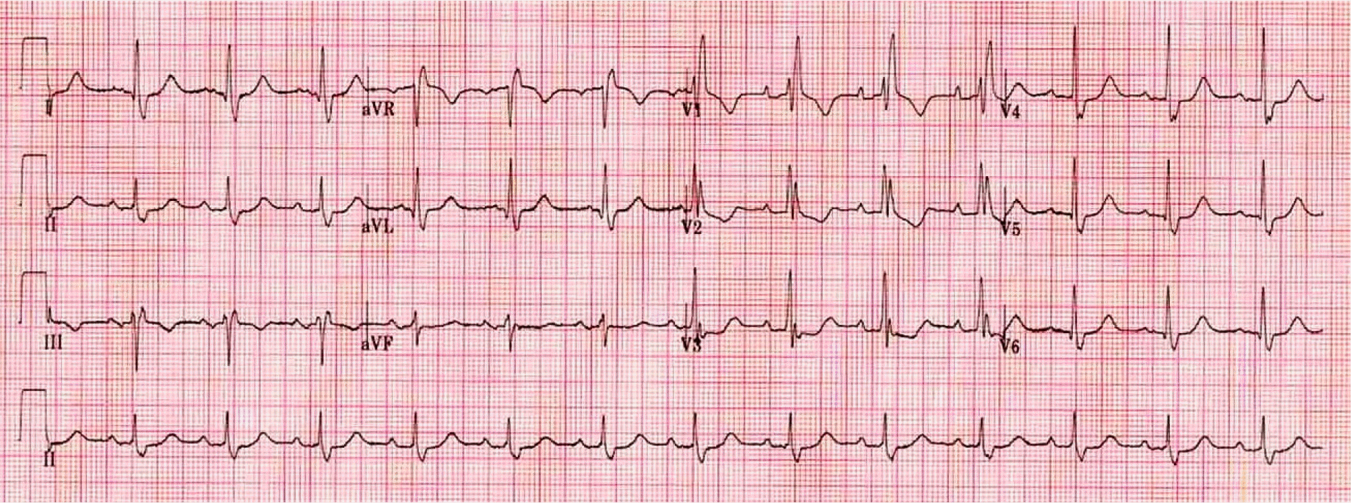 | Fig. 5.EKG showing cardiac M-wave pattern which represent RBBB in the chest leads (V1-V2). EKG, electroencephalogram; RBBB, right bundle branch block. |
Table 1.
Pertinent data in four patients with EEG bifid spikes
EEG, electroencephalogram; Neg., negative; GCS, generalized convulsive seizure; MR, mental retardation; VPA, valproic acid; N, normal; MTF, multifocal; LMT, lamotrigine; PCS, partial complex seizure; CBZ, carbamazepine; IED, interictal epileptiform discharge; R, right; T, temporal; SPS, simple partial seizure; DPH, phenytoin; L-OT, left occipital temporal region.


 PDF
PDF ePub
ePub Citation
 Print
Print


 XML Download