초록
Background
Previous studies of radial nerve conduction study (NCS) did not present how to measure the length of the radial nerve across the elbow, and did not even mention how to manage the spiral course of the nerve. This study aimed to applicate the most reliable method to measure the length of the radial nerve during NCS.
Methods
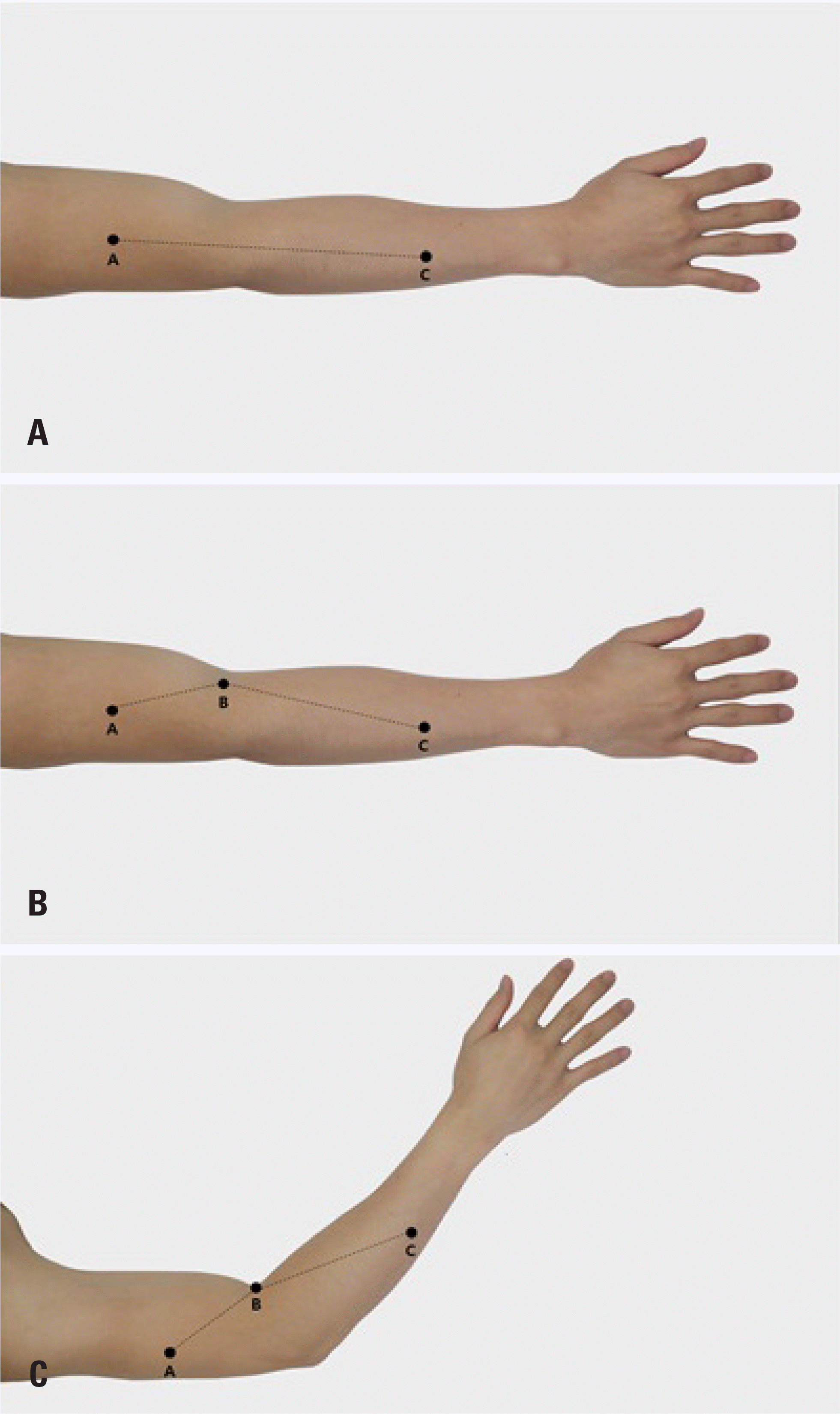
Three points (A, B, and C) were determined along the relatively straight course of the radial nerve. The distance was measured using three different methods: L1) straight distance corresponding to the A-C distance, L2) sum of the distances corresponding to the A-B-C distance, L3) based on the L2, but the elbow is flexed at a 45° angle. We compared the three methods of distance measurement and the calculated nerve conduction velocities (V1, V2, and V3) in normal healthy subjects.
Results
19 normal participants were enrolled. The mean value for method L1, L2 and L3 were 22.5 ± 1.8 cm, 24.0 ± 2.1 cm, and 23.2 ± 2.1 cm (p < 0.001). Calculated conduction velocities using those distance measurement methods as follows (p < 0.001): V1 (60.9 ± 2.7 m/s), V2 (64.6 ± 3.3 m/s), and V3 (63.4 ± 3.9 m/s). V2 was significantly greater than V1 and V3 (p < 0.001, p = 0.010, respectively).
Go to : 
REFERENCES
1.Kim BJ., Date ES., Lee SH., Yoon JS., Hur SY., Kim SJ. Distance measure error induced by displacement of the ulnar nerve when the elbow is flexed. Arch Phys Med Rehabil. 2005. 86:809–812.

2.Kim BJ., Koh SB., Park KW., Kim SJ., Yoon JS. Pearls & Oy-sters: false positives in short-segment nerve conduction studies due to ulnar nerve dislocation. Neurology. 2008. 70:e9–e13.
3.Checkles NS., Russakov AD., Piero DL. Ulnar nerve conduction velocity--effect of elbow position on measurement. Arch Phys Med Rehabil. 1971. 52:362–365.
4.Kimura J. Assessment of individual nerves. In: Electrodiagnosis in Diseases of Nerve and Muscle: Principles and Practice. 3rd ed.New York: Oxford University Press;2001. p. 130–177.
5.Apfelberg DB., Larson SJ. Dynamic anatomy of the ulnar nerve at the elbow. Plast Reconstr Surg. 1973. 51:79–81.

6.Harding C., Halar E. Motor and sensory ulnar nerve conduction velocities: effect of elbow position. Arch Phys Med Rehabil. 1983. 64:227–232.
7.Schubert H., Malin H. Radial nerve motor conduction. Am J Phys Med Rehabil. 1967. 46:1345–1350.
8.Strachan JC., Ellis BW. Vulnerability of the posterior interosseous nerve during radial head resection. J Bone Joint Surg Br. 1971. 53:320–323.

9.Kim KH., Park KD., Chung PW., Moon HS., Kim YB., Yoon WT, et al. The usefulness of proximal radial motor conduction in acute compressive radial neuropathy. J Clin Neurol. 2015. 11:178–182.

10.Jebsen RH. Motor conduction velocity of distal radial nerve. Arch Phys Med Rehabil. 1966. 47:12–16.
11.Di Benedetto M. Posterior interosseus branch of the radial nerve: conduction velocities. Arch phys Med Rehabil. 1972. 53:266–271.
12.Kim JG., Kim D., Seok HY., Kim Y., Yang KS., Rhyu IJ, et al. A method of radial nerve length measurement based on cadaveric investi-gation. Arch Phys Med Rehabil 2016 Sep 6 [Epub].http://dx.doi.org/10.1016/j.apmr.2016.08.464.
13.Koo YS., Cho CS., Kim BJ. Pitfalls in using electrophysiological studies to diagnose neuromuscular disorders. J Clin Neurol. 2012. 8:1–14.

14.Humphries R., Currier D. Variables in recording motor conduction of the radial nerve. Phys Ther. 1976. 56:809–814.

15.Date ES., Teraoka JK., Chan J., Kingery WS. Effects of elbow flexion on radial nerve motor conduction velocity. Electromyogr Clin Neurophysiol. 2002. 42:51–56.
16.Trojaborg W., Sindrup E. Motor and sensory conduction in different segments of the radial nerve in normal subjects. J Neurol Neurosurg Psychiatry. 1969. 32:354–359.

17.Ma DM., Liveson JA. Nerve conduction handbook. Philadelphia: FA Davis;1983.
18.Fernand VS., Young JZ. The sizes of the nerve fibres of muscle nerves. Proc R Soc Lond B Biol Sci. 1951. 139:38–58.
19.Gassel MM., Diamantopoulos E. Pattern of conduction times in the distribution of the radial nerve. A clinical and electrophysiological study. Neurology. 1964. 14:222–231.

20.Kalantri A., Visser BD., Dumitru D., Grant AE. Axilla to elbow radial nerve conduction. Muscle Nerve. 1988. 11:133–135.

21.Dorfman LJ., Bosley TM. Age-related changes in peripheral and central nerve conduction in man. Neurology. 1979. 29:38–44.

Go to : 
 | Fig. 1.Illustration of the measurement of L1, L2, and L3. (A) Measurement L1 based on the A-C distance represents a linear measurement between the proximal and distal stimulation point. The elbow is extended fully. (B) Measurement L2 based on the A-B-C distance has a segmented course as contrasted to the single segment of L1. Point B is added on the elbow crease between the biceps brachii tendon and the lateral epicondyle. The elbow angle is the same as that of L1. (C) Measurement L3 is also based on the A-B-C distance, but the elbow is flexed at a 45° angle. |
Table 1.
Summary of previous radial nerve motor conduction studies involving a segment across the elbow


 PDF
PDF ePub
ePub Citation
 Print
Print


 XML Download