초록
Background
Routine nerve conduction study (NCS) can only be used to evaluate the function of large fibers, and the results of NCS are often normal in patients with distal sensory polyneuropathy. The measurement of the current perception threshold (CPT) has been reported to represent a variety of peripheral nerve fiber functions. This study was performed to investigate the value of measuring CPT in patients with diabetic sensory polyneuropathy who have no abnormalities in routine NCS.
Methods
Twenty-seven diabetic patients with sensory polyneuropathy and normal routine NCS and 18 age-matched control subjects participated in this study. The CPT was measured on the unilateral index finger and great toe of each subject at frequencies of 5 Hz, 250 Hz, and 2,000 Hz.
Results
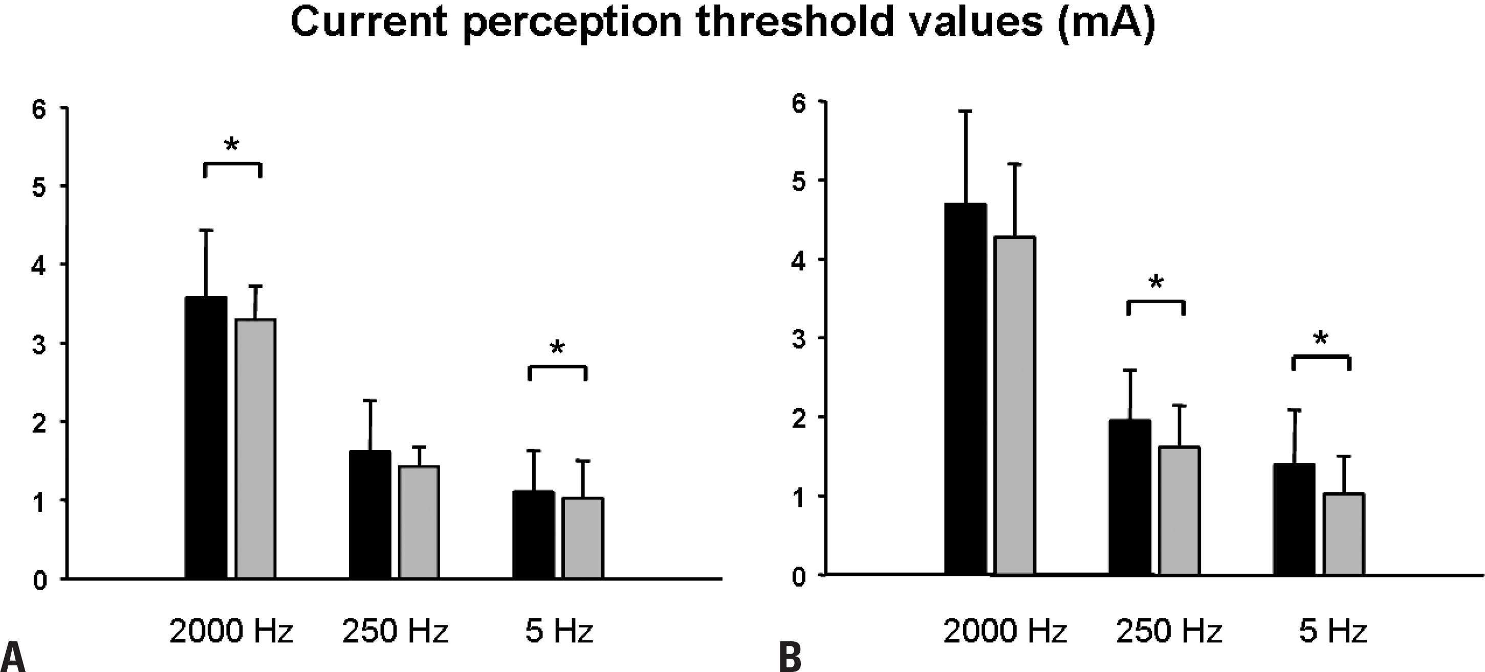
CPT values were significantly higher in the patient group than in the control group, especially with stimuli at the lowest frequency of 5 Hz (p < 0.05). There were significant correlations between the CPT values obtained at three different frequencies in the patient group, whereas the correlation was only significant in the pair of 250 Hz/5 Hz (both in the hands and feet), and in the pair of 2,000 Hz/250 Hz (in the feet) for the control group.
REFERENCES
1.Wolfe GI., Baker NS., Amato AA., Jackson CE., Nations SP., Saperstein DS, et al. Chronic cryptogenic sensory polyneuropathy: clinical and laboratory characteristics. Arch Neurol. 1999. 56:540–547.
2.Krarup C. An update on electrophysiological studies in neuropathy. Curr Opin Neurol. 2003. 16:603–612.

3.Periquet MI., Novak V., Collins MP., Nagaraja HN., Erdem S., Nash SM, et al. Painful sensory neuropathy: prospective evaluation using skin biopsy. Neurology. 1999. 53:1641–1647.

4.Nodera H., Logigian EL., Herrmann DN. Class of nerve fiber in-volvement in sensory neuropathies: clinical characterization and utility of the plantar nerve action potential. Muscle Nerve. 2002. 26:212–217.

5.Oh SJ., Melo AC., Lee DK., Cichy SW., Kim DS., Demerci M, et al. Large-fiber neuropathy in distal sensory neuropathy with normal routine nerve conduction. Neurology. 2001. 56:1570–1572.

6.Park KS., Lee SH., Lee KW., Oh SJ. Interdigital nerve conduction study of the foot for an early detection of diabetic sensory polyneuropathy. Clin Neurophysiol. 2003. 114:894–897.

7.Herrmann DN., Ferguson ML., Pannoni V., Barbano RL., Stanton M., Logigian EL. Plantar nerve AP and skin biopsy in sensory neuropathies with normal routine conduction studies. Neurology. 2004. 63:879–885.

8.McCarthy BG., Hsieh ST., Stocks A., Hauer P., Macko C., Cornblath DR, et al. Cutaneous innervation in sensory neuropathies: evaluation by skin biopsy. Neurology. 1995. 45:1848–1855.

9.Holland NR., Stocks A., Hauer P., Cornblath DR., Griffin JW., McArthur JC. Intraepidermal nerve fiber density in patients with painful sensory neuropathy. Neurology. 1997. 48:708–711.

10.Hossain P., Sachdev A., Malik RA. Early detection of diabetic peripheral neuropathy with corneal confocal microscopy. Lancet. 2005. 366:1340–1343.

11.Herrmann DN., Boger JN., Jansen C., Alessi-Fox C. In vivo confocal microscopy of Meissner corpuscles as a measure of sensory neuropathy. Neurology. 2007. 69:2121–2127.

12.Chong PS., Cros DP. Technology literature review: quantitative sensory testing. Muscle Nerve. 2004. 29:734–747.

13.Katims JJ., Naviasky EH., Rendell MS., Ng LK., Bleecker ML. Constant current sine wave transcutaneous nerve stimulation for the evaluation of peripheral neuropathy. Arch Phys Med Rehabil. 1987. 68:210–213.
14.Rendell MS., Katims JJ., Richter R., Rowland F. A comparison of nerve conduction velocities and current perception thresholds as correlates of clinical severity of diabetic sensory neuropathy. J Neurol Neurosurg Psychiatry. 1989. 52:502–511.

15.Masson EA., Veves A., Fernando D., Boulton AJ. Current perception thresholds: a new, quick, and reproducible method for the as-sessment of peripheral neuropathy in diabetes mellitus. Diabe-tologia. 1989. 32:724–728.

16.Masson EA., Boulton AJ. The Neurometer: validation and comparison with conventional tests for diabetic neuropathy. Diabet Med. 1991. 8(Spec No):S63–S66.

17.Pitei DL., Watkins PJ., Stevens MJ., Edmonds ME. The value of the Neurometer in assessing diabetic neuropathy by measurement of the current perception threshold. Diabet Med. 1994. 11:872–876.

18.Oishi M., Mochizuki Y., Suzuki Y., Ogawa K., Naganuma T., Nishijo Y, et al. Current perception threshold and sympathetic skin response in diabetic and alcoholic polyneuropathies. Intern Med. 2002. 41:819–822.

19.Matsutomo R., Takebayashi K., Aso Y. Assessment of peripheral neuropathy using measurement of the current perception threshold with the neurometer in patients with type 2 diabetes mellitus. J Int Med Res. 2005. 33:442–453.

20.Kim SH., Shim CS., Kim JH., Kim MH. Clinical usefulness of current perception thresholds in evaluating the diabetic neuropathy. J Korean Neurol Assoc. 1998. 16:666–671.
21.Lv SL., Fang C., Hu J., Huang Y., Yang B., Zou R, et al. Assessment of peripheral neuropathy using measurement of the current perception threshold with the Neurometer(R) in patients with type 1 diabetes mellitus. Diabetes Res Clin Pract. 2015. 109:130–134.
22.Technology review: the Neurometer Current Perception Thresh-old (CPT). AAEM Equipment and Computer Committee. Amer-ican Association of Electrodiagnostic Medicine. Muscle Nerve. 1999. 22:523–531.
23.Vinik AI., Suwanwalaikorn S., Stansberry KB., Holland MT., McNitt PM., Colen LE. Quantitative measurement of cutaneous perception in diabetic neuropathy. Muscle Nerve. 1995. 18:574–584.

24.Veves A., Malik RA., Lye RH., Masson EA., Sharma AK., Schady W, et al. The relationship between sural nerve morphometric findings and measures of peripheral nerve function in mild diabetic neuropathy. Diabet Med. 1991. 8:917–921.

25.Dyck PJ., Dyck PJ., Chalk CH. The 10 P's: a mnemonic helpful in characterization and differential diagnosis of peripheral neuropathy. Neurology. 1992. 42:14–18.
Fig. 1.
Current perception threshold (CPT) values in the hands (A) and feet (B) measured at three different frequencies in the patient (black bars) and control (grey bars) groups. * p < 0.05.

Table 1.
Clinical symptoms and signs in the patient group
Table 2.
Internal correlations of CPT values at different frequencies
| 250 Hz/5 Hz | 2,000 Hz/250 Hz | 2,000 Hz/5 Hz | |
|---|---|---|---|
| Hands | |||
| Patient group | r = 0.72 (p < 0.001 a) | r = 0.66 (p < 0.001 a) | r = 0.47 (p = 0.014 a) |
| Control group | r = 0.51 (p = 0.031 a) | r = 0.31 (p = 0.208) | r = 0.34 (p = 0.167) |
| Feet | |||
| Patient group | r = 0.66 (p < 0.001 a) | r = 0.80 (p < 0.001 a) | r = 0.56 (p = 0.002 a) |
| Control group | r = 0.84 (p < 0.001 a) | r = 0.62 (p = 0.007 a) | r = 0.34 (p = 0.165) |


 PDF
PDF ePub
ePub Citation
 Print
Print


 XML Download