

 PDF
PDF ePub
ePub Citation
 Print
Print


INTRODUCTION
Cholecystectomy is one of the most commonly performed procedure in abdominal surgery. Laparoscopic procedures continue to develop with improvements in surgical instruments and techniques. Since Erich Muhe of Germany performed the first procedure in 1985, laparoscopic cholecystectomy has become the gold standard for surgical treatment of gallbladder disease worldwide [12]. This procedure is minimally invasive and results in less postoperative pain, better cosmesis, shorter hospital stays, and less disability for work than large-incision open cholecystectomy [345]. Many surgeons have tried to reduce the number of ports and size of incisions [67]. In 1997, Navarra et al. [8] performed single-incision laparoscopic cholecystectomy (SILC) with transumbilical trocars and used transabdominal gallbladder traction sutures. Recently, many surgeons have shown interest in the feasibility of SILC [910].
There are many different SILC approaches with regard to transumbilical trocar position, liver retraction, and additional port insertion. However, many studies have reported that SILC is not suitable for acute complicated gallbladder disease because of technical difficulty and procedure risk. Moreover, there are still no definite indications and standard methods for SILC. Therefore, we introduced the Konyang Standard Method (KSM) for SILC since April 2010. Initially, we used 3-channel SILC which we called the KSM, and the KSM has been improved over the past 6 years. This report aimed to evaluate the adequacy and feasibility of the KSM for SILC, and the development of KSM is discussed.
METHODS
Patients
Between April 2010 and December 2016, 1,005 patients underwent SILC. Two hepatobiliary surgeons performed SILC in all patients, and all patients provided informed consent before the operation. Initially we excluded patients aged more than 70 years, and those with systemic medical problems, cystic duct abnormalities, and complications of acute cholecystitis. However, after 50 cases, all patients except for those with suspected malignancy were acceptable for SILC because of experience and improved technique. Between April 2010 and September 2012, 323 patients underwent SILC with the KSM using a hand-made 3-channel (3-trocar) single port. From October 2012 to August 2016, 645 patients underwent SILC with KSM using a modified 4-channel technique with a snake retractor for exposure of the hepatocystic triangle (Calot triangle); we called this a modified KSM (mKSM). Since September 2016, we have used a commercial 4-channel (Glove) port for simplicity in 37 patients; we call this a commercially modified KSM (C-mKSM).
Konyang Standard Method
Instruments and port
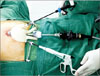
KSM is performed with a hand-made 3-channel port constructed with a 10-mm wound protector/retractor (Alexis, Applied Medical, Rancho Santa Margarita, CA, USA) covered with a size 7 sterile glove connected to two 5-mm ports and one 10-mm port (Laport, Sejong Medical, Paju, Korea). The wound retractor is inserted through a transumbilical incision and the 3-channel hand-made glove port is used as a working port for laparoscopic instruments. First, we cut the fingertips off the gloves and insert the trocar, which is tied and fixed. Then, the outer ring of the wound retractor is covered with the hand-made glove port. A CO2 pipe is connected to the 10-mm port for the pneumoperitoneum. The flexible videoscope (Olympus, Tokyo, Japan), long articulated Endo-Roticulator laparoscopic instrument (Coviden, Mansfield, MA, USA), a suction-hook bovie (Endopath Probe Plus II Pistol Grip handle, Ethicon, Bridgewater, NJ, USA) and 5- and 10-mm Hem-o-lok clips (Weck Closure Systems, Research Triangle Park, NC, USA) are used in the KSM (Fig. 1).
Patient and instrument positioning
The patients are placed in a supine position with both arms in extension, in a 15°–30° reverse Trendelenburg position, and with the right side tilted upward. The operator stands on the left side of the patient and the scopist stands below the operator. A 10- to 20-mm transumbilical incision is made and the 3-channel hand-made port is inserted. When the laparoscopic instrument is inserted, the operator's right hand controls the grasper for traction of the gallbladder and the left hand controls the dissector, scissors, and suction-hook Bovie for major procedures involving dissection, division, and ligation. The laparoscopic instrument is held with the hands crossed at the transumbilical incision, creating a triangular working area in the intraabdominal cavity. To secure adequate range of instrumental movement, the left hand should be located below the right hand instrument. Both instruments should be located on the left side of the telescope in the transumbilical incision to avoid conflict between the instruments.
Surgical technique
The operator uses the grasper in the right hand for cephalad traction on the fundus of the gallbladder in a supero-lateral direction. Then, the cystic duct and porta hepatis are exposed (gallbladder traction). If the cystic duct and porta hepatis are identified, the operator needs to establish the critical view of safety. The operator uses the grasper in the right hand to push the infundibulum of the gallbladder infero-laterally, and the dissector and suction-hook Bovie in the left hand to dissect the hepatocystic triangle boundary. If the gallbladder neck is dissected free of the liver surface, a “window” is created in the hepatocystic triangle crossed by the cystic duct and artery (dissection of the hepatocystic triangle). If the cystic duct and artery are fully dissected, they are ligated and resected by using 5- or 10-mm Hem-o-lok clips with the left hand of the operator (division of cystic artery and duct). After resection of the cystic artery and duct, the gallbladder is pulled cephalad using the grasper in the right hand. The connective tissues between the gallbladder bed and hepatic surface are dissected with electrocautery in the left hand (dissection of the gallbladder bed) [10].
Modified KSM
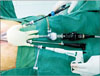
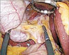
The KSM encountered some difficulty because of limited working range of the laparoscopic instrument in the hepatocystic triangle. Especially, in a patient with a large and heavy liver, it is difficult to widen the view of the subhepatic area. Therefore, we introduced a 4-channel, hand-made glove port by adding one more 5-mm port for a snake retractor to raise the under surface of the liver around the hepatocysctic triangle. The added snake retractor allowed wider exposure of the hepatocystic triangle, and improved the working range of the laparoscopic instrument. We called this the mKSM (Figs. 2, 3).
Commercially modified KSM
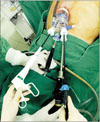
The mKSM is in standard use for SILC in Konyang University Hospital. Recently, we introduced a commercial 4-channel product port (Glove port, A-type, Nelis, Seoul, Korea) for simplicity (Fig. 4).
RESULTS
Clinicopathologic characteristics
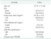
Among 1,005 patients who underwent SILC, the age range was 16–88 years and the median age was 52 years. There were 459 male patients (45.7%) and 549 female patients (54.3%). Of 246 patients with a history of previous abdominal surgery, 22 patients had upper abdominal surgery and 224 patients had lower abdominal surgery. Among the 1,005 patients, 928 (92.3%) had a body mass index < 30 kg/m2 and 925 (92.0%) had an American Society of Anesthesiologists physical status classification < III. The preoperative percutaneous transhepatic gallbladder drainage insertion rate was 12.0% (121 patients). The clinicopathologic information of the 1,005 patients is shown in Table 1.
Operative and postoperative outcomes
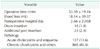
Mean operative time was 53.6 ± 19.2 minutes and blood loss was 18 ± 40 mL. Average postoperative hospital stay was 2.6 days. Among 1,005 cases, 24 (2.4%) needed insertion of an additional port. In 16 cases, gallbladder bed bleeding was controlled by laparoscopic suture ligation; 3 cases required dissection of intraabdominal adhesions, 3 required retraction of a heavy liver, 2 sustained a bile duct injury, and 1 had a duodenal injury treated with laparoscopic suture repair. In postoperative pathologic findings, 137 patients (13.6%) were diagnosed with acute cholecystitis or empyema and 868 (86.4%) were diagnosed with chronic or other benign diseases. A Jackon-Pratt drain tube was inserted in 18 cases (1.8%) and placed in the subhepatic area (Table 2).
Comparisons between KSM, mKSM, and C-mKSM
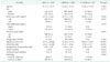
The KSM for SILC has undergone development for the past 6 years. There was no significant difference in preoperative clinicopathologic characteristics between patients undergoing these procedures. However, postoperative outcomes were improved and operative time was shortened. There was a significant difference in operative time in a comparison between the KSM, mKSM, and C-mKSM approaches (51.7 ± 13.8 minutes vs. 55.3 ± 15.2 minutes vs. 40.5 ± 13.2 minutes, respectively, P < 0.001), as well as in blood loss (24.6 ± 54.1 mL vs. 15.6 ± 30.7 mL vs. 4.9 ± 5.9 mL, P < 0.001) and mean postoperative hospital stay (2.9 ± 3.0 days vs. 2.6 ± 1.4 days vs. 2.4 ± 0.8 days, P = 0.051). There was no significant difference in drain insertion (3 cases [0.9%] vs. 15 cases [2.3%] vs. 0 cases, [0%], P = 0.214) and additional port insertion (8 cases [2.5%] vs. 16 cases [2.5%] vs. 0 cases [0%], P = 0.639). This improvement is shown in Table 3, which compare KSM, mKSM, and C-mKSM.
Postoperative complications
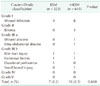
Postoperative complications were reported in 23 cases (2.3%). All complications were Clavien-Dindo classification grade I–V. Minor complications included 12 wound infections, 3 bilomas, and 3 wound or intraabdominal abscesses. Major complications included 2 biliary duct injuries treated with laparoscopic primary suture repair with T-tube insertion, 2 incisional hernias (followed after 6, 12 months), 1 duodenal perforation, and 1 small bowel injury. There was no significant difference between KSM and mKSM (P = 0.608). There were no postoperative complications with the C-mKSM procedure (Table 4).
DISCUSSION
Laparoscopic cholecystectomy is considered the gold standard for gallbladder disease, and minimally invasive surgery is preferred due to less postoperative pain and better cosmesis than in an open procedure. Thus, many surgeons have attempted to reduce the number of ports and size of the incisions [345]. Since Navarra et al. performed the first SILC in 1997, many studies have reported its feasibility and safety, but, there are still no standard methods for SILC [81112].
When Navarra et al. [8] first reported SILC, the procedure required two 10-mm trocars and 3 transabdominal holding sutures to elevate the gallbladder. Since then, many surgeons have introduced various techniques and instruments. Piskun and Rajpal [13] reported a method using two 5-mm trocars with 2 holding sutures in 1999. This method requires 2 transumbilical fascia incisions to insert the trocars. Thus, these used a single incision for skin but more than a single fascia incision. Podolsky et al. [14] reported a method using three 5-mm trocars and a rigid grasper through a single incision without percutaneous traction sutures. However, this method also used a separate fascial opening. Recently, Ceci et al. [15] reported using a commercial port with 3 working channel. In the KSM, we use a hand-made port, which has several advantages. It can be flexibly applied to many different situations: addition of a working port channel, and adjustment for umbilical wound size and use of bulky instruments such as a 10-mm Hem-o-lok clip. Moreover, a hand-made port offers a wide axis and space for instruments movement. However, the hand-made port is time-consuming to create during an operation. Accordingly, we began to use a commercial port in the KSM procedure (C-mKSM).
In the SILC procedure, liver retraction is needed to expose Calot triangle. Cuesta et al. [16] proposed a procedure using percutaneous Kirschner wires to expose Calot triangle. Tacchino et al. [17] directly performed SILC with 3 trocars through a single umbilical incision and used 2 holding sutures to suspend the gallbladder for exposure of Calot triangle. Since then, there have been ongoing attempts to perform SILC with minimal invasive hybrid procedure [1819] In the mKSM, we used a snake retractor to expose Calot triangle without suture elevation of the gallbladder or liver. This has the benefit of minimally invasive surgery and avoids gallbladder and liver injury from suture technique. Moreover, using a snake retractor enables flexible and immediate response in operative technique. This reduced conflict between laparoscopic instruments and offered a comfortable working range.
We used the Clavien-Dindo criteria to classify the complications of laparoscopic cholecystectomy. Minor complications included 12 wound infections, 3 bilomas, and 3 wound or intra-abdominal abscesses. Major complications included 2 biliary duct injuries treated with laparoscopic primary suture repair with T-tube insertion, 2 incisional hernias, 1 duodenal perforation, and 1 small bowel injury. There was no significant difference in complications between KSM and mKSM (P = 0.608). Biliary tract injury in laparoscopic cholecystectomy is the most severe complication, occurring in about 1% of cases and is reported to occur about 2.5 to 4 times more often than in open cholecystectomy. The Roux-en-Y hepaticojejunostomy procedure is reported to be successful when total resection of the bile duct and primary repair or T-tube insertion can be performed at the time of partial dissection [20212223]. Joseph et al. [24] reported a higher bile duct injury rate of 0.72% associated with SILC, even though most procedures were performed for nonacute cholecystitis. Moreover, many studies reported that SILC is not recommended in acute cholecystitis, because of technical difficulty and risk of complications [242526]. Our study only had 1 case of biliary tract injury in KSM (0.3%) and 1 in mKSM (0.15%), and both were treated with laparoscopic primary suture repair. In addition, we performed SILC in 137 cases of acute cholecystitis, comprising 13.6% of all SILC patients, without significant complications.
In conclusion, we described the evolution of the KSM for SILC. The findings suggest that the modified KSM using a 4-channel single port and snake retractor may be the safest and least invasive method for SILC.
 XML Download