Journal List > Korean Circ J > v.48(8) > 1099332

İkidağ: Retroaortic Course and Azygous Continuation of an Aberrant Left Brachiocephalic Vein
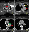
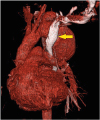
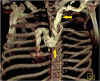
A 19 years old male student admitted to our hospital with a complaint of new onset headache. He had no previous history of surgery. There was blood pressure difference between right and left arm (130/80 mmHg at the left side and 170/95 at the right side). At computed tomography (CT) angiography, there was coarctation of the aortic arch, poststenotic bulbous dilatation of aorta and a variation regarding left brachiocephalic vein (LBV). Instead of joining with right brachiocephalic vein, it extended downwards at the left mediastinum, passed through left pulmonary artery and aorta, coursed behind the aorta at the carina level to the right side, and continued with the azygous vein (Figures 1, 2, 3). Elective surgery was planned for coarctation and aneurysmatic dilatation of the aorta.
Retrotracheal, circumaortic, subaortic or double LBV variations and coexistence with aortic coarctation or tetralogy of Fallot have been reported.1)2)3)4) Unlike our case, in none of these variations LBV courses downwards at left mediastinum or drains into azygous vein.
Embryological development of mediastinal venous system is well understood, and the previously reported variations have been attributed to embryological defects.1)2)3)4)5) Non regressing supracardinal vein that continues with an intersubcardinal anastomosis, and a defect in anastomosis between the anterior cardinal veins due to compression of poststenotic dilatation of the aorta may be responsible from this variation.
Consideration of this variation is clinically important especially in patients who will undergo mediastinal surgery, central venous line or pacemaker placement.

Figures and Tables

Figure 1

Axial CT images in arterial phase. Left subclavian and aberrant LBVs are brighter than the other vessels due to iodinated contrast medium that was injected from left antecubital vein. LBV (bright vessel, yellow arrow, A to D) courses inferior to left mediastinum, dives between left pulmonary artery (green arrow, C) and dilated descending aorta (red arrow, A to C), encircles the posterior part of aorta (D) at carina level and passes right to join azygous vein. Aortic coarctation is seen at the arcus level (blue arrow, B).

CT = computed tomography; LBV = left brachiocephalic vein.
kcj-48-763-g001
Figure 2

Volume rendered 3D image, left anterior view. The bright vessel is aberrant LBV (yellow arrow), courses through left pulmonary artery and aorta. Aortic coarctation is seen at the arcus level.

3D = three-dimensional; LBV = left brachiocephalic vein.
kcj-48-763-g002
Figure 3

Volume rendered 3D image of the aberrant LBV (yellow arrow), anterior view.

3D = three-dimensional; LBV = left brachiocephalic vein.
kcj-48-763-g003

Notes

Conflict of Interest The author has no financial conflicts of interest.

References

1. Konstantinov IE, Van Arsdell GS, O'Blenes S, et al. Retroaortic innominate vein with coarctation of the aorta: surgical repair and embryology review. Ann Thorac Surg. 2003; 75:1014–1016.
crossref
2. Morhy Borges Leal S, Andrade JL, de Souza M, et al. Anomalous subaortic course of the left brachiocephalic (innominate) vein: echocardiographic diagnosis and report of an unusual association. Cardiol Young. 2002; 12:159–163.
crossref
3. Topcuoglu OM, Atceken Z, Ariyurek OM. Circumaortic doubled left brachiocephalic vein: a rare confusing variation. Surg Radiol Anat. 2015; 37:315–318.
crossref
4. Yigit AE, Haliloglu M, Karcaaltincaba M, et al. Retrotracheal aberrant left brachiocephalic vein: CT findings. Pediatr Radiol. 2008; 38:322–324.
crossref
5. Chen SJ, Liu KL, Chen HY, et al. Anomalous brachiocephalic vein: CT, embryology, and clinical implications. AJR Am J Roentgenol. 2005; 184:1235–1240.
crossref
TOOLS
ORCID iDs

Mehmet Ali İkidağ
https://orcid.org/0000-0003-4004-9645

Similar articles