

 PDF
PDF ePub
ePub Citation
 Print
Print


INTRODUCTION
Chronic total occlusion (CTO) of the superficial femoral artery (SFA) is a commonly encountered target lesion in patients with symptomatic lower extremity arterial disease.1) Endovascular treatment of SFA CTO is often challenging because of the lesion length and presence of calcification. Bolia et al.2) introduced the subintimal angioplasty (SA) technique in 1989; a guidewire is intentionally placed between the intima and media to create a new channel for recanalization of the occluded arterial segment. Figure 1 shows a representative case of a long SFA occlusion treated with SA and stent implantation. Figure 2 shows intravascular ultrasound (IVUS) images taken before and after SA. Although this procedure has been widely adopted in the endovascular treatment of lower extremity arterial occlusions, the durability of the subintimal channel remains to be proved. No randomized clinical trial has compared SA with intraluminal angioplasty (IA) for SFA CTOs. The aim of this review was to assess the literature on the immediate and late outcomes of SA versus IA for SFA CTOs.
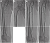
Figure 1
A representative subintimal angioplasty case. (A) Baseline angiogram showing superficial femoral artery long occlusion. (B) A hydrophilic wire was introduced into subintimal space, and a wire loop was formed by advancing the wire into the subintimal channel. (C) Angiogram after pre-dilation. (D) Final angiogram after stent implantation.

TECHNICAL SUCCESS
The early technical success rates of SA were between 74% and 85% in reports published before 2000 (Table 1).3)4)5) For lesions longer than 20 cm, the technical success rate was only 68%.3) At the time, an antegrade approach from the ipsilateral or contralateral femoral artery was the only way to tackle occluded SFA lesions with guidewires, and the subintimal channel was dilated with balloons without implanting stents. However, as operators accumulated experience and adopted retrograde approaches from distal arteries and using re-entry devices, the technical success rate gradually improved (Tables 1 and 2).6)7)8)9)10)11)12)13)14)15)16)17)18)19)20)21)22)23)24)
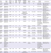
Table 1
Summary of published studies on the primary patency of SA for femoropopliteal artery occlusions
| First author | Study design | No. of patients/limbs | CLI (%) | Lesion length (cm) | Technical success (%) | Antegrade/retrograde approach | Reentry device | Stenting | Primary patency | Complications |
|---|---|---|---|---|---|---|---|---|---|---|
| London, 19943) | Retrospective, Single-center | 176/200 | 12 | 11 | 80 | Antegrade | No | No | 56% (1 year), 46% (3 years) | Major complication 1%, distal emboli 3.5%, arterial rupture 2%1) |
| Reekers, 19944) | Retrospective, single-center | 40/40 | 72.5 | 16.9 | 85 | Antegrade | No | No | 59% (1 year) | Overall 20%, distal emboli 0%, arterial rupture 0% |
| McCarthy, 20005) | Retrospective, single-center | 66/69 | 62 | 10 | 74 | Antegrade | No | No | 51% (6 months) | Overall 16%, distal emboli 1.5%, acute thrombosis 1.5% |
| Yilmaz, 20036) | Retrospective, single-center | 61/67 | 32.7 | 20.0 | 88 | Antegrade 18%/retrograde via popliteal artery 82% | No | 22.0%, BMS | 22.0% (1 year) | Overall 15%, distal emboli 4.5%, arterial rupture 6.0% |
| Laxdal, 20037) | Retrospective, single-center | 109/124 | 35 | 13 | 90 | Antegrade | No | No | 37% (1 year) | Operative mortality 1.8%, distal emboli 7.3% |
| Smith, 20058) | Retrospective, single-center | 43/48 | 35.4 | 6–10 | 92 | Antegrade/retrograde | No | No | 53% (1 year) | Overall 15%, distal emboli 10.4%, thrombosis 4.2% |
| Treiman, 20069) | Retrospective, single-center | 29/29 | 100 | N/R | 90 | Antegrade | No | All, BMS | 85% (1 year), 64% (2 years) | Overall 13.7%, distal emboli 3.4% |
| Schmieder, 200810) | Retrospective, single-center | 368/382 | 55.8 | N/R | 87 | Antegrade | No | 22.0%, BMS | 50% (1 year) for tent group, 45% (1 year) for non-stent group, (p=0.73) | Overall 3.2%, distal emboli 0.5%, arterial rupture 0.5% |
| Marks, 200811) | Retrospective, single-center | 103/116 | 42 | 12.7 | 85 | Antegrade | 6.9%, Outback | 77%, BMS | 59% (1 year) | “Intraoperative thrombosis” 6% |
| Setacci, 200912) | Retrospective, single-center | 145/145 | 100 | 17.1–22.5 | 83.5, 96.6 (using Outback) | Antegrade | 16.6%, Outback | 54.5%, BMS, spot stenting 43% | 70% (1 year), 34% (3 year) | Overall 6.2%, distal emboli 2.1%, arterial rupture 2.1% |
| Köcher, 201013) | Retrospective, single-center | 123/133 | 36.9 | 11.4 | 94.5 | Antegrade | No | 2.3%, BMS | 67.5% (1 year), 48.4% (3 year) | Overall 7.9%, distal emboli 6% |
| Siablis, 20112) | Prospective, single-center | 98/105 | 64.3 | 12.1 | 91.4 | Antegrade | 6.6%, Outback | 70.5%, BMS, spot stenting | 80.1% (1 year), 29.0% (3 year) | Overall 11.5%, distal emboli 2.9%, arterial rupture 3.8% |
| Hong, 201315) | Retrospective, single-center | 150/172 | 36.0 | 22.6 | 94.0 | Antegrade | 3.5%, Outback | All, BMS | 77.0% (1 year) | Major complication 0%, distal emboli 1.2%, arterial rupture 2.3 |
| Boufi, 201316) | Retrospective, single-center, comparison with bypass surgery | 58/58 | 53.4 | 16 | 93 | Antegrade | No | All, BMS, long stenting | 76.9% (1 year), 64.6% (3 year) | Major complication 5.5%, acute thrombosis 1.7% |
| Hong, 201517) | Retrospective, single-center, comparison between spot vs. long stenting | 163/196 | 32, 31 | 25.8 | 100 | Antegrade | 5.1%, Outback | All, BMS | 87% (1 year), 77% (2 years) for spot stenting; 56% (1 year), 47% (2 years) for long stenting (p<0.001) | Distal emboli 4.6%, arterial rupture 1.5% |
| Tatli, 201518) | Prospective, single-center | 74/74 | 22.0 | 13.4 | 97.0 | Antegrade | No | All, BMS, long stenting | 95% (6 months) | Major complication 0%, distal emboli 3%, arterial rupture 1% |
| Palena, 201719) | Prospective, single-center | 34 | 100 | 27.9 | 100 | Antegrade/retrograde (35.3%) | No | All, BMS (Supera) | 94.1% (1 year) | Pseudoaneurysm 14.7% |
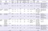
Table 2
Summary of published studies comparing SA and IA for femoropopliteal artery occlusions
| First author | Study design | Factor | No. of patients/limbs | CLI (%) | Lesion length (cm) | Technical success (%) | Retrograde or bidirectional approach (%) | Reentry device (%) | Stenting | Stented length (cm) | Primary patency | Complications |
|---|---|---|---|---|---|---|---|---|---|---|---|---|
| Ko, 20071) | Retrospective, single-center | SA | 52/61 | 32.8 | 22.7 | 95.1 | N/R | N/R | All, BMS | 8.0 | 76.4% (1 year) | Major complication 0%, distal emboli 0%, arterial ruptures 4.9% |
| IA | 54/60 | 31.6 | 22.0 | 86.7 | N/R | N/R | All, BMS | 7.5 | 59.2% (1 year) | Major complication 0%, distal emboli 6.7%, arterial rupture 1.7% | ||
| p value | 0.11 | 0.12 | 0.06 | |||||||||
| Antusevas, 200821) | Retrospective, single-center | SA | 71/73 | 47.9 | 12 | 87.7 | N/R | N/R | 4.1%, BMS | N/R | 68.5% (1 year), 65.8% (2 years) | Distal emboli 2.7%, arterial rupture 0% |
| IA | 75/75 | 61.3 | 6.3 | 81.3 | N/R | N/R | None | - | 42.7% (1 year), 38.7% (2 years) | N/R | ||
| p value | N/R | <0.001 | ||||||||||
| Soga, 201322) | Retrospective, multi-center | SA | 189/251 | 31 | 23.5 | 90 | 36 | N/R | All, BMS | 25.2 | 68% (1 year), 53% (3 years) | Overall 13%, distal emboli 1%, arterial rupture 0% |
| IA | 530/651 | 31 | 21.5 | 91 | 38 | N/R | All, BMS | 23.1 | 74% (1 year), 55% (3 years) | Overall 11%, distal emboli 1%, arterial rupture 0% | ||
| p value | 0.71 | 0.30 | ||||||||||
| Ishihara, 201623) | Prospective, multicenter, propensity- matched comparison, IVUS study | SA | 61 | 30 | 22.0 | N/R | N/R | N/R | All, DES (ZilverPTX) | N/R | 55% (1 year), 44% (2 years) | Overall 5% |
| IA | 61 | 25 | 21.0 | N/R | N/R | N/R | All, DES (ZilverPTX) | N/R | 65% (1 year), 49% (2 years) | Overall 3% | ||
| p value | 0.352 (1 year), 0.648 (2 years) | |||||||||||
| Kim, 201824) | Retrospective, multicenter | SA | 228/243 | 30.3 | 25.8 | 95.1 | 4.1 | 12.3 | 77.1%, BMS | 17.3 | 67.5% (1 year), 54.0% (2 years) | Major complication 4.1%, distal emboli 1.6%, arterial rupture 2.9% |
| IA | 233/244 | 34.8 | 24.5 | 89.8 | 20.1 | 0 | 70.3%, BMS | 16.5 | 73.4% (1 year), 61.3% (2 years) | Major complication 0.4%, distal emboli 0.8%, arterial rupture 1.6% | ||
| p value | 0.041 | 0.086 |
Yilmaz et al.6) reported an 88% success rate in long SFA occlusion (mean length 20.0 cm) and used a retrograde approach in 82% of study participants. Soga et al.22) employed a bidirectional approach in 37% of the SA group and achieved a technical success rate of 90% (mean occlusion length 23.5 cm). Gandini et al.25) attained a 100% success rate using the Outback Ltd re-entry catheter (Cordis, Bridgewater, NJ, USA) in patients with TransAtlantic Inter-Society Consensus for the Management of Peripheral Arterial Disease (TASC) II D femoropopliteal artery disease compared with a 42.3% success rate with a manual re-entry method. A multicenter retrospective study from Korea found a higher technical success rate for SA compared to IA.24) Other comparative studies found no difference in the technical success rates for the 2 strategies.21)22)24) However, Soga et al.22) reported that 25% of the IA cases in their study crossed over to a subintimal approach due to technical difficulties during IA. SA also had a significantly shorter procedure time, lesser use of guidewires, body surface echography, and IVUS.
PROCEDURAL COMPLICATIONS
Arterial rupture is the most concerning complication associated with SA, but the incidence is relatively low, ranging from 0% to 6%. Most arterial ruptures were managed conservatively or by endovascular therapy. Although distal embolization was thought to occur less frequently in SA than IA due to the absence of atherosclerotic plaques and thrombus in the subintimal channel, the incidence is similar to that of IA and varies from 0% to 7.3%. Overall, procure-related complication rates do not significantly differ between the 2 endovascular strategies.20)22) Only Kim et al.24) reported a higher major complication rate (4.1% vs. 0.4%) with SA. However, they still found similar incidence of distal embolizations and arterial ruptures for the 2 treatments.
PRIMARY PATENCY
In early studies, the 1-year patency rates for the SA using balloon angioplasty alone in SFA CTOs >20 cm varied from 22% to 56%.3)6)26) However, the introduction of self-expanding nitinol stents increased the 1-year SA patency rate from 65% to 80%.15)17)19)20)22)26) In a meta-analysis of 37 studies, the 1-year primary patency rate was 47.9% for SA without stent implantation, 61.6% for SA with provisional stenting, and 69.2% for SA with primary stenting.26)
To date, no randomized controlled trial has directly compared SA versus IA for femoropopliteal artery disease. One prospective registry study and four retrospective studies have compared the immediate and mid-term outcomes for both wire crossing techniques.20)21)22)23)24)
Antusevas et al.21) reported a higher primary patency rate for SA versus IA. However, the baseline clinical and lesion characteristics were different between the 2 groups, and stents were rarely used. Other studies found no significant differences between the 2 endovascular strategies. Surprisingly, Ishihara et al.23) reported relatively low 1-year patency rates for both techniques despite using drug-eluting stents (DESs; Zilver PTX, Cook Medical, Bloomington, IA, USA). This finding may be explained by the limited efficacy of DESs in long lesions. Long stenting may have also contributed to the lower patency rates. Hong et al.17) showed that primary patency was significantly lower with long stenting than with spot stenting following SA of long femoropopliteal occlusions. Thus, stenting strategies may play a more important role than the wire passage method in maintaining the patency of recanalized long arterial lesions.
FUTURE PERSPECTIVES
We need more clinical evidence including randomized control trial data to confirm the efficacy of SA in SFA occlusions. To date, just one study systematically evaluated guidewire location using IVUS to define SA or IA.23) In other reports, wiring strategies were judged by guidewire morphology. IVUS is essential to provide objective clinical evidence. Furthermore, we need more information regarding the effects of various drug-eluting balloons or stents on primary patency of the subintimal channel. The efficacy of dedicated spot stents for primary patency after SA will be also an interesting topic to investigate.
CONCLUSIONS
In the last several decades, new techniques and devices have improved the immediate and mid-term outcomes of SA. Based on the currently available data, SA is safe and has a higher technical success rate than IA despite its shorter procedure time. The mid-term primary patency of SA appears to be comparable with that of IA. An optimal stenting strategy and the role of drug-eluting technologies in SA remain to be defined.
 XML Download