

 PDF
PDF ePub
ePub Citation
 Print
Print


INTRODUCTION
Lung cancer is a leading malignancy in thoracic oncology that causes a majority of deaths both in China and worldwide.1 The prevalence of epidermal growth factor receptor (EGFR) mutations ranges from 5–10% in Caucasians to 60–70% in neversmoking Asian adenocarcinoma patients, indicating that EGFR mutation-positive non-small cell lung cancer (NSCLC) may have a unique disease course.2 In fact, NSCLC patients with sensitive EGFR mutations are highly responsive to EGFR inhibitors, including gefitinib and erlotinib, compared with standard chemotherapy.34 Because of inevitable EGFR-tyrosine kinase inhibitor (TKI) resistance, next-generation EGFR-TKIs have been developed, and clinical trials have demonstrated a higher response rate and longer progression-free survival (PFS) and overall survival (OS) among previously treated patients with EGFR-mutant NSCLC.56 Therefore, the precise detection of EGFR mutations plays a key role in the clinical management of EGFR mutation-positive NSCLC patients.
Currently, the methods for detecting EGFR mutations include Sanger sequencing,7 amplification refractory mutation system (ARMS),8 pyrosequencing,9 high resolution melting analysis,10 and genome sequencing.11 Sanger sequencing remains the gold standard for EGFR mutation detection in clinical practice and may detect unknown EGFR mutations. The ARMS method, which has also been approved by the China Food and Drug Administration (CFDA), is a highly sensitive and reliable method for detecting EGFR mutations. Due to limitations regarding labor, time, and expertise requirements, as well as low sensitivity, other methods, such as pyrosequencing, high resolution melting analysis, and whole genome sequencing, were excluded from the current clinical EGFR mutation analysis.
In this article, we compared patient outcomes based on EGFR mutation analysis by Sanger sequencing and ARMS in small specimens: both assays have been approved by the CFDA. Upon investigation of the survival data, we found that the curative effect of EGFR-TKIs may be better in lung cancer patients with a high abundance of EGFR mutations than in those with a low mutation abundance. Sanger sequencing could be useful for EGFR mutation detection, and our data support the implementation of secondary genetic testing of EGFR mutation-negative NSCLC patients with a promising response to EGFR-TKI treatment.
MATERIALS AND METHODS
Samples collection
A total of 200 NSCLC patients with an equal number of EGFR ARMS-positive and ARMS-negative cases at The First Affiliated Hospital of Guangzhou Medical University from August 2014 to August 2015 were selected as study participants (IRB number: 2016-29). The two main eligibility criteria were radiologically and pathologically confirmed NSCLC and patient consent. The other inclusion criteria were no previous chemotherapy or radiotherapy and no other severe systemic disease. We also included patients with stage I–III NSCLC who were EGFR ARMS-positive and self-medicated with an EGFR-TKI after refusing adjuvant chemotherapy and radiotherapy. There were 108 male and 92 female patients ranging in age from 48–87 years included in this study. Samples were obtained by CT-guided fine-needle aspiration (n=35) or surgery (n=165). All samples were confirmed to be adenocarcinoma. There were 113 stage I, 52 stage II, 29 stage III, and six stage IV cases.
DNA isolation
DNA was extracted from formalin-fixed, paraffin-embedded tumor tissue using the QIAamp DNA FFPE Tissue Kit (Qiagen, Hilsen, Germany) according to the manufacturer's recommendations. Genomic DNA was stored at -20±5℃ after measuring the concentration (ng/mL) thereof and absorbance (A260/280 ratio) using a NanoDrop 1000 Spectrophotometer (Thermo Fisher Scientific, Cleveland, OH, USA).
Sanger sequencing
Genomic DNA was amplified with four primer pairs targeting exons 18 to 21 and labeled using the EGFR Mutation Detection Kit (Guangzhou Life Technologies Daan Diagnostics Co., Ltd., Guangzhou, China). Sequencing and data collection were performed using an ABI 3100 Genetic Analyzer (Applied Biosystems). All sequence variations were confirmed by multiple independent PCR amplifications and repeat sequencing as previously described.12 The difference between high and low mutation abundance was as previously defined.13
ARMS qPCR
Common EGFR mutations (Del19, L858R and L861Q in exon 21, G719X in exon 18, S768I in exon 20, and three insertions in exon 20) were detected using an ADx-ARMS EGFR 21 Detection Kit (Amoy Diagnostics Co., Ltd., Xiamen, China). qRT-PCR was performed in a StepOne™ PCR System (Thermo Fisher Scientific) according to the manufacturer's instructions.14
Treatment and assessment
Treatment with EGFR-TKIs included oral administration of 250 mg/d gefitinib or 150 mg/d erlotinib, and efficacy was evaluated after treatment by chest CT of the thoracic lesion according to standard clinical practice. Patients with stage I–IIIA disease who self-purchased the targeted drugs after initial disease progression were included in our analysis. According to Response Evaluation Criteria in Solid Tumors, the effects were defined and categorized as complete response, partial response, stable disease, or progressive disease. OS and PFS were defined as the time interval from the beginning of treatment to documented disease progression or death from any cause censored at the last follow-up.15
Statistical analysis
All the analyses were performed using SPSS software, version 22.0 (IBM Corp., Armonk, NY, USA). The Kaplan-Meier method was used to compare median PFS after TKI therapy in the same follow-up group with different detection methods. p-values less than 0.05 were considered statistically significant.
RESULTS
Patient characteristics and samples
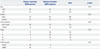
From August 2014 to August 2015, 200 patients were screened and met the enrollment criteria. The patient characteristics were as follows: 108 male and 92 female patients ranging in age from 48–87 years were included in this study. Samples were obtained by CT-guided fine-needle aspiration (n=35) or surgery (n=165). All samples were confirmed to be adenocarcinoma. Disease specimens of TNM stage I to IV were included. All patients with an EGFR-sensitive mutation who received a first-generation EGFR-TKI were also included. The patient characteristics are provided in Table 1. Age and TNM stage were well balanced among groups.
Comparison of mutation detection rates by direct sequencing and ARMS
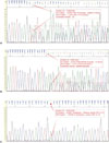
The EGFR mutation statuses of all patients detected by the two methods are summarized in Table 2. Among the 100 ARMS-positive EGFR samples, Sanger sequencing detected mutations in 90 samples; the other 10 were negative. Among the 100 ARMS-negative samples, three were positive for a mutation by the Sanger method, and 97 negative samples were confirmed. Based on the positive likelihood ratio (10.409) and the positive predictive value (96.77%), the ARMS-PCR method can detect EGFR mutations with high efficiency and specificity. Thus, the EGFR mutation rate was higher using ARMS than direct sequencing. Notably, the ARMS method covers only 29 EGFR mutation hotspots in exons 18–21, and Sanger sequencing detected three coding DNA sequence (CDS) mutations in ARMS-negative samples: c.2237_2251>TTC (complex), c.2231_2232ins18 (insertion), and c.2515G>A (substitution, position 2515, G→A) (Fig. 1).
EGFR mutation status and clinical outcomes
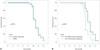
As a higher EGFR mutation abundance may yield better results with EGFR-TKI treatment,16 we compared patient outcomes after EGFR-TKI treatment based on ARMS and Sanger sequencing. In terms of EGFR-TKI treatment, the median PFSs among EGFR-positive patients detected by Sanger sequencing or ARMS were 11.1 months [95% confidence interval (CI), 10.6–11.4 months] and 10.9 months (95% CI, 10.7–11.3 months), respectively; this difference was not significant. The PFS was 12.4 months (95% CI, 11.6–12.4 months) for patients with a high EGFR mutation abundance (n=35), which was longer than that for patients with a low EGFR mutation abundance (95% CI, 10.7–11.3 months) (p<0.001) (Fig. 2). Interestingly, patients with the c.2237_2251>TTC (complex) or c.2231_2232ins18 (insertion) mutation who received EGFR-TKIs had a PFS of 3 months and 6 months, respectively. One patient with a c.2515G>A mutation (substitution, position 2515, G→A) was lost to follow-up after 4 months of EGFR-TKI treatment.
DISCUSSION
NSCLC accounts for over 80% of lung cancer cases and includes adenocarcinoma, large cell carcinoma, and squamous cell carcinoma.17 Similar to our results, patients who are female, never smokers, of Asian origin, and present with adenocarcinoma have a higher EGFR mutation frequency,1819 and this EGFR mutation rate is higher than that in non-adenocarcinoma patients, who have a rate of less than 10%.20 In recent years, NSCLC has been managed according to molecular subtype. In EGFR-mutant NSCLC patients, EGFR-TKI treatment has greatly increased survival compared to those with EGFR wild-type lung cancer.2122 The predominant EGFR mutations are in exons 18 through 21 and serve as predictors of the efficacy of EGFR-TKIs. Therefore, the identification of an EGFR mutation plays a critical role in NSCLC management.
Although it has been well recognized that EGFR mutations are associated with the therapeutic effect of TKIs in NSCLC patients, current methods do not provide the precision required for clinical practice. Currently, the two main detection methods are ARMS and Sanger sequencing. Although Sanger sequencing remains then gold standard, the ARMS method is considered an alternative because of its high sensitivity in detecting EGFR mutations;2324 EGFR mutations can be detected in small samples using ARMS. The reason for the high sensitivity with ARMS is its special primer design. One pair of primers amplifies a conserved region, and another primer pair targets the point mutation. ARMS is limited to the detection of known mutations; each reaction system can only detect the pre-specified gene mutation. Therefore, a large number of DNA samples and primer pairs are needed, making this method expensive, if an unknown region must be analyzed. Sanger sequencing can analyze unknown DNA sequences at relatively low cost; the biggest problem is the low sensitivity. Mutations are difficult to detect in specimens with a low content of tumor cells or mutant cells. Moreover, noise within peaks can affect calling EGFR mutations. Therefore, Sanger sequencing is suitable for detecting EGFR mutations in surgical specimens with a high proportion of tumor cells potentially harboring a mutation. The results of this study suggest that Sanger sequencing is recommended for EGFR redetection and for initial detection in surgical specimens.
At least 90% of EGFR mutations occur in exons 19 and 21; the remaining 10% of mutations are in less common sites, and these are called rare EGFR mutations. With the application of EGFR sequencing technology, the discovery of mutations in exons 18–21 is increasing.25 Few treatment strategies have been reported for less common EGFR mutations. For example, first-generation EGFR-TKIs could be used in patients with A763_Y764insFQEA, an exon 20 insertion.26 In our study, we detected 10 EGFR mutation-negative samples by Sanger sequencing among 100 ADx-ARMS-positive samples. Among the 100 ADx-ARMS-negative samples, three were positive for a mutation by Sanger sequencing. Of these, two harbored an exon 19 deletion, and one had an exon 21 c.2515G>A p.A839T mutation (Cosmic ID COSM13430), which might not have been detected by ARMS due to the assay design. The impact of these rare EGFR mutations on EGFR-TKI therapy are far from fully understood. Baek, et al.27 reported that the response to EGFR-TKI treatment and the survival of patients with rare or complex EGFR mutations is worse than those for patients with common mutations. In our study, only two cases with a PFS of 3 months and 6 months are not sufficient to reach a conclusion. Therefore, clinical trials, such as NCT01775943, involving a large number of patients with rare EGFR mutations are warranted to elucidate the efficacy of EGFR-TKIs in these patients.
In this analysis, we also determined that patients with a high EGFR mutation abundance have a better outcome after EGFRTKI treatment. For patients with a high EGFR mutation abundance, the PFS was 12.4 months (95% CI, 11.6–12.4 months), which was higher than that for those with a low EGFR mutation abundance (95% CI, 10.7–11.3 months) (p<0.001). In accordance with a previous report, the EGFR mutation abundance could predict the outcome of EGFR-TKI therapy for advanced NSCLC. Hence, in clinical practice, Sanger sequencing offers additional information for physicians to predict whether the patient may benefit from an EGFR-TKI.
In summary, our results suggest that Sanger sequencing can detect rare EGFR mutations and is applicable for redetermining EGFR status. NSCLC patients with a high mutation burden have a better response to EGFR-TKIs. A clinical trial evaluating the efficacy of EGFR-TKIs in patients with rare EGFR mutations is needed.

 XML Download