Abstract
Objective
To compare the quality of tissue from punch biopsy forceps (PB group) with round loop electrode (LE group) in colposcopically directed biopsy along with the evaluation of pain associated with each procedure.
Methods
Patients with abnormal cervical cytologic results and abnormal colposcopic findings were enrolled into a randomized trial into either a PB group or LE group. The quality of tissue was evaluated in regards to the size of tissue, site of tissue, and tissue damage. Each quality had 1 to 3 points and the sum of each quality contributed to the total tissue score that ranged from 3 to 9. Pain associated with each procedure was assessed by a visual analog scale (VAS). This was a clinical trial study and was registered at www.clinicaltrials.in.th (Identifier: TCTR20160404001).
Results
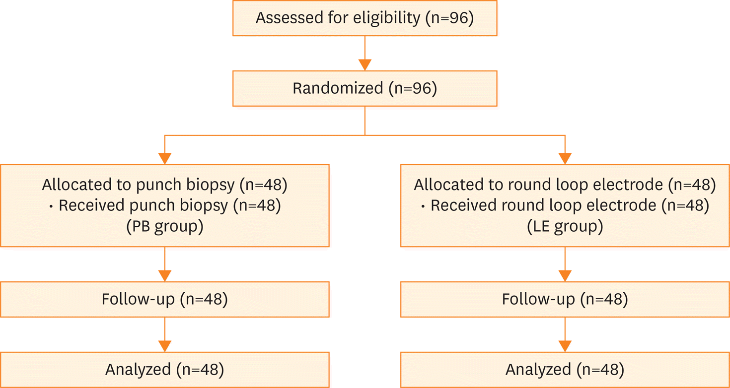
Ninety-six women who met all eligibility requirements were enrolled in the study. Forty-eight patients were randomly assigned to the PB group and 48 patients were randomized into the LE group. The characteristics of the patients were similar between the 2 groups with the exception of the median age. The median total tissue score was 8 points in the LE group which was more than the median of 7 points in the PB group with a statistically significant difference (p=0.014). However, the median VAS pain score in both groups was 3.4 (p=0.82).
Go to : 
References
1. Luesley D, Leeson S. Colposcopy and programme management. 2nd ed.Sheffield: NHS Cancer Screening Programme;2010.
2. Arbyn M, Kyrgiou M, Simoens C, Raifu AO, Koliopoulos G, Martin-Hirsch P, et al. Perinatal mortality and other severe adverse pregnancy outcomes associated with treatment of cervical intraepithelial neoplasia: meta-analysis. BMJ. 2008; 337:a1284.

3. Kyrgiou M, Koliopoulos G, Martin-Hirsch P, Arbyn M, Prendiville W, Paraskevaidis E. Obstetric outcomes after conservative treatment for intraepithelial or early invasive cervical lesions: systematic review and meta-analysis. Lancet. 2006; 367:489–98.

4. Anderson MC. Invasive carcinoma of the cervix following local destructive treatment for cervical intraepithelial neoplasia. Br J Obstet Gynaecol. 1993; 100:657–63.

5. Shumsky AG, Stuart GC, Nation J. Carcinoma of the cervix following conservative management of cervical intraepithelial neoplasia. Gynecol Oncol. 1994; 53:50–4.

6. Ouitrakul S, Udomthavornsuk B, Chumworathayi B, Luanratanakorn S, Supoken A. Accuracy of colposcopically directed biopsy in diagnosis of cervical pathology at Srinagarind Hospital. Asian Pac J Cancer Prev. 2011; 12:2451–3.
7. Bulten J, Horvat R, Jordan J, Herbert A, Wiener H, Ardyn M. European guidelines for quality assurance in cervical histopathology. Acta Oncol. 2011; 50:611–20.

8. Duesing N, Schwarz J, Choschzick M, Jaenicke F, Gieseking F, Issa R, et al. Assessment of cervical intraepithelial neoplasia (CIN) with colposcopic biopsy and efficacy of loop electrosurgical excision procedure (LEEP). Arch Gynecol Obstet. 2012; 286:1549–54.

9. Kietpeerakool C, Srisomboon J, Suprasert P, Cheewakriangkrai C, Charoenkwan K, Siriaree S. Routine prophylactic application of Monsel's solution after loop electrosurgical excision procedure of the cervix: is it necessary? J Obstet Gynaecol Res. 2007; 33:299–304.
10. Lee KE, Koh CF, Watt WF. Comparison of the grade of CIN in colposcopically directed biopsies with that in outpatient loop electrosurgical excision procedure (LEEP) specimens–a retrospective review. Singapore Med J. 1999; 40:694–6.
11. Church L, Oliver L, Dobie S, Madigan D, Ellsworth A. Analgesia for colposcopy: double-masked, randomized comparison of ibuprofen and benzocaine gel. Obstet Gynecol. 2001; 97:5–10.

12. Öz M, Korkmaz E, Cetinkaya N, Baş S, Özdal B, Meydanl MM, et al. Comparison of topical lidocaine spray with placebo for pain relief in colposcopic procedures: a randomized, placebo-controlled, double-blind study. J Low Genit Tract Dis. 2015; 19:212–4.
13. Campion MJ, Canfell K. Cervical cancer screening and preinvasive disease. In: Berek JS, Hacker NF, editors. Berek & Hacker's gynecologic oncology. 6th ed.Philadelphia, PA: Lippincott Williams & Wilkins;2015. p. 242–325.
14. Ferris DG, Mayeaux EJ. Colposcopic equipment, supplies, and data management. In: Mayeaux EJ, Cox JT, editors. Modern colposcopy: textbook & atlas. 3rd ed.Philadelphia, PA: Lippincott Williams & Wilkins Health;2012. p. 102–19.
15. Bulten J, Horvat R, Jordan J, Herbert A, Wiener H, Arbyn M. European guidelines for quality assurance in cervical histopathology. Acta Oncol. 2011; 50:611–20.

16. Srisomboon J, Kietpeerakool C, Suprasert P, Siriaunkgul S, Khunamornpong S, Prompittayarat W. Factors affecting residual lesion in women with cervical adenocarcinoma in situ after cone excisional biopsy. Asian Pac J Cancer Prev. 2007; 8:225–8.
Go to : 
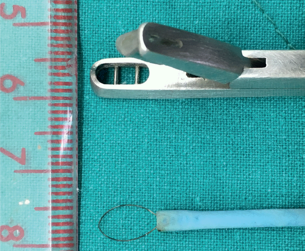
 | Fig. 1.Diagnostic equipment for cervical biopsy: Kevorkian biopsy forceps (upper) and the round loop electrode (lower). |
 | Fig. 2.Flow of patients through the study. LE group, round loop electrode group; PB group, punch biopsy forceps group. |
Table 1.
Quality of tissue scores
Table 2.
Patient characteristics
| Characteristics | PB group (n=48) | LE group (n=48) | p-value |
|---|---|---|---|
| Age (yr) | 38.9 (10.2) | 44.8 (12.2) | 0.011* |
| Reproductive status | 0.190† | ||
| Premenopause | 42 (87.5) | 36 (75) | |
| Menopause | 6 (12.5) | 12 (25) | |
| Parity | 0.350† | ||
| Nulliparous | 15 (31.2) | 10 (20.8) | |
| Multiparous | 33 (68.8) | 38 (79.2) | |
| History of pills used | 0.520† | ||
| Yes | 15 (31.2) | 19 (39.6) | |
| No | 33 (68.8) | 29 (60.4) | |
| History of chronic pelvic pain/dysmenorrhea | 1.000† | ||
| Yes | 21 (43.8) | 21 (43.8) | |
| No | 27 (56.2) | 27 (56.2) | |
| Previous sexually transmitted disease | 0.490‡ | ||
| Yes | 3 (6.2) | 6 (12.5) | |
| No | 45 (93.8) | 42 (87.5) | |
| Smoking | 1.000‡ | ||
| Yes | 1 (2.1) | 0 (0) | |
| No | 47 (97.9) | 48 (100) | |
| Cytology report | 0.720† | ||
| ASC-US | 17 (35.4) | 15 (31.2) | |
| ASC-H | 9 (18.8) | 8 (16.7) | |
| LSIL | 17 (35.4) | 16 (33.3) | |
| HSIL | 5 (10.4) | 9 (18.8) | |
| HPV status | 0.740† | ||
| Unknown | 26 (54.2) | 27 (56.2) | |
| HPV 16 or 18 positive | 14 (29.2) | 11 (22.9) | |
| HPV 16 and 18 negative | 8 (16.7) | 10 (20.8) | |
| Colposcopic assessment | 0.240† | ||
| Adequate | 39 (81.2) | 33 (68.8) | |
| Inadequate | 9 (18.8) | 15 (31.2) | |
| Reid colposcopic index score | 0.790† | ||
| 0–2 | 6 (12.5) | 8 (16.7) | |
| 3–5 | 33 (68.8) | 30 (62.5) | |
| 6–8 | 9 (18.8) | 10 (20.8) | |
| Histological report | 0.730‡ | ||
| No dysplasia, koilocytosis | 30 (62.5) | 28 (58.3) | |
| CIN1 | 6 (12.5) | 4 (8.3) | |
| CIN2 | 1 (2.1) | 3 (6.2) | |
| CIN3/CIS | 11 (22.9) | 13 (27.1) | |
| Post-operative complication | 1.000‡ | ||
| Yes | 1 (2.1) | 1 (2.1) | |
| No | 47 (97.9) | 47 (97.9) | |
| Further management | 0.250† | ||
| Yes | 12 (25) | 16 (33.3) | |
| No | 36 (75) | 32 (66.7) |
ASC-H, atypical squamous cells cannot rule out high-grade squamous intraepithelial lesion; ASC-US, atypical squamous cells of undetermined significance; CIN, cervical intraepithelial neoplasia; CIS, carcinoma in situ; HPV, human papillomavirus; HSIL, high-grade-squamous intraepithelial lesion; LE group, round loop electrode group; LSIL, low-grade squamous intraepithelial lesion; PB group, punch biopsy forceps group; SD, standard deviation.
Table 3.
Comparison of tissue scores
| Characteristics | PB group (n=48) | LE group (n=48) | p-value |
|---|---|---|---|
| Total tissue scores (median) | 7 | 8 | 0.014* |
| Size of tissue | <0.001† | ||
| 1 | 20 (41.7) | 5 (10.4) | |
| 2 | 16 (33.3) | 17 (35.4) | |
| 3 | 12 (25) | 26 (54.2) | |
| Site of tissue | 0.240‡ | ||
| 1 | 0 (0) | 0 (0) | |
| 2 | 3 (6.2) | 0 (0) | |
| 3 | 45 (93.8) | 48 (100) | |
| Tissue damage | 0.003† | ||
| 1 | 0 (0) | 0 (0) | |
| 2 | 36 (75) | 47 (97.9) | |
| 3 | 12 (25) | 1 (2.1) |
Table 4.
Comparison of VAS pain scores
| Comparisons | PB group (n=48) | LE group (n=48) | p-value* |
|---|---|---|---|
| VAS pain score (median) | 3.4 | 3.4 | 0.820 |


 PDF
PDF Citation
 Print
Print


 XML Download