

 PDF
PDF ePub
ePub Citation
 Print
Print


Introduction
In the brain of Mongolian gerbils (Meriones unguiculatus), the lack of posterior communicating arteries between the carotid and vertebral arteries constitutes an incomplete circle of Willis [31]. Due to this unique anatomical structure, gerbils have been widely used as an animal model for transient cerebral ischemia following bilateral common carotid artery occlusion (BCCAO) [11]. A transient episode of cerebral ischemia causes delayed neuronal death of Cornet d'Ammon 1 (CA1) pyramidal cells in the hippocampus, which takes 2–3 days to become morphologically obvious [1819], and delayed neuronal cell death in the hippocampal CA1 region at 4 to 7 days after an ischemic insult [1526]. In our previous study, we reported that CA1 pyramidal cells with pale cytoplasm are observed 1 day after BCCAO, and that atrophy of those pyramidal cells is first observed 3 days post-operation; moreover, it worsens over time [27]. In rats, there is a correlation between hippocampal damage and a deficit in spatial learning following global cerebral ischemia [6]. In gerbils, working memory is largely dependent on the hippocampus and is impaired after global ischemia [13]. In many studies investigating the relationship between ischemia and behavior [837], the study animals are examined several days after the ischemic insult, when they are already exhibiting signs of extensive neuronal cell death. However, it is difficult to decipher the relationship between neuronal cell death and memory impairment from studies in which behavioral analysis is performed after evidence of marked neuronal cell death is observed. Further, limited information is available on the changes in memory function that occur immediately after ischemia. At present, there is only one report that examined cognitive performance immediately after ischemia; that is, on days 1 and 10 after ischemic surgery [1]. Another report examined cognitive performance by using a win-shift strategy every other day from day 1 to day 7 after ischemic surgery [14]. Olton and Schlosberg [28] have reported that rats predominantly follow a win-shift strategy when searching for food. Another study on gerbils has reported that the animals had more difficulty learning the win-stay rule than the win-shift rule [38]. In most of these studies, the win-shift task, not the win-stay task, was used to evaluate cognitive impairment via examination of food-searching behavior after ischemia [2330]. Further, the passive avoidance test has also been used to evaluate cognitive impairment after ischemia [42933]. The present study was designed to clarify the cognitive performance in Mongolian gerbils immediately after ischemia, when neurons are undergoing delayed cell death, by testing their win-stay behavior in radial maze and passive avoidance tests.
Materials and Methods
Animals and operation
Six-month-old (body weight, 65–75 g) Mongolian gerbils (Jms/Hos) were used for all experiments. The gerbils were reared under the following conditions: 23℃ ± 2℃, 55% ± 10%, humidity and a 12-h light/12-h dark cycle. The animals were kept on a commercial diet (CE2; Japan Clea, Japan) and given water ad libitum. Some gerbils exhibit seizure-like convulsions; however, the present study was carried out without considering the occurrence of such convulsions, as it has been reported by Herrmann et al. [12] that these inherent epileptic seizures do not influence the outcome of brain injury after ischemia, making the gerbils a reliable model for studies on transient brain ischemia. BCCAO was performed as previously described [27]. Briefly, the animals were anesthetized with isoflurane. During the operation, gerbil rectal temperature was measured by using a thermocouple probe in the anus, and the body temperature was maintained at 37.0℃ to 37.5℃. Both common carotid arteries were exposed by a midline neck incision and clamped with surgical clips (Sugita aneurysm clips; Mizuho Ikakogyo, Japan) inducing transient forebrain ischemia. After 5 min of occlusion, the clips were removed. The restoration of blood flow was visually confirmed, and the skin was sutured. Sham operations were performed by using similar procedures to those in a BCCAO operation, except that neither common carotid artery was clamped. Animals were placed under a heating lamp to maintain normothermia until full recovery from anesthesia. Animals subjected to BCCAO and sham operations were designated as the Isch group and the Sham group, respectively. All experiments in the present study were performed in accordance with the Guidelines for Animal Experimentation of Osaka Prefecture University, Japan and approved by the Animal Experiment Committee of Osaka Prefecture University (approval No. 23-14).
Histological examinations
At 1, 2, 3, 4, 5, 6, and 10 days after the operation, 5 or 6 gerbils in each group were anesthetized via isoflurane and perfused transcardially with heparinized physiological saline followed by 10% phosphate-buffered formalin. The fixed brains were removed, immersed in the same fixative for two days, dehydrated in a graded series of ethanol, soaked in toluene, and embedded in paraffin (TissuePrep; Fisher Scientific, USA). The brains were coronally sectioned at 6 µm and the sections were stained with H&E. Neuronal death analysis was performed according to the modified methods of Colbourne and Corbett [7]. Briefly, viable-looking neurons were counted in three 500 µm long fields in the CA1 region of the hippocampus. Subsequently, the number of neurons per millimeter of the CA1 was calculated.
Behavioral tests
Radial maze test
Using a modification of the method by Levin et al. [22], a radial maze test was performed to evaluate cognitive impairment. The maze consists of a central platform, 8 arms, and guillotine doors between the platform and each arm. Each arm was 40 cm long and 7 cm wide, with 15 cm high walls. The maze was placed on a table at 110 cm above the floor. The gerbils could not enter 4 of the 8 arms due to obstruction by guillotine doors (closed arms) but could enter the other 4 arms through a hole in their doors (open arm). The shape of each hole varied with the door (circle, triangle, quadrangle, and hexagon). Learning and memory were evaluated by giving a win-stay task as follows. In the 4-arm radial maze test, a reward (20 mg piece of cheese) was provided in 1 arm and not provided in the other 3 arms; the gerbil's arm choice (an entry) was recorded after all four paws crossed completely into an arm. Initially, the gerbil was placed on the central platform, and its first entry into one of the 3 arms without the reward was counted as 1 error (due to the loss of reference memory). Re-entry into the same arm that does not contain the reward was counted as 2 errors (due to the loss of reference and working memories). When the trial time reached 5 min, or when the number of arm entries reached 20, the trial was concluded. Seven days prior to radial maze testing, food consumption was restricted to reduce the gerbil's body weight to 80% to 85% of the free-feeding weight, and this weight was maintained throughout the testing. For two days prior to the test period, the gerbils were adapted to the radial maze and handled daily for 10 min. The test period consisted of acquisition memory and retention memory sessions. During the acquisition memory task, the gerbils underwent three trials per day for 10 days. The gerbils were then subjected to BCCAO or a sham operation. One day after the operation, the gerbils underwent 10 days of retention memory tasks.
Passive avoidance test
Used to evaluate memory function based on learning to avoid an aversive stimulus, the 2-compartment step-through passive avoidance test was conducted according to the modified method of Lee et al. [21]. The shuttle box of the passive avoidance test apparatus (Neurosicence, Japan) consists of bright and dark compartments separated by a wall with a guillotine door. Floor rods in the dark compartment were connected to an electrical stimulator. An initial training trial of the passive avoidance task was performed. Gerbils were placed in the bright compartment and when the gerbil turned in the opposite direction of the guillotine door, the door was raised to allow the gerbil to enter the dark compartment. When the gerbil entered the dark compartment, an electrical foot shock (1.0 mA) was delivered for 3 sec. When the gerbil stayed in the bright compartment for 180 sec after moving from the dark compartment, the training session was ended. The training trial consisted of 3 sessions with 30 min rests between the sessions. The day after the training trial, the gerbils were subjected to BCCAO or a sham operation. The retention tests were performed for three days (days 1, 3, and 4–6 post-operation). For the retention test, gerbils were placed in the bright compartment and the latency to re-enter the dark compartment was recorded up to a maximum of 180 sec. In the same period, freezing time was also evaluated.
Statistics
The data were expressed as mean ± SEM. Analysis of data between the Isch and Sham groups was performed by using Student's t-test. Analysis of data between the trial days of the pre- or post-operative periods for the behavioral tests was performed by undertaking one-way ANOVA followed by Tukey's honest significant difference (HSD) test. A p value of less than 0.05 was considered statistically significant. Statistical analysis was performed using IBM SPSS Statistics (ver. 22; IBM, Japan).
Results
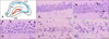
A schematic of the various hippocampal regions is shown in panel A in Fig. 1. In the sham-operated gerbils, the pyramidal cells in the CA1 region were densely distributed and exhibited normal morphologies (panel B in Fig. 1). One day after BCCAO, the neurons in the CA1 region of the hippocampus were sparsely distributed, many of the cells had clumped chromatin, and some of the cells had pale cytoplasm (panel C in Fig. 1). Three days after BCCAO, cells with pale cytoplasm were more frequently observed, and some of the pyramidal cells displayed condensed nuclei (panel D in Fig. 1). Five days after the operation, cells with condensed nuclei were more frequently observed (panel E in Fig. 1). Ten days after BCCAO, most of the pyramidal cells had condensed nuclei (panel F in Fig. 1).
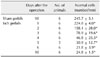
The number of CA1 pyramidal neurons with normal morphologies for both sham and BCCAO gerbils are shown in Table 1. On all days after BCCAO, the numbers of normal cells in ischemic gerbils were significantly lower than those in sham-operated ones.
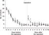
In the radial maze test, the number of errors was significantly higher on the 1st and 2nd days after BCCAO than after the sham operation. The number of errors in the BCCAO group tended to decrease with each consecutive training day and became comparable to that of the sham-operated gerbils at post-operative day 3 and thereafter (Fig. 2). The ANOVA and Tukey's HSD test results revealed a significant decrease in the number of errors from 8 to 7 days before the operation in both the Isch and Sham groups. In the Isch group, the number of errors significantly increased from pre-operative day 1 to post-operative day 1 and significantly decreased from 1 day to 2 days after the operation.
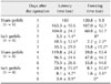
In the passive avoidance test, on the 1st and 2nd days and the 4th to 6th days after the operation, the latency time and freezing times were significantly lower in Isch group than in the Sham group (Table 2). ANOVA and Tukey's HSD test results revealed no significant difference in latency and freezing times among post-operative days in the Isch group. However, there was a significant decrease in latency and freezing times from the 2nd to 3rd post-operative day in the Sham group.
Discussion
The present study demonstrated that cognitive performance is impaired by transient forebrain ischemia before any remarkable neuronal cell death, and that memory impairment can recover in spite of the onset of significant neuronal cell death. These results, obtained by examining the animals immediately after ischemia onset, may be pertinent in the development of therapies to prevent cognitive impairment in patients during the days immediately after transient brain ischemia in the event of a stroke.
Kirino [19] has previously reported that the nuclear chromatin of pyramidal cells is slightly clumped at 1 day after bilateral carotid occlusion. In the present study, pyramidal cells with clumped chromatin were found in the CA1 region at one day after BCCAO. These observations are the first signs of neuronal cell death in the hippocampus after transient forebrain ischemia. The present study identified that some pyramidal cells have condensed nuclei at 3 days post-BCCAO and nuclei-condensed cells were more frequent at 5 days post-BCCAO; as well, most pyramidal cells had condensed nuclei at 10 days post-BCCAO. These results suggest that there is progressive neurodegeneration after BCCAO, which supports the results in a previous report that showed widespread necrotic changes in the CA1 region 3 days after the common carotid occlusion [35].
It seems unlikely that only a single foraging strategy is used by a species, and such animals likely use both win-stay and win-shift strategies [38]. It has been reported that normal rats predominantly use the win-shift strategy rather than the win-stay strategy in food-searching situations [10]. Moreover, rats learn the win-shift strategy more rapidly than the win-stay strategy [28]. Similarly, Mongolian gerbils have more difficulty learning the win-stay strategy than the win-shift strategy [8]. Therefore, in the present study, we chose to evaluate the effect of BCCAO on cognitive performance by using the more difficult to learn win-stay test.
In many studies, maze tests after several days of post-ischemia rest have been used to relate neuronal cell death to cognitive impairment. However, it is difficult to decipher the relationship between the process of neuronal cell death and memory impairment from studies in which behavioral analysis is performed several days after an ischemic operation, since it has been reported that significant neuronal cell death has already occurred at 4 days after ischemia [1526]. Therefore, we investigated cognitive performance in Mongolian Gerbils immediately after ischemia.
In the present study, gerbils showed a memory defect on days 1 and 2 post-BCCAO when clumped chromatin in the CA1 pyramidal cells was visible histologically. However, the number of erroneous selections in the maze test decreased with each trial after surgery, and the influence of ischemia was no longer evident on the final test day in the maze. These results suggest that memory impairment occurs before the onset of significant neural cell death in the CA1 region and that ischemia-induced impairment in spatial learning and memory shows recovery with repeated experience. This notion is supported by the following observations: Delayed neuronal death began to appear 3 days after occlusion, but memory deficit induced by a 5 min ischemia period began to recover at 3 days after the occlusion [14]; memory impairment in the radial maze at 10 days after occlusion was easily overcome, and that performance recovered to the normal level after additional training trials [1]; rats are able to perform cued-learning and discrimination-learning tasks, suggesting some degree of spontaneous behavioral recovery after ischemic injury [36].
There are two possible reasons that this study showed recovery of cognitive impairment starting 3 days after ischemia. One possibility for this recovery is the experimental schedule to which the gerbils in the present study were subjected (i.e., the acquisition memory task for 10 days before ischemic surgery) and another is the potential for compensation by another region of the brain at an early stage. The former is supported by a report by Babcock and Graham-Goodwin [3] that demonstrated that pre-operative training can reduce working memory impairment following ischemia in gerbils subjected to a win-stay task, while the latter is supported by the present results that show no severe pathological change in regions other than the CA1 region of the hippocampus (e.g., CA3, dentate gyrus, and striatum). Thus, the present study, examining memory impairment over the several days immediately after ischemia and associated pathological changes of the brain, points to the importance of developing therapeutics that could prevent cognitive impairment during the initial several days after transient brain ischemia in the event of a stroke.
Fujimoto et al. [9] used the passive avoidance test to evaluate the effect of bisphenol A on learning and memory (avoidance learning) of rats. Stubley-Weatherly et al. [34] used the passive avoidance test as an associative/contextual paradigm when they evaluated the effects of an excitatory amino acid lesion within the rat hippocampus. Kohara et al. [20] examined cognitive dysfunction in glucose-loaded rats by using the step-through passive avoidance test, which assesses fear-motivated learning and memory, as well as the maze test, which measures spatial learning and memory. Thus, we used the maze test to evaluate spatial memory performance and the passive avoidance test to evaluate associative/contextual memory performance. In the present study, latency and freezing times in the passive avoidance test were significantly lower in BCCAO gerbils than in sham-operated gerbils on the 1st and 2nd days after the operation. The increase in locomotor activity in BCCAO gerbils after 5 min in the bright period of light and dark cycle was most remarkable at 1 day after the BCCAO operation, compared to that in the sham-operated animals [2]. Post-ischemic hyperactivity is a well-known behavioral change in a transient forebrain ischemic model using gerbil [17]. Based upon these results, the decreased times in latency and freezing at least on the 1st day after operation in the present study may imply hyperactivity rather than impairment of associative/contextual memory performance by transient forebrain ischemia. Nishino et al. [25] reported that damage to the striatum leads to hyperactivity; moreover, Kawaguchi et al. [16] reported that the striatum may be involved in fear-motivated learning and emotionality. Therefore, the hyperactivity seen in this study may have been induced by striatum impairment due to transient forebrain ischemia. However, Block [5] has reported that such hyperactivity was attributed to hippocampal neuronal damage. Further, we have been unable to detect impairments (neuronal cell death) in the striatum of gerbils induced by the BCCAO (unpublished data). It is thought that the significant decrease in latency and freezing times on the 1st day after BCCAO in the present study implies impairment of the hippocampus by the operation. This is supported by a report by Mileson and Schwartz [24] that indicated that the increase in gerbil locomotor activity following a 5-min bilateral carotid artery occlusion can be used as a predictor of CA1 damage but not as a predictor of striatal or cortical damage.
Since the hyperactivity level induced by ischemia becomes comparable to that in sham-operated animals at 4 days after occlusion [2], the present observation that the decreased times in latency and freezing were still observable on the 4th to 6th days after operation indicates that associative/contextual memory was damaged by the transient forebrain ischemia, suggesting that the defects in associative/contextual memory are related to the hippocampal neuronal cell death. This notion is supported by the report by Soeda et al. [32] that showed that the step-through passive avoidance response requires hippocampus-dependent learning.
In the Sham group, latency time decreased with each trial over the 1st to 3rd and 4th to 6th post-operative days (the difference between the 2nd and 3rd post-operative day was significant); however, in the Isch group, latency time increased over the 1st to 3rd post-operation days and decreased over the 4th to 6th post-operation days. Since no electrical stimulation was given during passive avoidance tests performed on post-operation days, it seems that the Sham group gerbils recognized that the dark compartment was not dangerous. However, among the Isch group gerbils, there was no such recognition.
In conclusion, the present study has revealed that transient forebrain ischemia induces an early defect in cognitive performance that occurs prior to the onset of significant neuronal cell death, and that associative/contextual memory is strongly affected by transient forebrain ischemia.
 XML Download