

 PDF
PDF ePub
ePub Citation
 Print
Print


Abstract
Purpose
To prevent excessive sliding and subsequent fixation failures in unstable intertrochanteric fractures with posteromedial comminution, extramedullary reduction through overlapping of the anteromedial cortices of both proximal and distal fragments as a buttress has been introduced. The purpose of this study was to compare the biomechanical properties between two reduction methods–intramedullary reduction and extramedullary reduction–in treating unstable intertrochanteric fractures with posteromedial comminution (AO/OTA classification 31-A2.2).
Materials and Methods
Eight pairs of frozen human cadaveric femora were used. The femora of each pair were randomly assigned to one of two groups: the intramedullary reduction group or the extramedullary reduction group. A single axial load-destruction test was conducted after cephalomedullary nailing. Axial stiffness, maximum load to failure, and energy absorbed to failure were compared between the two groups. Moreover, the pattern of mechanical failure was identified.
Results
The mean axial stiffness in the extramedullary reduction group was 27.3% higher than that in the intramedullary reduction group (422.7 N/mm vs. 332.0 N/mm, p=0.017). Additionally, compared with the intramedullary reduction group, the mean maximum load to failure and mean energy absorbed to failure in the extramedullary group were 44.9% and 89.6% higher, respectively (2,848.7 N vs. 1,966.5 N, p=0.012 and 27,969.9 N·mm vs. 14,751.0 N·mm, p=0.012, respectively). In the intramedullary reduction group, the mechanical failure patterns were all sliding and varus deformities. In the extramedullary reduction group, sliding and varus deformities after external rotation were noted in 3 specimens, sliding and varus deformities after internal rotation were noted in 3 specimens, and medial slippage was noted in 2 specimens.
Conclusion
In unstable intertrochanteric fractures with posteromedial comminution, the biomechanical properties of extramedullary reduction are superior to those of intramedullary reduction. Anteromedial cortex could be the proper buttress, despite a comminuted posteromedial cortex. It could help enhance the stability of the bone-nail construct.
Go to : 
References
1. Zlowodzki M, Jönsson A, Paulke R, Kregor PJ, Bhandari M. Shortening after femoral neck fracture fixation: is there a solution? Clin Orthop Relat Res. 461:213–218. 2007.
2. Zlowodzki M, Brink O, Switzer J, et al. The effect of shortening and varus collapse of the femoral neck on function after fixation of intracapsular fracture of the hip: a multicentre cohort study. J Bone Joint Surg Br. 90:1487–1494. 2008.
3. Song HK, Yoon HK, Yang KH. Presence of a nail in the medullary canal; is it enough to prevent femoral neck shortening in trochanteric fracture? Yonsei Med J. 55:1400–1405. 2014.

4. Gilat R, Lubovsky O, Atoun E, et al. Proximal femoral shortening after cephalomedullary nail insertion for intertrochanteric fractures. J Orthop Trauma. 31:311–315. 2017.
5. Filipov O. Biplane double-supported screw fixation (F-technique): a method of screw fixation at osteoporotic fractures of the femoral neck. Eur J Orthop Surg Traumatol. 21:539–543. 2011.
6. Mir H, Collinge C. Application of a medial buttress plate may prevent many treatment failures seen after fixation of vertical femoral neck fractures in young adults. Med Hypotheses. 84:429–433. 2015.
7. Haidukewych GJ. Intertrochanteric fractures: ten tips to improve results. J Bone Joint Surg Am. 91:712–719. 2009.
8. Weiser L, Ruppel AA, Nüchtern JV, et al. Extra- vs. intramedullary treatment of pertrochanteric fractures: a biomechanical in vitro study comparing dynamic hip screw and intramedullary nail. Arch Orthop Trauma Surg. 135:1101–1106. 2015.
9. Carr JB. The anterior and medial reduction of intertrochanteric fractures: a simple method to obtain a stable reduction. J Orthop Trauma. 21:485–489. 2007.
10. Tsukada S, Okumura G, Matsueda M. Postoperative stability on lateral radiographs in the surgical treatment of pertrochanteric hip fractures. Arch Orthop Trauma Surg. 132:839–846. 2012.
11. Kozono N, Ikemura S, Yamashita A, Harada T, Watanabe T, Shirasawa K. Direct reduction may need to be considered to avoid postoperative subtype P in patients with an unstable trochanteric fracture: a retrospective study using a multivariate analysis. Arch Orthop Trauma Surg. 134:1649–1654. 2014.
12. Chang SM, Zhang YQ, Ma Z, Li Q, Dargel J, Eysel P. Fracture reduction with positive medial cortical support: a key element in stability reconstruction for the unstable pertrochanteric hip fractures. Arch Orthop Trauma Surg. 135:811–818. 2015.
13. Ito J, Takakubo Y, Sasaki K, Sasaki J, Owashi K, Takagi M. Prevention of excessive postoperative sliding of the short femoral nail in femoral trochanteric fractures. Arch Orthop Trauma Surg. 135:651–657. 2015.
14. Park SY, Park J, Rhee DJ, Yoon HK, Yang KH. Anterior or posterior obliquity of the lag screw in the lateral view-does it affect the sliding characteristics on unstable trochanteric fractures? Injury. 38:785–791. 2007.
15. Kuzyk PR, Zdero R, Shah S, Olsen M, Waddell JP, Schemitsch EH. Femoral head lag screw position for cephalomedullary nails: a biomechanical analysis. J Orthop Trauma. 26:414–421. 2012.
16. Rybicki EF, Simonen FA, Weis EB Jr. On the mathematical analysis of stress in the human femur. J Biomech. 5:203–215. 1972.
17. Nicayenzi B, Shah S, Schemitsch EH, Bougherara H, Zdero R. The biomechanical effect of changes in cancellous bone density on synthetic femur behaviour. Proc Inst Mech Eng H. 25:1050–1060. 2011.
19. Rüedi TP, Buckley RE, Moran CG. AO principles of fracture management. 2nd ed.New York, Thieme: 249–256;2007.
20. Teramoto H, Uchida K, Kaneko S, Koshimune K, Ise M, Murakami T. The change of anatomical reposition cases in femoral trochanteric fracture with lateral view X ray. Fracture. 38:650–652. 2016.
21. Parker MJ. Valgus reduction of trochanteric fractures. Injury. 24:313–316. 1993.
22. Andruszkow H, Frink M, Frömke C, et al. Tip apex distance, hip screw placement, and neck shaft angle as potential risk factors for cut-out failure of hip screws after surgical treatment of intertrochanteric fractures. Int Orthop. 36:2347–2354. 2012.
Go to :
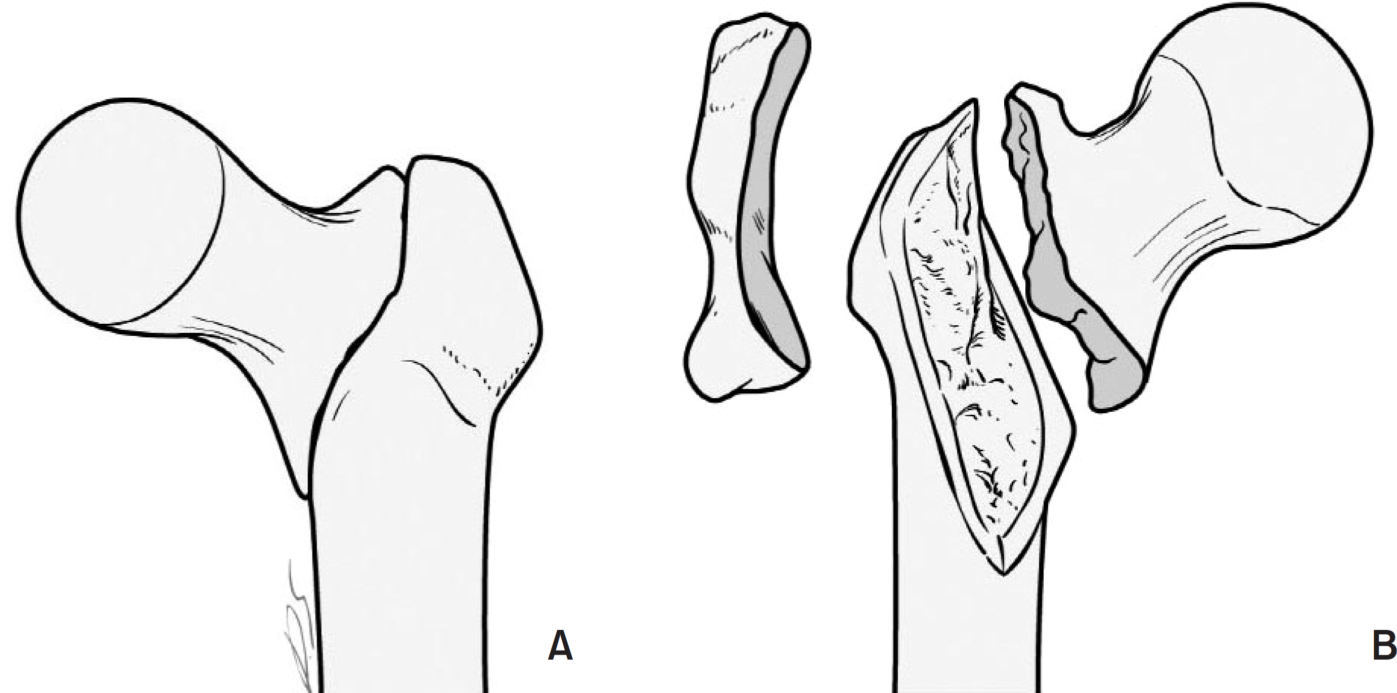 | Fig. 1.Schematic drawing of fracture creation. Unstable intertrochanteric fracture with posteromedial defect including the lesser and greater trochanters (AO/OTA classification 31-A2.2); anterior (A) and posterior (B) views. |
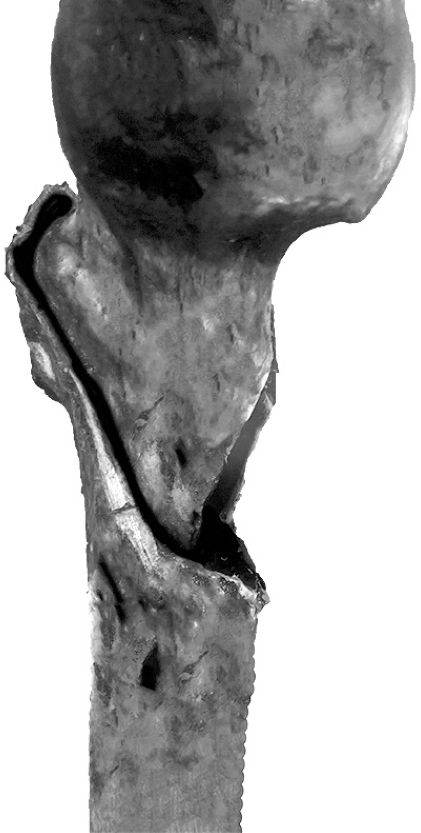 | Fig. 2.Intramedullary reduction. The anteromedial cortex of the proximal fragment is positioned inside the distal shaft fragment (anteromedial aspect of femur). |
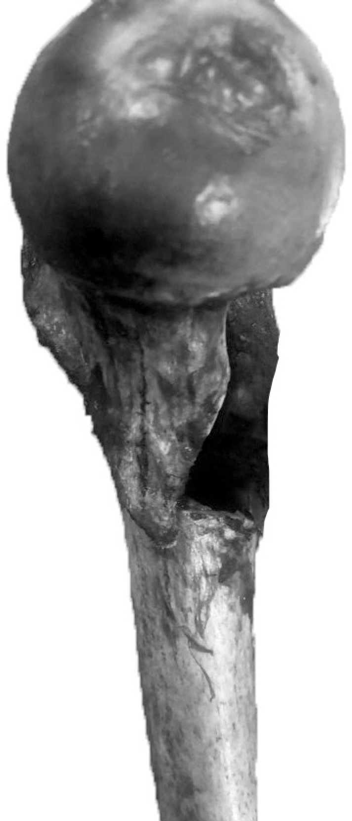 | Fig. 3.Extramedullary reduction. The anteromedial cortex of the proximal fragment is positioned outside the distal shaft fragment (anteromedial aspect of femur). |
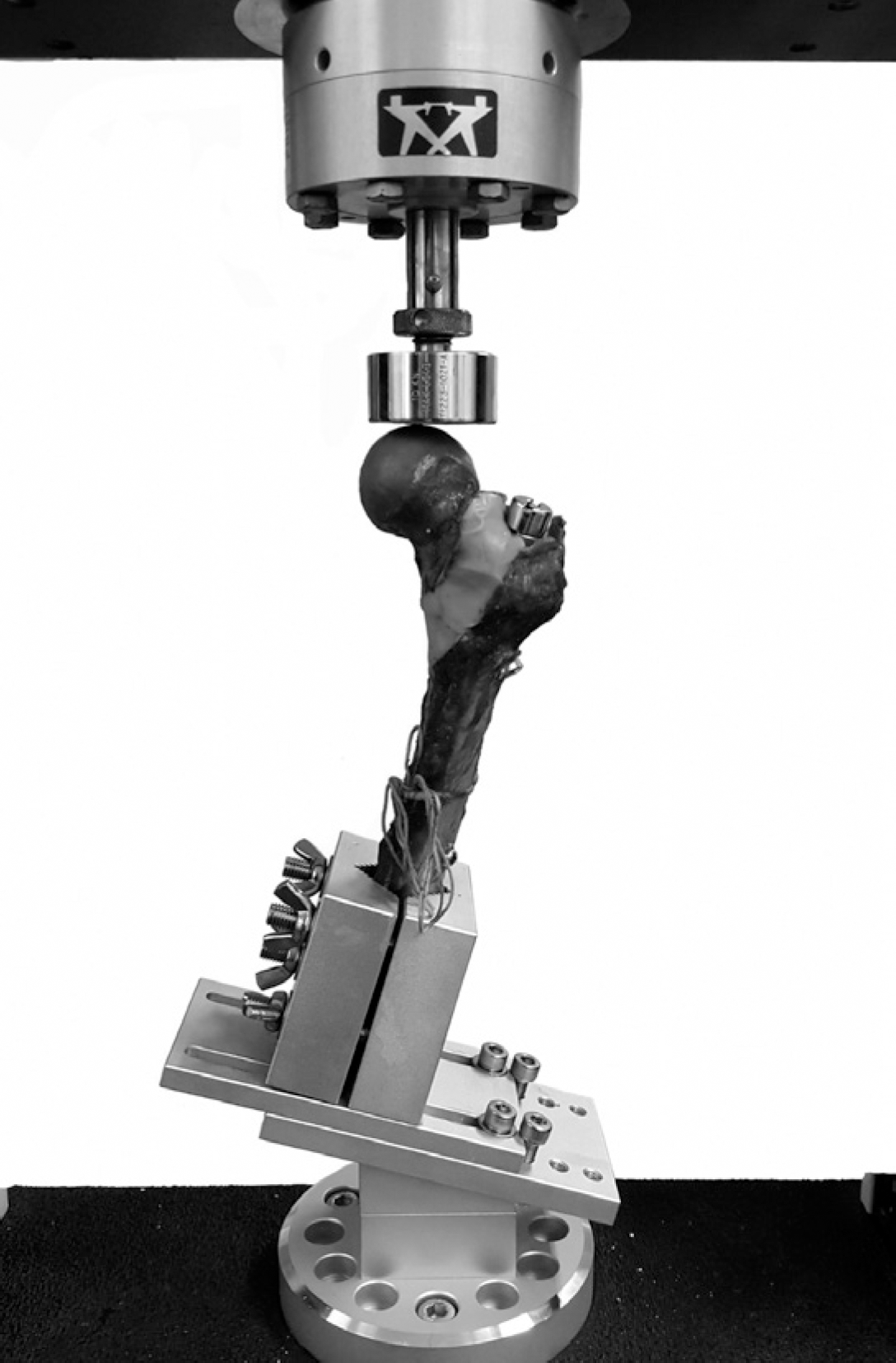 | Fig. 4.Setup of the mechanical test. The specimen is embedded at 15 degrees in the varus position. |
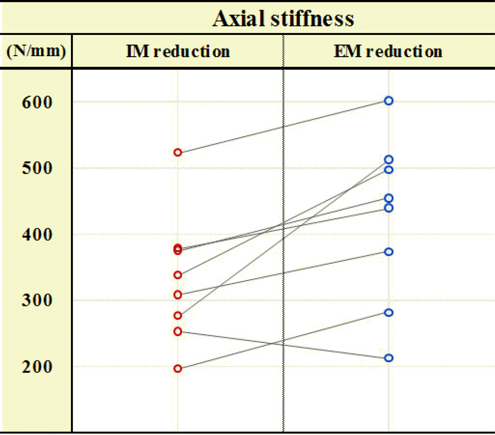 | Fig. 5.Paired graph showing differences in axial stiffness between the intramedullary (IM) reduction and extramedullary (EM) reduction groups. |
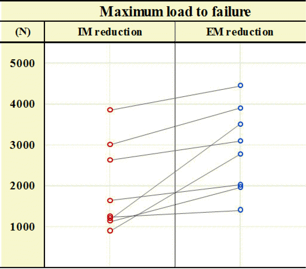 | Fig. 6.Paired graph showing differences in maximum load to failure between the intramedullary (IM) reduction and extramedullary (EM) reduction groups. |
Table 1.
Axial Stiffness, Maximum Load to Failure, Energy Absorbed to Failure, and Axial Displacement for the Intramedullary Reduction and Extramedullary Reduction Groups
Table 2.
Mechanical Failure Patterns for the Intramedullary Reduction and Extramedullary Reduction Groups
 XML Download