Journal List > Ann Lab Med > v.38(6) > 1099049

Nam, Shin, Park, Kim, Yoon, Kwon, and Song: Association of FOXP3 Single Nucleotide Polymorphisms With Clinical Outcomes After Allogenic Hematopoietic Stem Cell Transplantation

Abstract

Background

Forkhead box P3 (FOXP3) is an important marker of regulatory T cells. FOXP3 polymorphisms are associated with autoimmune diseases, cancers, and allograft outcomes. We examined whether single nucleotide polymorphisms (SNPs) at the FOXP3 locus are associated with clinical outcomes after allogenic hematopoietic stem cell transplantation (HSCT).

Methods

Five FOXP3 SNPs (rs5902434, rs3761549, rs3761548, rs2232365, and rs2280883) were analyzed by PCR-sequencing of 172 DNA samples from allogenic HSCT patients. We examined the relationship between each SNP and the occurrence of graft-versus-host disease (GVHD), post-HSCT infection, relapse, and patient survival.

Results

Patients with acute GVHD (grades II-IV) showed higher frequencies of the rs3761549 T/T genotype, rs5902434 ATT/ATT genotype, and rs2232365 G/G genotype than did patients without acute GVHD (P=0.017, odds ratio [OR]=5.3; P=0.031, OR=2.4; and P=0.023, OR=2.6, respectively). Multivariate analysis showed that the TT genotype of rs3761549 was an independent risk factor for occurrence of acute GVHD (P=0.032, hazard ratio=5.6). In contrast, the genotype frequencies of rs3761549 T/T, rs5902434 ATT/ATT, and rs2232365 G/G were lower in patients with post-HSCT infection than in patients without infection (P=0.026, P=0.046, and P=0.031, respectively).

Conclusions

rs3761549, rs5902434, and rs2232365 are associated with an increased risk of acute GVHD and decreased risk of post-HSCT infection.

INTRODUCTION

Allogenic hematopoietic stem cell transplantation (HSCT) is a curative treatment for patients with hematologic malignancy, bone marrow failure, and congenital immunologic diseases. However, despite the increasing use of allogenic HSCT, it still has severe complications with high morbidity and mortality, such as acute and chronic graft-versus-host disease (GVHD) and post-HSCT infections [1]. GVHD is a donor T cell-mediated immune response, which results in tissue damage of the recipient [2]. The removal of donor T cells can prevent GVHD, but contributes to a delayed immune constitution and thus increased opportunistic infections [34].
During immune responses, regulatory T cells (Tregs) not only play essential roles in maintaining immune homeostasis [5] but also establish tolerance after transplantation [6]. Recent studies have implicated Tregs in the development of GVHD and suggested that the immunosuppressive potential of Tregs can be used therapeutically to reduce the incidence and/or severity of GVHD [7]. The use of Tregs in transplantation has garnered substantial interest. Many researchers have attempted to identify new markers related to Tregs and to develop therapeutic strategies to improve graft survival and avoid post-transplant complications [89].
Forkhead box P3 (FOXP3) is a master regulator of Treg development and function. FOXP3 expression is important to regulate CD4+CD25+ Treg development and function, but the molecular mechanisms that are involved in the regulation of the FOXP3 expression remain unclear [10]. Many studies have shown that the regulatory mechanism of gene expression is controlled by genomic polymorphisms. Single nucleotide polymorphisms (SNPs) in FOXP3 have been associated with various diseases [11], including asthma [12], preeclampsia [13], systemic lupus erythematosus [14], autoimmune thyroid disease [15], lung cancer [16], breast cancer [17], and colorectal cancer [18]. Recently, several studies reported that FOXP3 SNPs are associated with allograft outcomes after renal transplantation, but there is ongoing debate over whether FOXP3 SNPs have a positive or negative association with allograft outcomes [19202122]. Moreover, few studies have investigated whether there is an association between SNPs in FOXP3 and clinical outcomes after allogenic HSCT [23]. We examined the association between five SNPs (rs5902434, rs3761549, rs3761548, and rs2232365 located in the promoter region and rs2280883 located in the intronic region) in FOXP3 and different clinical outcomes after allogenic HSCT: the occurrence of GVHD, post-HSCT infection, relapse, and patient survival. These five SNPs were selected among those directly or potentially associated with diseases by a literature search and analysis using the HaploReg v. 4.1 database.

METHODS

1. Study population

Our retrospective study included 172 patients with hematologic malignancy or bone marrow failure who received allogenic HSCT between April 2006 and August 2014 at Seoul National University Hospital, Seoul, Korea. Written informed consent was obtained from all patients. Baseline clinical characteristics, including age and sex of patients and donors, underlying diagnosis, stem cell source, the number of HLA mismatches, cytomegalovirus (CMV) IgG seropositivity, conditioning regimen, and European Society for Blood and Marrow Transplantation (EBMT) risk score [24], were obtained from medical records (Table 1).
Patients' median age was 37 years (range 17–67 years), and the majority were male (60.5%). Most patients had fewer than two mismatched HLA alleles, except for six patients who had three or more allele mismatches. In addition, most patients were diagnosed as having acute leukemia before allogenic HSCT, and the dominant stem cell source was peripheral blood (92.4%). Conditioning chemotherapy was performed before transplantation for patients who received HSCT from an unrelated donor. The regimen varied according to the type of underlying disease and the condition of the patient, but included the following: busulfan plus (cyclophosphamide or anti-thymocyte globulin plus fludarabine), fludarabine plus (cyclophosphamide, melphalan, cyclophosphamide plus anti-thymocyte globulin, or melphalan plus anti-thymocyte globulin), total body irradiation plus cyclophosphamide, and total lymphocyte irradiation plus anti-thymocyte globulin. Patients were treated with cyclosporine or tacrolimus with or without a short course of methotrexate (days 1, 3, 6, and 10) as GVHD prophylaxis and treated with ciprofloxacin, itraconazole, acyclovir, sulfamethoxazole/trimethoprim, or intravenous immune globulins as infection prophylaxis. Our study was approved by the Institutional Review Board for Human Research of Seoul National University (IRB No. 1702-024-829).
Acute (grades II–IV) and chronic GVHD were diagnosed based on published criteria [2526]. To reduce potential bias of the acute GVHD group, 13 patients who died within 28 days after allogenic HSCT were excluded from our analysis of acute GVHD, but they were included in our analysis of infection. Infection was defined as the isolation of a certain pathogen, such as virus, bacteria, fungus, and tuberculosis (TB), from microbial cultures or positive results from nucleic acid amplification or antigen tests. Disease relapse was determined based on the bone marrow examination of patients. Overall survival was defined as the time from graft infusion to death from any cause at time of analysis (March 1, 2017). For event-free survival, death or relapse was considered events.

2. DNA preparation and FOXP3 SNP genotyping

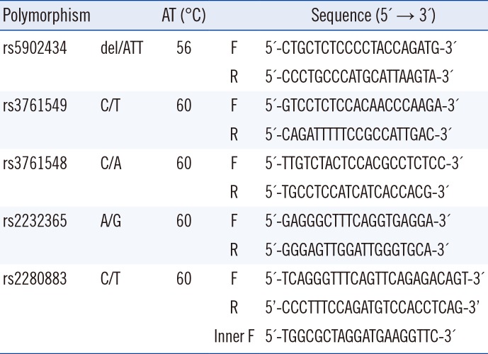
We collected DNA samples from 172 patients whose pre-transplant HLA typing test had been requested. DNA was extracted from peripheral blood or bone marrow using the QuickGene-Mini80 DNA isolation system (Fujifilm, Tokyo, Japan) when pre-transplant HLA typing was done. After HLA typing, DNA stored at −80℃ were genotyped for the selected five FOXP3 SNPs (rs5902434, rs3761549, rs3761548, and rs2232365 in promoter region, and rs2280883 in intronic region) by PCR sequencing. Among our cohort of 172 patients, we excluded two to five cases depending on the SNP, because they could not be accurately genotyped due to poor DNA sample quality. PCR was performed with 40 µL reaction mixtures containing 40 ng DNA, 0.8 µL dNTP mix (10 mM of each dNTP), 2 µL of each primer at a concentration of 10 pmol/µL, 2.0 mM MgCl2, 1.0 U Taq DNA polymerase (Roche, Basel, Switzerland), and 4 µL of 10X reaction buffer. Five SNPs were analyzed following the identical PCR protocol. Initial denaturation was performed at 95℃ for 5 minutes, followed by 30 cycles of denaturation at 95℃ for 30 seconds, annealing at the respective annealing temperature for 30 seconds, and extension at 72℃ for 30 seconds with a final extension at 72℃ for 5 minutes. Next, 2 µL ExoSAP-IT PCR Clean Up (Affymetrix, Santa Clara, CA, USA) was added to 5 µL of PCR product, followed by incubation at 37℃ for 15 minutes and at 80℃ for 15 minutes. We then added 1 µL of 5 pmol/µL sequencing primer, 4 µL of deionized water, and 4 µL of BigDye Terminator Ready Reaction Mix (Life Technologies, Grand Island, NY, USA) to 1 µL of purified PCR product. Following 30 thermal cycles of 96℃ for 10 seconds, 50℃ for 5 seconds, and 60℃ for 4 minutes, 25 µL of absolute ethanol (EtOH) and 2 µL of 3 M sodium acetate/ EDTA buffer (pH 4.6) were added. After vortexing and centrifugation at 2,000×g for 30 minutes, the supernatant was discarded, following by the addition of 50 µL of 80% EtOH and centrifugation at 2,000×g for 5 minutes. Finally, 15 µL of Hi-Di Formamide (Life Technologies) was added, and the sample was heated at 95℃ for 4 minutes. Samples were then analyzed on an ABI 3730XL DNA analyzer (Applied Biosystems, Foster City, CA, USA), and electropherograms were analyzed using Chromas Lite 2.1.1 (Technelysium, South Brisbane, Australia) (Table 2).

3. Statistical analysis

Differences in genotype distributions between each group were calculated using Pearson's chi-squared test or Fisher's exact test. Univariate and multivariate analyses were performed using logistic regression. The following variables were included in our analysis: recipient age and sex, the number of HLA allelic matches for HLA-A, B, Cw, DR, and DQ locus, the type of disease at transplantation, the conditioning regimen, stem cell source, the EBMT risk score, and donor age and sex. Estimates of acute GVHD and relapse were calculated using cumulative incidence rates. Overall survival and event-free survival were calculated using the Kaplan-Meier method and compared using the log-rank test. Odds ratios (ORs) and 95% confidence intervals (CIs) were estimated for genotypes showing a significant P value. P<0.05 was considered statistically significant. Statistical analysis was performed using SPSS Statistics for Windows Version 23.0 (IBM Corp., Armonk, NY, USA).

RESULTS

1. Association between FOXP3 SNPs and acute GVHD and post-HSCT infection

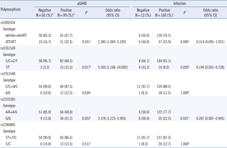
Acute GVHD patients showed an increased frequency of the rs3761549 T/T genotype (15.5% vs 3.3%, respectively, OR=5.3, P=0.017), rs5902434 ATT/ATT (32.3% vs 16.7%, OR=2.4, P=0.031), and rs2232365 G/G (31.2% vs 15.0%, OR=2.6, P=0.023), whereas patients with post-HSCT infection had lower frequencies of these genotypes (8.9% vs 33.3%, respectively, OR=0.2, P=0.026; 23.9% vs 50.0%, OR=0.3, P=0.046; and 22.3% vs 50.0%, OR=0.3, P=0.031, respectively) (Table 3).
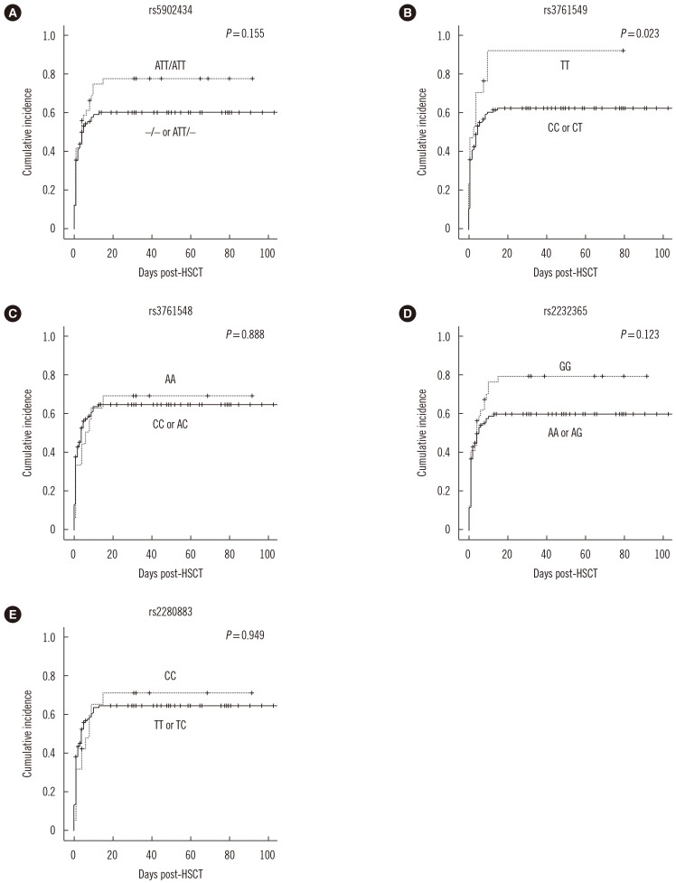
No association was observed between genotype frequency of these five FOXP3 SNPs and infection of a given pathogen, such as bacteria, CMV, Epstein-Bar virus, fungus, and TB (data not shown). Fig. 1 shows that patients with the T/T genotype of rs3761549 showed higher cumulative incidence of acute GVHD than those patients with the C/C or C/T genotype (P=0.023), while patients with the ATT/ATT genotype of rs5902434 or the G/G genotype of rs2232365 showed a tendency of higher cumulative incidence of acute GVHD, although these findings were not statistically significant (P=0.155 and P=0.123, respectively) (Fig. 1).
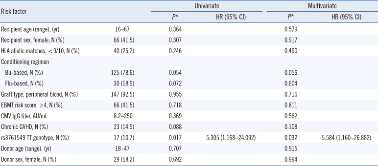
In the multivariate analysis, the TT genotype of FOXP3 rs3761549 was an independent risk factor for the occurrence of acute GVHD (P=0.032, hazard ratio=5.584, 95% CI=1.160–26.882; Table 4). The five FOXP3 SNPs were not associated with chronic GVHD (data not shown).

2. Association between FOXP3 SNPs and relapse and patient survival

Based on the 50 patients in our cohort who relapsed, we found no evidence of an effect of FOXP3 SNP genotype on the incidence of relapse. Genotypes at these five FOXP3 SNPs did not influence their overall survival and event-free survival (data not shown).

DISCUSSION

We evaluated the clinical impact of five different FOXP3 SNPs on outcomes of allogenic HSCT. A previous study found that patients with the rs3761548 CC genotype showed higher incidences of hepatic veno-occlusive disease and CMV infection, but found no evidence of an association with GVHD or relapse [23]. We also found no evidence of an association between rs3761548 and clinical outcomes in allogenic HSCT, although rs3761548 has been associated with various immune-related disorders [11] and renal allograft outcomes [1920].
The T/T genotype of rs3761549 has been associated with the development and progression of endometriosis [27], endometriosis-related infertility [28], and Graves' disease [15]; thus, this genotype may be involved in regulation of inflammatory or autoimmune responses. However, no study has investigated whether there is an association between rs3761549 and clinical outcomes in organ transplantation or HSCT.
Wu et al [29] reported an association between the del genotype of rs5902434 and an increased risk of unexplained recurrent spontaneous abortion. However, other studies did not find a significant association with renal allograft outcomes [21] or psoriasis susceptibility [30]. Wu et al [29] also reported an association between the G/G genotype of rs2232365 and increased risk of unexplained recurrent spontaneous abortion. Misra et al [20] reported that renal allograft patients with the rs2232365 G/G genotype showed lower overall survival and 5-year survival. In contrast, Qui et al [19] found no association between rs2232365 and renal allograft rejection in their cohort.
Because of these conflicting findings, the impact of rs3761548, rs3761549, rs5902434, and rs2232365 at the FOXP3 locus on regulatory immune responses, which are related to the development of acute GVHD after allogenic HSCT, remains unclear. These four SNPs are all located in the FOXP3 promoter region, which contains DNA binding sites for transcription factors (TFs). It can be hypothesized that depending on the alleles of these SNPs in FOXP3, FOXP3 expression are modulated by altering the binding affinity of transcription factors to their binding element and by modifying the kinetics of transcription regulation. A variant(s) is likely to contribute to a decrease in the quantity or quality of FOXP3, resulting in a sequential decrease of Tregs, which play a critical role in suppressing autoreactive lymphocytes and regulating hyperactive immune responses. To clarify the putative functional relevance of FOXP3 SNP genotypes in detail, we utilized the P-match program (http://gene-regulation.com/pub/programs.html#pmatch) to search TF binding sites [31]. Interestingly, we found that rs3761549 is situated in a DNA binding site for activating enhancer binding protein 4 (AP4), and it is predicted that AP4 cannot bind to DNA with the T allele of rs3761549. AP4 is a TF regulated by IL-2R signaling involved in a Myc-dependent gene expression program that sustains the rapid clonal expansion of antigen-specific CD8+ T cells by encoding components of the glycolysis pathways [32]. Therefore, although our understanding of the molecular mechanism of AP4 and FOXP3 in Tregs is limited, the modification of AP4 binding affinity in individuals homozygous for rs3761549T may induce less clonal expansion of Tregs and relative proliferation of effector T lymphocytes. We speculate further that these changes may cause the destruction of tissues and organs, thus leading to an increased incidence and/or severity of acute GVHD [33].
Importantly, the rs3761549 T/T genotype, rs5902434 ATT/ATT genotype, and rs2232365 G/G genotype were associated with decreased risk of post-HSCT infection. Tregs have been related to immune responses for pathogens and outcomes of some infectious diseases [3435]. More specifically, Tregs inhibit the proliferation of effector cells, such as CD4+ helper T cells or CD8+ cytotoxic T cells, and the production of cytokines such as interferon-γ, as well as directly secrete immunosuppressive cytokines, such as IL-10 and TGF-β [3637]. Further, a decreased number of Tregs confers resistance to viral infection [38]. Therefore, in our study, the association of FOXP3 SNPs with a lower infection rate after HSCT may reflect down-regulation of Tregs, which inhibit helper T cells, cytotoxic T cells, and cytokine production, and may contribute in preventing pathogen proliferation by effector T cells and cytokine release.
Few studies have investigated associations between FOXP3 SNPs and infection after organ transplantation or HSCT. Piao et al [23] reported an association between rs3761548 and CMV infection after allogenic HSCT. SNPs of other immune-regulating genes, such as IL28B, TLR9, DC-SIGN (also known as CD209), and IFNL3/4 (interferon lambda 3/interferon lambda 4), have been associated with CMV infection in solid organ transplantation [3940]. In our study, the lack of an association between FOXP3 SNPs and post-HSCT infection in subgroup analysis including CMV infection may have been caused by the small number of cases.
The present study has some limitations. The number of patients was relatively small, and patients were recruited from a single center, so there may be some selection bias. Further studies with a larger number of patients are needed to confirm our hypothesis. In addition, we did not confirm whether FOXP3 SNP genotypes actually contributed to the decrease in the FOXP3 expression, although we checked which TFs were able to bind to FOXP3 SNPs using software.
Nevertheless, our findings provide the first evidence that allogenic HSCT patients harboring the rs3761549 T/T genotype, the rs5902434 ATT/ATT genotype, and the rs2232365 G/G genotype have an increased risk of acute GVHD and a decreased risk of post-HSCT infection. These SNPs can be used as potential markers to predict clinical outcomes of allogenic HSCT and provide personalized care for high-risk patients. Furthermore, these findings may also contribute toward the development of new treatment modalities targeting immune reconstitution or enhancing Tregs population in allogenic HSCT.

Acknowledgment

This research was supported by a grant from the Basic Science Research Program through the National Research Foundation of Korea (NRF), funded by the Ministry of Education, Science, and Technology (grant number 2015R1D1A1A01060641).

Notes

Authors' Disclosures of Potential Conflicts of Interest: The authors declare that no conflicts of interest exist.

References

1. Arnaout K, Patel N, Jain M, El-Amm J, Amro F, Tabbara IA, et al. Complications of allogeneic hematopoietic stem cell transplantation. Cancer Invest. 2014; 32:349–362. PMID: 24902046.
2. Beres AJ, Drobyski WR. The role of regulatory T cells in the biology of graft versus host disease. Front Immunol. 2013; 4:163. PMID: 23805140.
3. Gratama JW, van Esser JW, Lamers CH, Tournay C, Löwenberg B, Bolhuis RL, et al. Tetramer-based quantification of cytomegalovirus (CMV)-specific CD8+ T lymphocytes in T-cell-depleted stem cell grafts and after transplantation may identify patients at risk for progressive CMV infection. Blood. 2001; 98:1358–1364. PMID: 11520783.
4. van Esser JW, van der Holt B, Meijer E, Niesters HG, Trenschel R, Thijsen SF, et al. Epstein-Barr virus (EBV) reactivation is a frequent event after allogenic stem cell transplantation (SCT) and quantitatively predicts EBV-lymphoproliferative disease following T-cell--depleted SCT. Blood. 2001; 98:972–978. PMID: 11493441.
5. Sakaguchi S, Yamaguchi T, Nomura T, Ono M. Regulatory T cells and immune tolerance. Cell. 2008; 133:775–787. PMID: 18510923.
6. von Herrath MG, Harrison LC. Antigen-induced regulatory T cells in autoimmunity. Nat Rev Immunol. 2003; 3:223–232. PMID: 12658270.
7. Le NT, Chao N. Regulating regulatory T cells. Bone Marrow Transplant. 2007; 39:1–9. PMID: 17057725.
8. Elbadry MI, Noreldin AKA, Hassanein HA. After moving of regulatory T-cell therapy to the clinic: will we need a new Tregs source? Hematol Transfus Int J. 2017; 5:00117.
9. Singer BD, King LS, D'Alessio FR. Regulatory T cells as immunotherapy. Front Immunol. 2014; 5:1–10. PMID: 24474949.
10. Curiel TJ. Regulatory T-cell development: is Foxp3 the decider? Nat Med. 2007; 13:250–253. PMID: 17342117.
11. Oda JM, Hiraata BK, Guembarovski RL, Watanabe MA. Genetic polymorphism in FOXP3 gene: imbalance in regulatory T-cell role and development of human diseases. J Genet. 2013; 92:163–171. PMID: 23640423.
12. Marques CR, Costa RS, Costa GNO, da Silva TM, Teixeira TO, de Andrade EMM, et al. Genetic and epigenetic studies of FOXP3 in asthma and allergy. Asthma Res Pract. 2015; 1:10. PMID: 27965764.
13. Chen X, Gan T, Liao Z, Chen S, Xiao J. Foxp3 (-/ATT) polymorphism contributes to the susceptibility of preeclampsia. PLoS One. 2013; 8:e59696. PMID: 23560055.
14. Lin YC, Lee JH, Wu AS, Tsai CY, Yu HH, Wang LC, et al. Association of single-nucleotide polymorphisms in FOXP3 gene with systemic lupus erythematosus susceptibility: a case-control study. Lupus. 2011; 20:137–143. PMID: 21078762.
15. Bossowski A, Borysewicz-SaXMLLink_XYZczyk H, Wawrusiewicz-Kurylonek N, Zasim A, Szalecki M, Wikiera B, et al. Analysis of chosen polymorphisms in FoxP3 gene in children and adolescents with autoimmune thyroid diseases. Autoimmunity. 2014; 47:395–400. PMID: 24784317.
16. Fazelzadeh Haghighi M, Ali Ghayumi M, Behzadnia F, Erfani N. Investigation of FOXP3 genetic variations at positions -2383 C/T and IVS9+459 T/C in southern Iranian patients with lung carcinoma. Iran J Basic Med Sci. 2015; 18:465–471. PMID: 26124932.
17. Jiang LL, Ruan LW. Association between FOXP3 promoter polymorphisms and cancer risk: a meta-analysis. Oncol Lett. 2014; 8:2795–2799. PMID: 25364468.
18. Mojtahedi Z, Erfani N, Haghshenas MR, Hosseini SV, Ghaderi A. Association of FoxP3/Scurfin germline polymorphism (C-2383T/rs3761549) with colorectal cancer. Ann Colorectal Res. 2013; 1:12–16.
19. Qiu XY, Jiao Z, Zhang M, Chen JP, Shi XJ, Zhong MK. Genetic association of FOXP3 gene polymorphisms with allograft rejection in renal transplant patients. Nephrology (Carlton). 2012; 17:423–430. PMID: 22239151.
20. Misra MK, Mishra A, Pandey SK, Kapoor R, Sharma RK, Agrawal S. Association of functional genetic variants of transcription factor Forkhead Box P3 and Nuclear Factor-κB with end-stage renal disease and renal allograft outcome. Gene. 2016; 581:57–65. PMID: 26794449.
21. Engela AU, Boer K, Roodnat JI, Peeters AM, Eilers PH, Kal-van Gestel JA, et al. Genetic variants of FOXP3 influence graft survival in kidney transplant patients. Hum Immunol. 2013; 74:751–757. PMID: 23459079.
22. Song EY, Park MH, Kang SJ, Park HJ, Kim BC, Tokunaga K, et al. HLA class II allele and haplotype frequencies in Koreans based on 107 families. Tissue Antigens. 2002; 59:475–486. PMID: 12445317.
23. Piao Z, Kim HJ, Choi JY, Hong CR, Lee JW, Kang HJ, et al. Effect of FOXP3 polymorphism on the clinical outcomes after allogeneic hematopoietic stem cell transplantation in pediatric acute leukemia patients. Int Immunopharmacol. 2016; 31:132–139. PMID: 26735609.
24. Gratwohl A. The EBMT risk score. Bone Marrow Transplant. 2012; 47:749–756. PMID: 21643021.
25. Przepiorka D, Weisdorf D, Martin P, Klingemann HG, Beatty P, Hows J, et al. 1994 Consensus Conference on Acute GVHD Grading. Bone Marrow Transplant. 1995; 15:825–828. PMID: 7581076.
26. Filipovich AH, Weisdorf D, Pavletic S, Socie G, Wingard JR, Lee SJ, et al. National Institutes of Health consensus development project on criteria for clinical trials in chronic graft-versus-host disease: I. Diagnosis and staging working group report. Biol Blood Marrow Transplant. 2005; 11:945–956. PMID: 16338616.
27. Barbosa CP, Teles JS, Lerner TG, Peluso C, Mafra FA, Vilarino FL, et al. Genetic association study of polymorphisms FOXP3 and FCRL3 in women with endometriosis. Fertil Steril. 2012; 97:1124–1128. PMID: 22341374.
28. André GM, Barbosa CP, Teles JS, Vilarino FL, Christofolini DM, Bianco B, et al. Analysis of FOXP3 polymorphisms in infertile women with and without endometriosis. Fertil Steril. 2011; 95:2223–2227. PMID: 21481380.
29. Wu Z, You Z, Zhang C, Li Z, Su X, Zhang X, et al. Association between functional polymorphisms of Foxp3 gene and the occurrence of unexplained recurrent spontaneous abortion in a Chinese Han population. Clin Dev Immunol. 2012; 2012:896458. PMID: 21876709.
30. Gao L, Li K, Li F, Li H, Liu L, Wang L, et al. Polymorphisms in the FOXP3 gene in Han Chinese psoriasis patients. J Dermatol Sci. 2010; 57:51–56. PMID: 19880293.
31. Chekmenev DS, Haid C, Kel AE. P-Match: transcription factor binding site search by combining pattern and weight matrices. Nucleic Acids Res. 2005; 1:432–437.
32. Chou C, Pinto AK, Curtis JD, Persaud SP, Cella M, Lin CC, et al. c-Myc-induced transcription factor AP4 is required for host protection mediated by CD8+ T cells. Nat Immunol. 2014; 15:884–893. PMID: 25029552.
33. Zorn E, Kim HT, Lee SJ, Floyd BH, Litsa D, Arumugarajah S, et al. Reduced frequency of FOXP3+ CD4+CD25+ regulatory T cells in patients with chronic graft-versus-host disease. Blood. 2005; 106:2903–2911. PMID: 15972448.
34. Mittrucker HW, Kaufmann SH. Mini-review: regulatory T cells and infection: suppression revisited. Eur J Immunol. 2004; 34:306–312. PMID: 14768034.
35. Belkaid Y. Regulatory T cells and infection: a dangerous necessity. Nat Rev Immunol. 2007; 7:875–888. PMID: 17948021.
36. Mills KH. Regulatory T cells: friend or foe in immunity to infection? Nat Rev Immunol. 2004; 4:841–855. PMID: 15516964.
37. Belkaid Y, Rouse BT. Natural regulatory T cells in infectious disease. Nat Immunol. 2005; 6:353–360. PMID: 15785761.
38. Aandahl EM, Michaëlsson J, Moretto WJ, Hecht FM, Nixon DF. Human CD4+ CD25+ regulatory T cells control T-cell responses to human immunodeficiency virus and cytomegalovirus antigens. J Virol. 2004; 78:2454–2459. PMID: 14963140.
39. Fernández-Ruiz M, Corrales I, Arias M, Campistol JM, Giménez E, Crespo J, et al. Association between individual and combined SNPs in genes related to innate immunity and incidence of CMV infection in seropositive kidney transplant recipients. Am J Transplant. 2015; 15:1323–1335. PMID: 25777542.
40. Manuel O, Wójtowicz A, Bibert S, Mueller NJ, van Delden C, Hirsch HH, et al. Influence of IFNL3/4 polymorphisms on the incidence of cytomegalovirus infection after solid-organ transplantation. J Infect Dis. 2015; 211:906–914. PMID: 25301956.
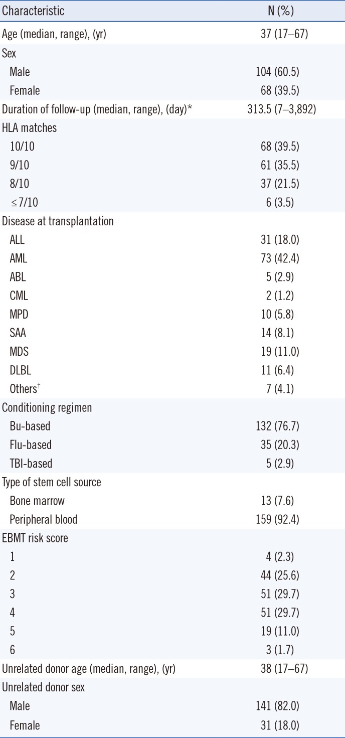
Fig. 1

Impact of the FOXP3 polymorphism on acute GVHD occurrence after allogenic HSCT. (A) rs5902434, (B) rs3761549, (C) rs3761548, (D) rs2232365, and (E) rs2280883. Recipients with rs3761549 TT genotype showed higher cumulative incidence of acute GVHD than recipients with rs3761549 CC or CT genotype (P=0.023).

Abbreviations: FOXP3, Forkhead box P3; GVHD, graft-versus-host disease; HSCT, hematopoietic stem cell transplantation.
alm-38-591-g001
Table 1

Characteristics of the study population (N=172)

alm-38-591-i001
Characteristic N (%)
Age (median, range), (yr) 37 (17–67)
Sex
 Male 104 (60.5)
 Female 68 (39.5)
Duration of follow-up (median, range), (day)* 313.5 (7–3,892)
HLA matches
 10/10 68 (39.5)
 9/10 61 (35.5)
 8/10 37 (21.5)
 ≤ 7/10 6 (3.5)
Disease at transplantation
 ALL 31 (18.0)
 AML 73 (42.4)
 ABL 5 (2.9)
 CML 2 (1.2)
 MPD 10 (5.8)
 SAA 14 (8.1)
 MDS 19 (11.0)
 DLBL 11 (6.4)
 Others 7 (4.1)
Conditioning regimen
 Bu-based 132 (76.7)
 Flu-based 35 (20.3)
 TBI-based 5 (2.9)
Type of stem cell source
 Bone marrow 13 (7.6)
 Peripheral blood 159 (92.4)
EBMT risk score
 1 4 (2.3)
 2 44 (25.6)
 3 51 (29.7)
 4 51 (29.7)
 5 19 (11.0)
 6 3 (1.7)
Unrelated donor age (median, range), (yr) 38 (17–67)
Unrelated donor sex
 Male 141 (82.0)
 Female 31 (18.0)

*The cut-points for duration of follow-up were defined as the time of analysis (March 1, 2017).

Others include NK cell lymphoma (N=5), Hodgkin lymphoma (N=1), and blastic plasmacytoid dendritic cell neoplasm (N=1).

Abbreviations: ALL, acute lymphoblastic leukemia; AML, acute myeloid leukemia; ABL, acute biphenotypic leukemia; CML, chronic myelogenous leukemia; MPD, myeloproliferative disease; SAA, severe aplastic anemia; MDS, myelodysplastic syndrome; DLBL, diffuse large B-cell lymphoma; NK, natural killer; Bu, busulfan; Flu, fludarabine; TBI, total body irradiation; EBMT, European Group for Blood and Marrow Transplantation.

Table 2

Primers for PCR-sequencing

alm-38-591-i002
Polymorphism AT (℃) Sequence (5′ → 3′)
rs5902434 del/ATT 56 F 5′-CTGCTCTCCCCTACCAGATG-3′
R 5′-CCCTGCCCATGCATTAAGTA-3′
rs3761549 C/T 60 F 5′-GTCCTCTCCACAACCCAAGA-3′
R 5′-CAGATTTTTCCGCCATTGAC-3′
rs3761548 C/A 60 F 5′-TTGTCTACTCCACGCCTCTCC-3′
R 5′-TGCCTCCATCATCACCACG-3′
rs2232365 A/G 60 F 5′-GAGGGCTTTCAGGTGAGGA-3′
R 5′-GGGAGTTGGATTGGGTGCA-3′
rs2280883 C/T 60 F 5′-TCAGGGTTTCAGTTCAGAGACAGT-3′
R 5′-CCCTTTCCAGATGTCCACCTCAG-3′
Inner F 5′-TGGCGCTAGGATGAAGGTTC-3′

Abbreviations: Del, deletion; AT, annealing temperature; F, forward primer; R, reverse primer.

Table 3

Association between genotype frequencies of FOXP3 SNPs and incidence of acute GVHD (grades II–IV) and infection

alm-38-591-i003
Polymorphism aGVHD Infection
Negative N=60 (%)* Positive N=99 (%)* P Odds ratio (95% CI) Negative N = 12 (%) Positive N = 160 (%)† P Odds ratio (95% CI)
rs5902434
 Genotype
  del/del+del/ATT 50 (83.3) 65 (67.7) 6 (50.0) 118 (76.1)
  ATT/ATT 10 (16.7) 31 (32.3) 0.031 2.385 (1.069–5.320) 6 (50.0) 37 (23.9) 0.046 0.314 (0.095–1.031)
rs3761549
 Genotype
  C/C+C/T 58 (96.7) 82 (84.5) 8 (66.7) 144 (91.1)
  T/T 2 (3.3) 15 (15.5) 0.017§ 5.305 (1.168–24.092) 4 (33.3) 14 (8.9) 0.026§ 0.194 (0.052–0.728)
rs3761548
 Genotype
  C/C+A/C 54 (90.0) 84 (87.5) 11 (91.7) 139 (88.5)
  A/A 6 (10.0) 12 (12.5) 0.634 1 (8.3) 18 (11.5) 1.000§
rs2232365
 Genotype
  A/A+A/G 51 (85.0) 66 (68.8) 6 (50.0) 122 (77.7)
  G/G 9 (15.0) 30 (31.2) 0.023 2.576 (1.123–5.905) 6 (50.0) 35 (22.3) 0.031 0.287 (0.087–0.945)
rs2280883
 Genotype
  T/T+T/C 54 (90.0) 83 (86.5) 11 (91.7) 137 (87.3)
  C/C 6 (10.0) 13 (13.5) 0.511 1 (8.3) 20 (12.7) 1.000§

*Among a cohort of 172 patients, of which 13 patients who died within 28 days after allogenic HSCT were excluded and two to five cases, depending on the SNP, failed to provide genotypes because of unavailable DNA samples after quality control.

Among a cohort of 172 patients, two to five cases failed to provide genotypes because of unavailable DNA samples after quality control.

Chi-square test used to analyze the data.

§Fisher's exact test used to analyze the data.

Abbreviations: FOXP3, Forkhead box P3; GVHD, graft-versus-host disease; CI, confidence interval; del, deletion; SNP, single nucleotide polymorphism.

Table 4

Multivariate analysis of risk factors for acute GVHD (grades II–IV)

alm-38-591-i004
Risk factor Univariate Multivariate
P* HR (95% CI) P* HR (95% CI)
Recipient age (range), (yr) 16–67 0.364 0.579
Recipient sex, female, N (%) 66 (41.5) 0.307 0.917
HLA allelic matches, < 9/10, N (%) 40 (25.2) 0.246 0.490
Conditioning regimen
 Bu-based, N (%) 125 (78.6) 0.054 0.056
 Flu-based, N (%) 30 (18.9) 0.072 0.604
Graft type, peripheral blood, N (%) 147 (92.5) 0.955 0.716
EBMT risk score, ≥ 4, N (%) 66 (41.5) 0.718 0.811
CMV IgG titer, AU/mL 8.2–250 0.369 0.562
Chronic GVHD, N (%) 23 (14.5) 0.088 0.108
rs3761549 TT genotype, N (%) 17 (10.7) 0.017 5.305 (1.168–24.092) 0.032 5.584 (1.160–26.882)
Donor age (range), (yr) 18–47 0.707 0.915
Donor sex, female, N (%) 29 (18.2) 0.692 0.994

*Univariate and multivariate analyses were performed using logistic regression.

Abbreviations: GVHD, graft-versus-host disease; HR, hazard ratio; CI, confidence interval; Bu, busulfan; Flu, fludarabine; EBMT, European Group for Blood and Marrow Transplantation; CMV, cytomegalovirus.

TOOLS
Similar articles