Journal List > Ann Lab Med > v.38(6) > 1099042

Choi, Kim, Sim, Hong, Kim, Jeong, Kim, Shin, and Ahn: Serum Copeptin Levels Predict Clinical Outcomes After Successful Percutaneous Coronary Intervention in Patients With Acute Myocardial Infarction

Abstract

Background

Serum copeptin has been demonstrated to be useful in early risk stratification and prognostication of patients with acute myocardial infarction (AMI). However, the prognostic value of copeptin after percutaneous coronary intervention (PCI) for clinical outcomes remains uncertain. We investigated the prognostic role of serum copeptin levels immediately after successful PCI as a prognostic marker for major adverse cardiac events (MACE; comprising death, repeat PCI, recurrent MI, or coronary artery bypass grafting) in patients with AMI.

Methods

A retrospective study was performed in 149 patients with AMI who successfully received PCI. Serum copeptin levels were analyzed in blood samples collected immediately after PCI. The association between copeptin levels and MACE during the follow-up period was evaluated.

Results

MACE occurred in 34 (22.8%) patients during a median follow-up of 30.1 months. MACE patients had higher copeptin levels than non-MACE patients did. Multiple logistic regression analysis showed that the increase in serum copeptin levels was associated with increased MACE incidence (odds ratio=1.6, P=0.005).

Conclusions

A high level of serum copeptin measured immediately after PCI was associated with MACE in patients with AMI during long-term follow-up. Serum copeptin levels can serve as a prognostic marker in patients with AMI after successful PCI.

INTRODUCTION

Coronary artery disease remains a progressive disease after successful percutaneous coronary intervention (PCI) and stent implantation; it can progress up to three to five years after PCI [12]. Therefore, accurate management decisions with comprehensive evaluation may improve the outcomes of high-risk patients. Periprocedural myocardial infarction (MI) or even a small increase in cardiac biomarker levels such as troponin or creatine kinase-myocardial band (CK-MB) fraction after PCI is associated with a significantly higher risk of late mortality [345].
Copeptin, a novel marker of arginine-vasopressin (AVP) activity, is an antidiuretic hypothalamo-pituitary hormone mainly regulated by changes in plasma osmolality, blood volume, and blood pressure via three AVP receptors [67]. The V1a receptor mediates vasoconstriction and platelet aggregation in blood vessels, as well as glycogenolysis and gluconeogenesis in the liver. The V1b receptor is expressed in the anterior hypophysis and in the Langerhans islets of the pancreas, where it mediates the secretion of adrenocorticotrophic hormone, insulin, and glucagon. AVP exerts various effects on the kidneys, such as an antidiuretic effect, by stimulating the V2 receptor. Copeptin is the C-terminal section of pro-AVP [8]; thus, copeptin mirrors AVP release because it is cleaved from pro-AVP in equimolar amounts. Most assays measuring AVP levels have relatively limited sensitivity because AVP is a small and short-lived peptide. Recently, an assay has been developed to measure blood copeptin, which is more stable and easier to measure than AVP [9]. The physiological function of copeptin has long remained unknown. Recently, many studies have shown that blood copeptin levels are associated with adverse clinical outcomes in various vascular diseases including acute MI (AMI), heart failure, and stroke [101112]. However, the prognostic value of blood copeptin levels after PCI for clinical outcomes remains uncertain. Therefore, we aimed to investigate the association between blood copeptin levels immediately after successful PCI and the incidence of major adverse cardiac events (MACE) in patients with AMI.

METHODS

1. Study population

A retrospective study was performed for 149 patients, including 37 (24.9%) female patients, with AMI who successfully received PCI with or without coronary stenting between February 2013 and December 2014 at Chonnam National University Hospital, Gwangju, Korea. Patients were followed up by visits to the outpatient department or via telephone contact. Data on clinical characteristics, laboratory characteristics, and procedural findings for patients with and without MACE were obtained through a retrospective review of clinical records. The median follow-up duration was 30.1 months (interquartile range [IQR], 22.9–36.8 months). The median age was 67 years. Complete blood counts (CBC), glucose, creatinine, and high-sensitivity C-reactive protein (hsCRP) levels were measured at the time of admission. Lipid profiles were obtained after at least 9 hours of fasting within 24 hours of hospitalization. All parameters were measured using an automated chemistry analyzer (AU5832, Beckman Coulter Inc., Brea, CA, USA), except for CBC, which were analyzed using the XE-2100 system (Sysmex Corp., Kobe, Japan). Patients received a loading dose of 300 mg aspirin and other antiplatelet medication (600 mg clopidogrel, 60 mg prasugrel, or 180 mg ticagrelor) prior to PCI. Unfractionated heparin (50–70 U/kg) was administered prior to or during PCI to maintain an activated clotting time of 250–300 seconds. After PCI, 100–300 mg aspirin and 75 mg clopidogrel (10 mg prasugrel or 180 mg ticagrelor) were prescribed daily as a maintenance dose. This study was conducted in accordance with the Declaration of Helsinki and approved by the ethics committee of Chonnam National University Hospital, Gwangju, Korea. We used serum samples that were already collected in another prospective registry of our institution (IRB number: CNUH-2016-048). The requirement for informed consent was waived because of the retrospective nature of the current study.

2. Serum copeptin measurement

Blood samples were obtained from 149 patients immediately after successful PCI and collected in serum-separating tubes (Becton Dickinson, Franklin Lakes, NJ, USA). All samples were immediately centrifuged at 2,465 g for 10 minutes at room temperature (20–25℃) and processed according to a standardized operating procedure [13]. Serum samples were stored at −80℃ until analyzed. Copeptin levels were measured using a time-resolved amplified cryptate emission immunoassay (Thermo Fisher Scientific Clinical Diagnostics BRAHMS GmbH, Hennigsdorf, Germany). This assay had a limit of detection of 0.9 pmol/L and a functional sensitivity of 1.9 pmol/L, assessed as an inter-assay precision of 20%. Intra- and inter-assay precision was 2.9% and 2.4%, respectively, at low-level quality control and 2% and 2.2%, respectively, at high-level quality control. The measuring range with automatic dilution was 1.9–2,000 pmol/L.
For reference interval verification of serum copeptin levels, 206 healthy controls (109 males and 97 females) aged 35–87 years were selected among the people who visited the Health Promotion Center; these controls had normal routine laboratory parameters such as renal and liver function tests.

3. Study definitions and endpoints

ST-segment elevation MI (STEMI) and non-STEMI (NSTEMI) diagnoses were based on the third universal definition of MI [14]; diagnoses were made by at least two interventional cardiologists at our institution. STEMI diagnosis was also based on recent guidelines for the management of STEMI, with a 12-lead electrocardiogram evaluating infarct-related arteries as determined by coronary angiography with increased cardiac-specific biomarkers such as troponin-I (TnI) [15]. Baseline left ventricular ejection fraction was measured by two-dimensional echocardiography prior to or immediately after PCI. The extent of coronary blood flow prior to and post PCI was graded using thrombolysis in MI (TIMI) flow grade. Complexity of coronary lesions was based on the definitions of the American College of Cardiology/American Heart Association [1617]. Multivessel disease was defined as coronary lesions associated with 50% or more stenosis in at least two coronary arteries, including the culprit artery, by quantitative coronary analysis. The culprit vessel was determined by 12-lead electrocardiogram and coronary angiography in STEMI. For NSTEMI, the culprit vessel was determined by coronary angiography. Coronary stenting was performed for individual patients at the discretion of the operators.
The study endpoint was MACE, a term that comprises all-cause death, any repeat PCI, recurrent MI, and coronary artery bypass graft. Nonfatal recurrent MI was defined as the development of recurrent angina symptoms accompanied by changes in 12-lead electrocardiogram or increased levels of cardiac-specific biomarkers. Repeat PCI included target lesion revascularization, target vessel revascularization, and non-target vessel revascularization [18].

4. Statistical analysis

Continuous variables were presented as mean±SD or as median with interquartile range (IQR). These were compared using the unpaired t-test or Mann-Whitney rank-sum test. Discrete variables were expressed as counts with percentages and were analyzed using Pearson's chi-squared test or Fisher's exact test. As copeptin levels exhibited a right-skewed distribution, data were subjected to a natural log transformation for statistical analysis. Multivariate logistic regression analysis was used to estimate predictors of MACE occurrence. The following variables with P<0.05 in univariate logistic regression were included in multivariate analysis: age, sex, diabetes mellitus, body mass index, previous history of PCI, use of beta-blocker at discharge, log-transformed copeptin (lnCopeptin), white blood cell counts, hemoglobin, low density lipoprotein cholesterol (LDL-C), high density lipoprotein cholesterol (HDL-C), and hsCRP. All patients were divided equally into three groups according to copeptin levels. Kaplan-Meier curves were constructed to illustrate MACE incidence over time according to copeptin level tertiles. Differences were assessed with a log-rank test.
All analyses were two-tailed and performed using SPSS for Windows ver. 23.0 (SPSS Inc., Chicago, IL, USA). P<0.05 was considered statistically significant.

RESULTS

1. Clinical, laboratory, and procedural characteristics

Patients' demographic and clinical characteristics are presented in Table 1. Age was significantly higher in MACE patients (N= 34) than in non-MACE patients (N=115), and a higher percentage of MACE patients had type 2 diabetes mellitus and a history of PCI. However, body mass index was higher in non-MACE patients than in MACE patients. In addition, fewer MACE patients than non-MACE patients received beta-blockers as a discharge medication. However, there were no significant differences in atherosclerotic risk factors such as hypertension, smoking, or dyslipidemia. Further, there were no significant differences in the prevalence of multivessel disease, distribution of culprit vessels, lesion complexity, procedural success rate, mean implanted stent number, or coronary stent implantation between MACE and non-MACE patients.
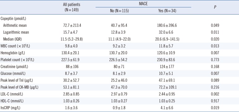
The laboratory characteristics of all 149 patients are presented in Table 2. MACE patients had higher serum copeptin levels (P=0.020), hsCRP (P=0.019), serum glucose levels (P=0.007), and WBC count (P=0.013) and lower serum LDL-C levels (P=0.002) and hemoglobin (P=0.007) than non-MACE patients. lnCopeptin was also higher (P=0.011) in MACE patients than in non-MACE patients. Peak levels of cardiac enzyme (TnI and CK-MB) and serum creatinine were similar in both groups.

2. Serum copeptin levels

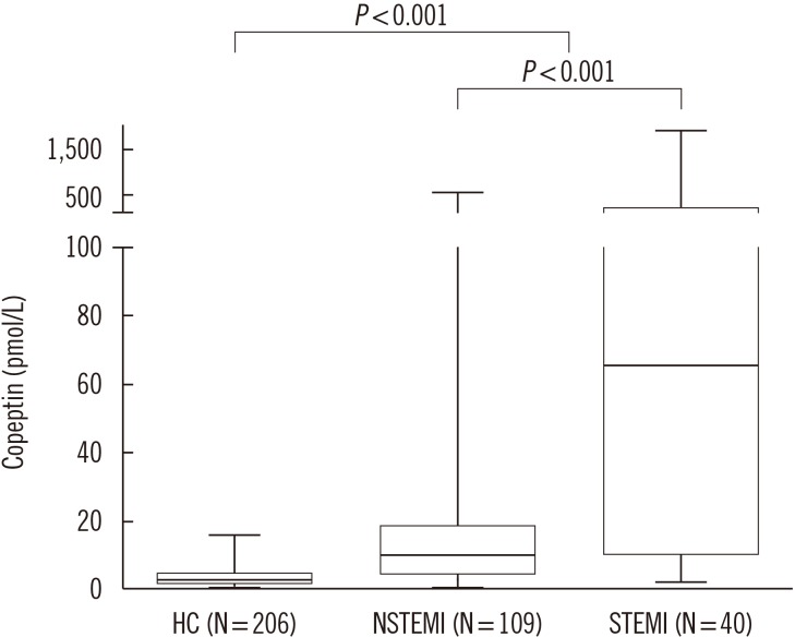
The median copeptin level of all 206 healthy controls was 2.9 pmol/L. The 97.5th percentile copeptin level of the healthy control group was 9.6 pmol/L, and the 2.5th percentile was 0.9 pmol/L. The median copeptin level was higher in men than in women (men, 3.4 pmol/L; women, 2.3 pmol/L). The median copeptin level in the patient group was higher than in the healthy control group (11.5 pmol/L vs 2.9 pmol/L). Patients with STEMI had higher copeptin levels than NSTEMI patients (65.6 pmol/L vs 10.0 pmol/L; Fig. 1).

3. Independent predictors of MACE occurrence

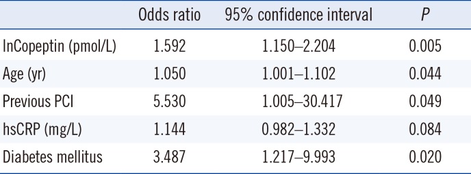
Overall, 34 (22.8%) patients experienced MACE during a median follow-up period of 30.1 months (IQR, 22.9–36.8 months). Multiple logistic regression analysis demonstrated that an approximate 2.72-fold increase in copeptin levels was associated with increased MACE (odds ratio, 1.6; 95% confidence interval, 1.15–2.20; P=0.005) after adjusting the following confounders: age, sex, diabetes mellitus, body mass index, previous history of PCI, use of beta-blocker at discharge, lnCopeptin, white blood cell counts, hemoglobin, LDL-C, HDL-C, and hsCRP (Table 3).

4. Survival analysis

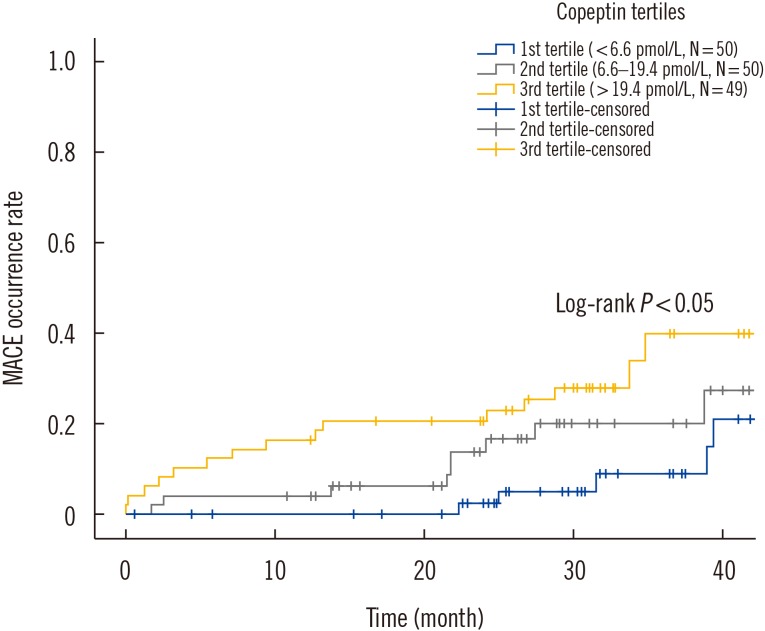
The MACE occurrence among the patients in copeptin level tertiles is shown in Fig. 2. There was a statistically significant separation among the estimated Kaplan-Meier survival curves of the copeptin tertiles (P=0.045, log-rank test). Serum copeptin levels were associated with MACE in a dose-dependent manner.

DISCUSSION

We demonstrated that serum copeptin levels were higher in STEMI or NSTEMI patients after PCI than in healthy controls, MACE patients showed higher serum copeptin levels than non-MACE patients, and high serum copeptin levels were associated with increased risk of MACE in patients with AMI during long-term follow-up.
A previous study reported that copeptin levels are higher in STEMI patients than in NSTEMI patients at presentation to the emergency department [19]. The main reason is the difference in peak time of copeptin level between STEMI and NSTEMI [20]. Peak copeptin levels are detected during ambulance transport in NSTEMI patients and at admission in STEMI patients [20]. In the current study, blood sampling for copeptin measurement was performed immediately after PCI, and all STEMI patients received primary PCI within 90 minutes of admission, unlike the NSTEMI patients, whose PCI time varied. Therefore, copeptin levels might be higher in STEMI patients.
Copeptin is a prognostic marker of various cardiovascular diseases [21222324252627]. High copeptin levels predict poorer long-term clinical outcomes in patients with AMI and are associated with the development of ischemic heart failure after AMI [212223]. High copeptin levels are also related to worse clinical outcomes in patients who underwent cardiovascular surgery, cerebrovascular accidents, and those who survived after cardiac arrest [24252627]. Furthermore, PCI can cause additional vessel injury or ischemia/reperfusion injury in AMI [28]; thus, it may cause more stress to patients with AMI. As a result, periprocedural MI or ischemic heart failure can develop despite good recanalization of diseased coronary arteries [29]. In the current study, copeptin levels obtained immediately after PCI predicted long-term MACE in AMI patients in a dose-dependent manner. This finding suggests that AMI patients with higher copeptin levels after successful PCI are more prone to develop future cardiovascular events than are those with lower copeptin levels.
The current study had some limitations. First, it was a retrospective, single-center study with a relatively small number of patients. Second, copeptin levels were not examined serially during hospitalization. Third, symptom or admission-to-balloon time was not considered for the study population. As copeptin levels rapidly decrease after symptom onset, the time interval between symptom onset and balloon might be associated with measured copeptin levels. Finally, the possibility of residual confounders, due to the presence of unmeasured confounders or measurement errors in the included factors, cannot be ruled out completely.
In conclusion, high copeptin levels measured immediately after successful PCI were associated with MACE in patients with AMI during long-term clinical follow-up. Our results strongly indicate that serum copeptin levels can serve as a prognostic marker for risk stratification in patients with AMI after successful PCI.

Acknowledgment

This study was supported by a grant from the Korea Health Technology R&D Project through the Korea Health Industry Development Institute (HI15C0498), grants from the National Research Foundation of Korea (NRF) funded by the Ministry of Education (2015M3A9B4051063, 2015M3A9B4066496, 2015R1D1A1A01057910, and 2016R1D1A1A09917796), and a grant from the Chonnam National University Hospital Biomedical Research Institute (CRI14035-1).

Notes

Authors' Disclosures of Potential Conflicts of Interest: No potential conflicts of interest relevant to this article were reported.

References

1. Stone GW, Maehara A, Lansky AJ, de Bruyne B, Cristea E, Mintz GS, et al. A prospective natural-history study of coronary atherosclerosis. N Engl J Med. 2011; 364:226–235. PMID: 21247313.
2. Zellweger MJ, Kaiser C, Jeger R, Brunner-La Rocca HP, Buser P, Bader F, et al. Coronary artery disease progression late after successful stent implantation. J Am Coll Cardiol. 2012; 59:793–799. PMID: 22361397.
3. Akkerhuis KM, Alexander JH, Tardiff BE, Boersma E, Harrington RA, Lincoff AM, et al. Minor myocardial damage and prognosis: are spontaneous and percutaneous coronary intervention-related events different? Circulation. 2002; 105:554–556. PMID: 11827918.
4. Ioannidis JP, Karvouni E, Katritsis DG. Mortality risk conferred by small elevations of creatine kinase-MB isoenzyme after percutaneous coronary intervention. J Am Coll Cardiol. 2003; 42:1406–1411. PMID: 14563583.
5. Jang JS, Jin HY, Seo JS, Yang TH, Kim DK, Kim DS, et al. Prognostic value of creatine kinase-myocardial band isoenzyme elevation following percutaneous coronary intervention: a meta-analysis. Catheter Cardiovasc Interv. 2013; 81:959–967. PMID: 22744792.
6. Bolignano D, Cabassi A, Fiaccadori E, Ghigo E, Pasquali R, Peracino A, et al. Copeptin (CTproAVP), a new tool for understanding the role of vasopressin in pathophysiology. Clin Chem Lab Med. 2014; 52:1447–1456. PMID: 24940718.
7. Morgenthaler NG, Struck J, Jochberger S, Dünser MW. Copeptin: clinical use of a new biomarker. Trends Endocrinol Metab. 2008; 19:43–49. PMID: 18291667.
8. Land H, Schütz G, Schmale H, Richter D. Nucleotide sequence of cloned cDNA encoding bovine arginine vasopressin-neurophysin II precursor. Nature. 1982; 295:299–303. PMID: 6276766.
9. Morgenthaler NG, Struck J, Alonso C, Bergmann A. Assay for the measurement of copeptin, a stable peptide derived from the precursor of vasopressin. Clin Chem. 2006; 52:112–119. PMID: 16269513.
10. Tasevska I, Enhörning S, Persson M, Nilsson PM, Melander O. Copeptin predicts coronary artery disease cardiovascular and total mortality. Heart. 2016; 102:127–132. PMID: 26661323.
11. Tu WJ, Dong X, Zhao SJ, Yang DG, Chen H. Prognostic value of plasma neuroendocrine biomarkers in patients with acute ischaemic stroke. J Neuroendocrinol. 2013; 25:771–778. PMID: 23701638.
12. Voors AA, von Haehling S, Anker SD, Hillege HL, Struck J, Hartmann O, et al. C-terminal provasopressin (copeptin) is a strong prognostic marker in patients with heart failure after an acute myocardial infarction: results from the OPTIMAAL study. Eur Heart J. 2009; 30:1187–1194. PMID: 19346228.
13. CLSI. Procedures for the Handling and Processing of Blood Specimen for Common Laboratory Tests. 4th ed. Wayne, PA: Clinical and Laboratory Standards Institute;2010. H18-A4.
14. Thygesen K, Alpert JS, Jaffe AS, Simoons ML, Chaitman BR, White HD, et al. Third universal definition of myocardial infarction. J Am Coll Cardiol. 2012; 60:1581–1598. PMID: 22958960.
15. Ibanez B, James S, Agewall S, Antunes MJ, Bucciarelli-Ducci C, Bueno H, et al. 2017 ESC Guidelines for the management of acute myocardial infarction in patients presenting with ST-segment elevation: The Task Force for the management of acute myocardial infarction in patients presenting with ST-segment elevation of the European Society of Cardiology (ESC). Eur Heart J. 2018; 39:119–177. PMID: 28886621.
16. Manginas A, Gatzov P, Chasikidis C, Voudris V, Pavlides G, Cokkinos DV. Estimation of coronary flow reserve using the Thrombolysis In Myocardial Infarction (TIMI) frame count method. Am J Cardiol. 1999; 83:1562–1565. PMID: 10363873.
17. Myler RK, Shaw RE, Stertzer SH, Hecht HS, Ryan C, Rosenblum J, et al. Lesion morphology and coronary angioplasty: current experience and analysis. J Am Coll Cardiol. 1992; 19:1641–1652. PMID: 1593061.
18. Cutlip DE, Windecker S, Mehran R, Boam A, Cohen DJ, van Es GA, et al. Clinical end points in coronary stent trials: a case for standardized definitions. Circulation. 2007; 115:2344–2351. PMID: 17470709.
19. Reichlin T, Hochholzer W, Stelzig C, Laule K, Freidank H, Morgenthaler NG, et al. Incremental value of copeptin for rapid rule out of acute myocardial infarction. J Am Coll Cardiol. 2009; 54:60–68. PMID: 19555842.
20. Slagman A, Searle J, Müller C, Möckel M. Temporal release pattern of copeptin and troponin T in patients with suspected acute coronary syndrome and spontaneous acute myocardial infarction. Clin Chem. 2015; 61:1273–1282. PMID: 26341999.
21. von Haehling S, Papassotiriou J, Morgenthaler NG, Hartmann O, Doehner W, Stellos K, et al. Copeptin as a prognostic factor for major adverse cardiovascular events in patients with coronary artery disease. Int J Cardiol. 2012; 162:27–32. PMID: 22284271.
22. Khan SQ, Dhillon OS, O'Brien RJ, Struck J, Quinn PA, Morgenthaler NG, et al. C-terminal provasopressin (copeptin) as a novel and prognostic marker in acute myocardial infarction: Leicester Acute Myocardial Infarction Peptide (LAMP) study. Circulation. 2007; 115:2103–2110. PMID: 17420344.
23. Kelly D, Squire IB, Khan SQ, Quinn P, Struck J, Morgenthaler NG, et al. C-terminal provasopressin (copeptin) is associated with left ventricular dysfunction, remodeling, and clinical heart failure in survivors of myocardial infarction. J Card Fail. 2008; 14:739–745. PMID: 18995178.
24. Katan M, Nigro N, Fluri F, Schuetz P, Morgenthaler NG, Jax F, et al. Stress hormones predict cerebrovascular re-events after transient ischemic attacks. Neurology. 2011; 76:563–566. PMID: 21228295.
25. Katan M, Fluri F, Morgenthaler NG, Schuetz P, Zweifel C, Bingisser R, et al. Copeptin: a novel, independent prognostic marker in patients with ischemic stroke. Ann Neurol. 2009; 66:799–808. PMID: 20035506.
26. Jarai R, Mahla E, Perkmann T, Jarai R, Archan S, Tentzeris I, et al. Usefulness of pre-operative copeptin concentrations to predict post-operative outcome after major vascular surgery. Am J Cardiol. 2011; 108:1188–1195. PMID: 21798504.
27. Geri G, Dumas F, Chenevier-Gobeaux C, Bouglé A, Daviaud F, Morichau-Beauchant T, et al. Is copeptin level associated with 1-year mortality after out-of-hospital cardiac arrest? Insights from the Paris registry*. Crit Care Med. 2015; 43:422–429. PMID: 25599466.
28. Ibáñez B, Heusch G, Ovize M, Van de Werf F. Evolving therapies for myocardial ischemia/reperfusion injury. J Am Coll Cardiol. 2015; 65:1454–1471. PMID: 25857912.
29. McMurray JJ. Clinical practice. Systolic heart failure. N Engl J Med. 2010; 362:228–238. PMID: 20089973.
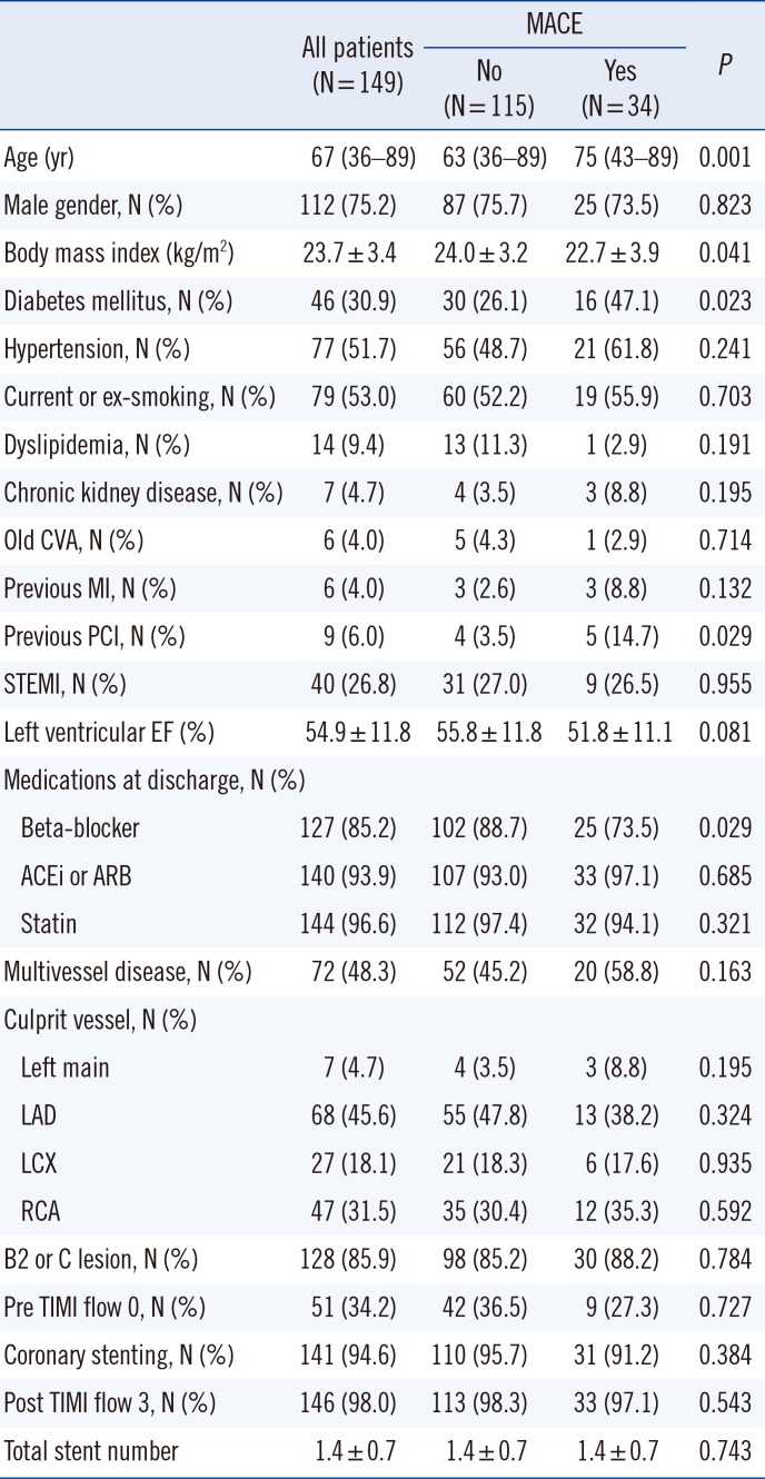
Fig. 1

Copeptin levels in healthy controls and patients with STEMI or NSTEMI obtained immediately after successful PCI. Copeptin levels were higher in patients with STEMI than in patients with NSTEMI. Each box plot shows the median (horizontal line within each box) with the third quartile (upper border) and first quartile (lower border).

*P<0.001.
Abbreviations: HC, healthy controls; STEMI, ST-segment elevation myocardial infarction.
alm-38-538-g001
Fig. 2

Kaplan-Meier curves for MACE according to copeptin level tertiles obtained immediately after successful percutaneous coronary intervention.

Abbreviation: MACE, major adverse cardiac events.
alm-38-538-g002
Table 1

Demographic, clinical, and procedural characteristics of patients with and without MACE

alm-38-538-i001
All patients (N=149) MACE P
No (N=115) Yes (N=34)
Age (yr) 67 (36–89) 63 (36–89) 75 (43–89) 0.001
Male gender, N (%) 112 (75.2) 87 (75.7) 25 (73.5) 0.823
Body mass index (kg/m2) 23.7±3.4 24.0±3.2 22.7±3.9 0.041
Diabetes mellitus, N (%) 46 (30.9) 30 (26.1) 16 (47.1) 0.023
Hypertension, N (%) 77 (51.7) 56 (48.7) 21 (61.8) 0.241
Current or ex-smoking, N (%) 79 (53.0) 60 (52.2) 19 (55.9) 0.703
Dyslipidemia, N (%) 14 (9.4) 13 (11.3) 1 (2.9) 0.191
Chronic kidney disease, N (%) 7 (4.7) 4 (3.5) 3 (8.8) 0.195
Old CVA, N (%) 6 (4.0) 5 (4.3) 1 (2.9) 0.714
Previous MI, N (%) 6 (4.0) 3 (2.6) 3 (8.8) 0.132
Previous PCI, N (%) 9 (6.0) 4 (3.5) 5 (14.7) 0.029
STEMI, N (%) 40 (26.8) 31 (27.0) 9 (26.5) 0.955
Left ventricular EF (%) 54.9±11.8 55.8±11.8 51.8±11.1 0.081
Medications at discharge, N (%)
 Beta-blocker 127 (85.2) 102 (88.7) 25 (73.5) 0.029
 ACEi or ARB 140 (93.9) 107 (93.0) 33 (97.1) 0.685
 Statin 144 (96.6) 112 (97.4) 32 (94.1) 0.321
Multivessel disease, N (%) 72 (48.3) 52 (45.2) 20 (58.8) 0.163
Culprit vessel, N (%)
 Left main 7 (4.7) 4 (3.5) 3 (8.8) 0.195
 LAD 68 (45.6) 55 (47.8) 13 (38.2) 0.324
 LCX 27 (18.1) 21 (18.3) 6 (17.6) 0.935
 RCA 47 (31.5) 35 (30.4) 12 (35.3) 0.592
B2 or C lesion, N (%) 128 (85.9) 98 (85.2) 30 (88.2) 0.784
Pre TIMI flow 0, N (%) 51 (34.2) 42 (36.5) 9 (27.3) 0.727
Coronary stenting, N (%) 141 (94.6) 110 (95.7) 31 (91.2) 0.384
Post TIMI flow 3, N (%) 146 (98.0) 113 (98.3) 33 (97.1) 0.543
Total stent number 1.4±0.7 1.4±0.7 1.4±0.7 0.743

Data are presented as median (range), mean±SD or N (%).

Abbreviations: ACEi, angiotensin-converting enzyme inhibitor; ARB, angiotensin-II receptor blocker; CVA, cardiovascular accident; EF, ejection fraction; LAD, left anterior descending; LCX, left circumflex; MACE, major adverse cardiac events; MI, myocardial infarction; PCI, percutaneous coronary intervention; RCA, right coronary artery; STEMI, ST-segment elevation MI.

Table 2

Laboratory characteristics of patients with and without MACE

alm-38-538-i002
All patients (N=149) MACE P
No (N = 115) Yes (N = 34)
Copeptin (pmol/L)
 Arithmetic mean 72.7 ± 213.4 40.7 ± 95.4 180.6 ± 396.6 0.049
 Logarithmic mean 15.7 ± 4.7 12.8 ± 3.9 32.0 ± 6.6 0.011
 Median (IQR) 11.5 (5.2–29.8) 11.1 (4.9–22.0) 20.6 (6.9–141.5) 0.020
WBC count ( × 109/L) 9.8 ± 4.0 9.2 ± 3.2 11.8 ± 5.7 0.013
Hemoglobin (g/L) 130.4 ± 20.1 130.7 ± 20.0 120.6 ± 10.9 0.007
Platelet count ( × 109/L) 227.5 ± 61.9 226.5 ± 54.2 230.9 ± 83.6 0.773
Creatinine (µmol/L) 88 ± 106 80 ± 71 124 ± 177 0.168
Glucose (mmol/L) 8.7 ± 3.7 8.1 ± 2.9 10.7 ± 5.1 0.007
Peak level of TnI (µg/L) 30.2 ± 52.7 25.2 ± 46.0 47.1 ± 69.1 0.089
Peak level of CK-MB (µg/L) 53.1 ± 81.1 47.3 ± 70.0 72.2 ± 109.1 0.216
LDL-C (mmol/L) 2.85 ± 0.85 2.97 ± 0.79 2.44 ± 0.95 0.002
HDL-C (mmol/L) 1.03 ± 0.26 1.03 ± 0.27 1.03 ± 0.25 0.917
hsCRP (mg/L) 1.6 ± 3.6 0.9 ± 1.8 4.1 ± 6.6 0.019

Data are presented as mean±SD or median (interquartile range [IQR]).

Abbreviations: CK-MB, creatine kinase-myocardial band isoenzyme; hsCRP, high-sensitivity C-reactive protein; HDL-C, high density lipoprotein cholesterol; LDL-C, low density lipoprotein cholesterol; MACE, major adverse cardiac events; TnI, troponin I; WBC, white blood cell.

Table 3

Predictors of MACE by multivariate analysis

alm-38-538-i003
Odds ratio 95% confidence interval P
lnCopeptin (pmol/L) 1.592 1.150–2.204 0.005
Age (yr) 1.050 1.001–1.102 0.044
Previous PCI 5.530 1.005–30.417 0.049
hsCRP (mg/L) 1.144 0.982–1.332 0.084
Diabetes mellitus 3.487 1.217–9.993 0.020

Abbreviations: hsCRP, high-sensitivity C-reactive protein; MACE, major adverse cardiac events; PCI, percutaneous coronary intervention.

TOOLS
Similar articles