

 PDF
PDF ePub
ePub Citation
 Print
Print


INTRODUCTION
Petersen's hernia was first described in 1900 by German surgeon, Dr. Walther Petersen. It is a specific type of internal hernia in which the small bowel moves into a potential space bounded by the caudal surface of the transverse mesocolon, the retroperitoneum and the mesentery of the gastrojejunostomy's limb (1). This type of internal hernia after laparoscopic gastrectomy with Roux-en-Y reconstruction for the treatment of obesity have been reported frequently (23). However, reports about Petersen's hernia after gastrectomy with Billroth II gastrojejunostomy for gastric cancer are a few.
We report a case of Petersen's hernia after radical subtotal gastrectomy with Billroth II gastrojejunostomy through antecolic route for gastric cancer and demonstrate an unreported specific computed tomography (CT) finding as a predictor of Petersen's hernia.
CASE REPORT
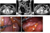
A 37-year-old woman presented with intermittent abdominal pain. She had undergone a robot-assisted radical subtotal gastrectomy with Billroth II gastrojejunostomy for advanced gastric cancer 8 months prior to her presentation. After undergoing gastrectomy, she was followed up with outpatient visits and received XELOX for 6 months. She did not have any other noteworthy symptoms and her physical examination results were also unremarkable. Laboratory investigations revealed nonspecific findings (C-reactive protein < 0.3 mg/L, white blood cell 6240/µL). Abdominal contrast-enhanced CT images showed the whirl sign, which indicates mesenteric vessel rotation, with mesenteric fat haziness (Fig. 1A, arrow). On a coronal image, the vessels showed engorgement (Fig. 1B, arrow) and the small bowel showed diffuse dilatation without evidence of strangulation (Fig. 1B, arrowheads). Furthermore, the interposed small bowel appeared (Fig. 1C, asterisk) between the transverse colon (Fig. 1C, arrowheads) and the afferent limb (Fig. 1C, arrow).
A diagnostic laparoscopy was performed, and revealed internal herniation of the efferent limb (Fig. 1D, arrow) through the Petersen defect, a space between the transverse mesocolon (Fig. 1D, E, asterisks) and the afferent limb (Fig. 1D, E, arrowheads), confirming the diagnosis of Petersen's hernia. A dilated small bowel loop was observed without any sign of strangulation. The herniated small bowel was successfully reduced and the Petersen defect was closed by running absorbable sutures (Fig. 1E). The patient was followed up and no recurrence of intestinal obstruction was observed thereafter.
DISCUSSION
Petersen's hernia is a rare specific type of internal hernia in which the small bowel moves into a potential space bounded by the caudal surface of the transverse mesocolon, the retroperitoneum and the mesentery of the gastrojejunostomy's limb (1). This type of internal hernia usually occurs within 1 year after bariatric surgery or (subtotal) gastrectomy performed to treat cancer, although it can develop several years after surgery (2). Internal hernias more frequently develop after laparoscopic surgery than laparotomy. This might be because more adhesion occurs after laparotomy which can reduce the mobility of the small bowel, and this in turn, prevents the internal herniation of the small bowel (23). Loss of body weight and abdominal fat after gastrectomy which may widen potential spaces is another predisposing factor (3). Closure of all the mesenteric defects with running non-absorbable sutures can reduce the risk of an internal hernia developing after gastrectomy (2). Petersen's hernia can lead to the small intestinal obstruction and necrosis of the small bowel necessitating intestinal resection. When Petersen's hernia is diagnosed, the herniated intestinal loop should be reduced with the closure of all the mesenteric defect (2456).
The diagnosis of internal hernia including Petersen's hernia is challenging due to nonspecific clinical symptoms, laboratory findings and radiological findings (23). The most common clinical symptoms are abdominal pain, nausea and vomiting, and these might be transient and self-resolving (23). On CT, rotation of the mesenteric vessels, mesenteric fat haziness, intestinal distention in the upper abdomen, and herniated intestinal loop above the gastric level can be seen (7). The whirl sign has been reported to be the most sensitive single predictor of internal hernia (37). However, even if these aforementioned signs are present on CT, it is hard to diagnose a specific type of internal hernia. In this case report, we demonstrate a specific CT finding, which is an interposed small bowel between the transverse colon and the gastrojejunostomy's limb as a predictor of Petersen's hernia. It is a reasonable finding considering that Petersen's hernia is an internal hernia through a space between the transverse mesocolon and the gastrojejunostomy's limb.
 XML Download