

 PDF
PDF ePub
ePub Citation
 Print
Print


INTRODUCTION
Glomus tumors are perivascular neoplasms, derived from modified smooth muscle cells of a neuromyoarterial glomus-commonly termed a glomus body-with the function of temperature regulation through arteriovenous shunting of blood (1). Glomus tumors are rare and have been reported in approximately 1.6% of patients with primary soft tissue tumors of upper and lower extremities (2). The lesion characteristically occurs in a digital subungual location, presenting with well-localized pain that is exacerbated by temperature changes. Although tumors are most often located on the digits, up to 45% of glomus tumors are extradigital, with common locations being the hands, arms, legs, and trunk (34). Glomus tumors may also develop in sites where normal glomus bodies may be sparse or even absent, such as the gastrointestinal tract, liver, lung, and trachea, as well as intraosseous and intraneural locations (3). In the musculoskeletal system, extradigital glomus tumors have been described around the shoulder, elbow, wrist, hip, knee, and ankle (5). There have been several reported cases of extradigital glomus tumors in the thigh. In the majority of these cases, the tumors were located superficially, such as the subcutaneous and intramuscular tissues (6). To our knowledge, there have been only three reported cases of periosteal glomus tumors adjacent to the femoral cortex (789). We report a rare case of a histologically proven glomus tumor found in the periosteal location of the distal femur.
CASE REPORT
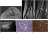
A 34-year-old woman presented with a 5-year history of chronic pain in her left thigh. The patient had no history of trauma, other medical, or surgical history. On physical examination, she reported pinpoint tenderness on palpation of the anteromedial distal thigh. The range of motion of the knee was normal. Radiographs did not demonstrate any abnormality. Magnetic resonance imaging (MRI) of the left thigh revealed a 10- × 8-mm, well-circumscribed mass at the anteromedial aspect of the femoral cortex that appeared hyperintense on T2-weighted images and isointense relative to muscle on T1-weighted images (Fig. 1A, B). The mass was strongly and homogeneously enhanced after contrast agent administration (Fig. 1C). The adjacent femoral cortex was normal. Ultrasonography (US) was performed for preoperative skin marking, this procedure showed that the mass was solid and hypoechoic, and there was no obvious Doppler signal (Fig. 1D). The differential diagnosis included vascular lesions such as hemangioma.
Surgery was performed to remove the lesion at the anteromedial aspect of the femoral cortex and a palpable mass, approximately 1 cm in size, was excised. Histologic examination revealed solid sheets of tumor cells that were interrupted by vessels of variable size. The round nuclei were centrally placed with lightly eosinophilic cytoplasm, and no atypical mitotic figures were observed (Fig. 1E). Immunohistochemistry revealed that the tumor cells were strongly positive for smooth muscle actin (Fig. 1F). The histology was characteristic of a solid-type glomus tumor. At the 4-month follow-up, the patient reported relief from pain, and there was no evidence of residual tumor on the follow-up US.
DISCUSSION
Glomus bodies are located in the stratum reticularis of the dermis and are particularly numerous in the hands and feet. They are composed of an afferent arteriole, an arteriovenous complex with a neurovascular reticulum, and efferent venules. Glomus tumors usually occur in areas rich in glomus bodies, such as the subungual regions of digits or the deep dermis of the palm, wrist, and forearm (1). However, these tumors have also been observed in extra-cutaneous locations not known to contain glomus bodies, where they presumably arise from perivascular smooth muscle cells that differentiate into glomoid cells. Recently reported review articles suggest that extradigital tumor distribution may be more frequent than has been generally assumed (34). When glomus tumors occur outside the fingers, they are found in a different demographic group, with a higher male to female ratio of 4:1 as compared to 1:2 in the fingers (4). The most common symptoms are pain and localized tenderness. Glomus tumors in extradigital locations contain nerve bundles that correlate with the presenting symptoms of pain. However, the classic clinical features of well-localized pain and cold hypersensitivity may not be present in extradigital sites, and preoperative diagnosis can be difficult. Moreover, the unusual locations of these tumors make application of diagnostic tests cumbersome. The average duration from symptom onset to diagnosis is 7 years (4).
On radiographs, erosive changes may be present in a minority of subungual glomus tumors. However, radiography is less useful in diagnosing extradigital lesions. On US, glomus tumors present as circumscribed, solid hypoechoic masses with small cystic- appearing spaces. Marked blood flow within the masses and adjacent feeding vessels can be observed on Doppler examinations (10). However, similar to our case, a case with juxtacortical glomus tumor of the distal femur was identified on US with no Doppler signal (8). MRI has proved to be the most sensitive modality for diagnosis, especially for smaller tumors. Most often, a characteristic, well-circumscribed, T2 high and T1 intermediate signal intensity lesion is found with homogeneous enhancement (10). Although MRI and US were not shown to provide a specific diagnosis of the extradigital glomus tumor, they did show its precise location and size. The imaging findings in our case are consistent with the findings in previous studies of extradigital glomus tumor.
On histopathological analysis, glomus tumors are typically composed of three components: vascular, smooth muscle, and neural components (3). The classic histological features of the glomus tumor include angiocentric uniform sheets of cells with oval nuclei, forming a perivascular “collar” around vessels. The histologic features differentiate the three tumor variants. The solid form has poor vasculature and scant smooth muscle components, while glomangiomas have a prominent vascular component, and glomangiomyomas are composed of prominent vascular and smooth muscle components (1). Solid glomus tumors, the subtype observed in our case, are the most common (about 75%), followed by glomangiomas and glomangiomyomas. Although the exact etiopathogenesis and cellular origins of this tumor are poorly understood, several lines of evidence suggest a modified pericytic/modified smooth muscle phenotype for glomus tumor.
In conclusion, this case details the clinical presentation of exaggerated pinpoint tenderness on palpation and a well-enhanced periosteal mass at the distal femur on MRI. Extradigital glomus tumors should be considered in the differential diagnosis when characteristic clinical features and compatible imaging findings are present, even if the tumor is found within deep tissues.
 XML Download