

 PDF
PDF ePub
ePub Citation
 Print
Print


Abstract
Purpose
To report a case of polypoidal choroidal vasculopathy in the right eye which improved after intravitreal injection of anti-vascular endothelial growth factor, and serous retinal detachment (SRD) in the left eye which improved spontaneously in a patient with a bilateral dome-shaped macula (DSM) with a tilted optic disc and inferonasal posterior staphyloma.
Case summary
A 50-year-old female visited our clinic with visual disturbance of the right eye for 5 days. A tilted optic disc with inferonasal posterior staphyloma and DSM were observed in both eyes by fundus examination and spectral domain optical coherence tomography (SD-OCT), and there was no specific finding in the left eye, but pigment epithelial detachment (PED) with subretinal hemorrhage was observed in the right eye. Polyps and branching vascular networks were found using indocyanine green angiography. We performed intravitreal C3F8 gas and aflibercept injection. After 3 months, SD-OCT of the right eye showed no subretinal hemorrhage and diminished PED. SD-OCT of the left eye showed SRD but the SRD disappeared after 1 month. SD-OCT of the left eye showed no recurrence of the SRD.
Figures and Tables
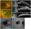 | Figure 1Fundus photography, optical coherence tomography (OCT) and fluorescein angiography at first visit. Fundus photography of right eye (A) and left eye (B) show bilateral tilted optic disc with inferonasal staphyloma and submacular hemorrhage in the right eye. Spectral domain optical coherence tomography (SD-OCT) of right eye (C: horizontal scan, D: vertical scan) shows subretinal hemorrhage and pigment epithelial detachment (PED) with dome-shape macula. SD-OCT of left eye (E) shows dome-shaped macula. Fluorescein angiography of right eye (F) shows blocked fluorescence due to subretinal hemorrhage. Indocyanine green angiography of right eye (G) shows branching vascular network (arrow) and circular polypoidal hyperfluorescent lesions (arrowheads) which corresponds with PED in SD-OCT (D).
|
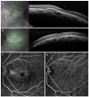 | Figure 2Optical coherence tomography (OCT) and fluorescein angiography after intravitreal injection of C3F8 gas and aflibercept. After intravitreal injection of C3F8 gas and aflibercept and two additional monthly intravitreal injections of aflibercept, spectral domain optical coherence tomography (SD-OCT) of right eye (A) shows decreased subretinal hemorrhage and pigment epithelial detachment. At that time, SD-OCT of left eye (B) shows serous retinal detachment. Fluorescein angiography (C) and indocyanine green angiography (D) of left eye show no fluorescein leakage but show multiple hyperfluorescent dots and window defect due to atrophy of retinal pigment epithelium.
|
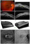 | Figure 3Mltiomodal imaging of dome shaped macula. At 4 months after first visit, fundus photography (A) and spectral domain optical coherence tomography (SD-OCT) of right eye (B) show recurrence of subretinal fluid (arrowhead) and elevated pigment epithelial detachment (arrow). After 1 month from additional intravitreal injection of aflibercept in the right eye, SD-OCT (C) shows slightly improved subretinal fluid and pigment epithelial detachment. SD-OCT of left eye (D) shows improved serous retinal detachment without treatment. 3D SD-OCT of right eye (E) and left eye (F) show dome-shaped macula. After 1 year from first visit, fluorescein angiography (G) and indocyanine green angiography (H) of right eye show that the branching vascular network is still the same but the circular polypoidal hyperfluorescent lesions have decreased.
|
References
1. Gaucher D, Erginay A, Lecleire-Collet A, et al. Dome-shaped macula in eyes with myopic posterior staphyloma. Am J Ophthalmol. 2008; 145:909–914.

2. Ohsugi H, Ikuno Y, Oshima K, et al. Morphologic characteristics of macular complications of a dome-shaped macula determined by swept-source optical coherence tomography. Am J Ophthalmol. 2014; 158:162–170.e1.
3. Errera MH, Michaelides M, Keane PA, et al. The extended clinical phenotype of dome-shaped macula. Graefes Arch Clin Exp Ophthalmol. 2014; 252:499–508.
4. Ellabban AA, Tsujikawa A, Matsumoto A, et al. Three dimensional tomographic features of dome-shaped macula by swept-source optical coherence tomography. Am J Ophthalmol. 2013; 155:320–328.e2.
5. Imamura Y, Iida T, Maruko I, et al. Enhanced depth imaging optical coherence tomography of the sclera in dome-shaped macula. Am J Ophthalmol. 2011; 151:297–302.
6. Witmer MT, Margo CE, Drucker M. Tilted optic disks. Surv Ophthalmol. 2010; 55:403–428.
7. Pardo-López D, Gallego-Pinazo R, Mateo C, et al. Serous macular detachment associated with dome-shaped macula and tilted disc. Case Rep Ophthalmol. 2011; 2:111–115.
8. Byeon SH, Chu YK. Dome-shaped macula. Am J Ophthalmol. 2011; 151:1101. author reply 1101-2.
9. Nakanishi H, Tsujikawa A, Gotoh N, et al. Macular complications on the border of an inferior staphyloma associated with tilted disc syndrome. Retina. 2008; 28:1493–1501.
10. Mauget-Faÿsse M, Cornut PL, Quaranta El-Maftouhi M, Leys A. Polypoidal choroidal vasculopathy in tilted disk syndrome and high myopia with staphyloma. Am J Ophthalmol. 2006; 142:970–975.
11. Furuta M, Iida T, Maruko I, et al. Submacular choroidal neovascularization at the margin of staphyloma in tilted disk syndrome. Retina. 2013; 33:71–76.
12. Cohen SY, Dubois L, Nghiem-Buffet S, et al. Spectral domain optical coherence tomography analysis of macular changes in tilted disk syndrome. Retina. 2013; 33:1338–1345.
13. Maruko I, Iida T, Sugano Y, et al. Morphologic choroidal and scleral changes at the macula in tilted disc syndrome with staphyloma using optical coherence tomography. Invest Ophthalmol Vis Sci. 2011; 52:8763–8768.
14. Mehdizadeh M, Nowroozzadeh MH. Dome-shaped macula in eyes with myopic posterior staphyloma. Am J Ophthalmol. 2008; 146:478. author reply 478-9.
15. Kang HM, Koh HJ. Lack of polypoidal lesions in patients with myopic choroidal neovascularization as evaluated by indocyanine green angiography. Am J Ophthalmol. 2014; 157:378–383.e1.
 XML Download