

 PDF
PDF ePub
ePub Citation
 Print
Print


Abstract
Purpose
To report a case of isolated conjunctival lymphangioma mimicking a recurrent conjunctival cyst.
Case summary
A 39-year-old male with a conjunctival cyst in the right eye lasted for 1 month visited our hospital. He had previously undergone aspiration of the cyst at another hospital 1 week before visiting our hospital. However, the cyst recurred, and he was referred to our hospital. On slit lamp biomicroscopy, yellow-colored turbid fluid and a hemorrhage were observed in the conjunctival cyst, but no specific finding was found in the fundus photography. The patient was initially treated with topical antibiotics and steroids. Three weeks later, absorption of the hemorrhage was noted, but there was no change in the size of the cyst. Therefore, surgical removal and histological examination of the cyst were performed. The histological examination revealed that the lesion was positive for CD 31 and D2-40, and the cyst was diagnosed as a cystic conjunctival lymphangioma. Thereafter, brain magnetic resonance imaging was performed to screen for orbital lymphangioma and systemic disease that could accompany a conjunctival lymphangioma. However, no specific findings were observed. There was no recurrence of the conjunctival cyst at 1 year and 6 months after surgical removal, and no other ophthalmic or systemic complication was observed.
Figures and Tables
Figure 1
Slit-lamp photography of right eye. (A) Preoperative photography after 3 weeks of medical treatment. Yellow-colored turbid fluid was observed in the conjunctival cyst at inferonasal area. (B) On postoperative day 2, stable operation site with subconjunctival hemorrhage was noted. (C) On 1 year and 6 months after the operation, stable surface of operative site was noted with no recurrence of conjunctival cyst.
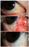
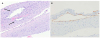
Figure 2
Cross section planes of immunohistochemical staining of the cyst. (A) In Hematoxylin-Eosin stain (×200), lymphatic proliferation and ectasia with a network of empty bloodless channels lined by flattened endothelium was seen (arrows). And large dilated lymphatics within the substantia propria of conjunctiva with the presence of many lymphocytes infiltrates were seen. (B) In D2-40 stain (×200), the dilated lymphatics stained positively with special immunohistochemical D2-40 staining (brown color).
References
1. Thatte S, Jain J, Kinger M, et al. Clinical study of histologically proven conjunctival cysts. Saudi J Ophthalmol. 2015; 29:109–115.

2. Tunç M, Sadri E, Char DH. Orbital lymphangioma: an analysis of 26 patients. Br J Ophthalmol. 1999; 83:76–80.
3. Kalisa P, Van Zieleghem B, Roux P, Meire F. Orbital lymphangioma: clinical features and management. Bull Soc Belge Ophtalmol. 2001; (282):59–68.
4. Jastrzebski A, Brownstein S, Manusow J, Rubab S. Isolated conjunctival lymphangioma. Can J Ophthalmol. 2011; 46:369–370.
5. Seca M, Borges P, Reimão P, et al. Conjunctival lymphangioma: a case report and brief review of the literature. Case Rep Ophthalmol Med. 2012; 2012:836573.
6. Rootman J, Hay E, Graeb D, Miller R. Orbital-adnexal lymphangiomas. A spectrum of hemodynamically isolated vascular hamartomas. Ophthalmology. 1986; 93:1558–1570.
7. Jones IS. Lymphangiomas of the ocular adnexa: an analysis of 62 cases. Trans Am Ophthalmol Soc. 1959; 57:602–665.
8. Weigand S, Eivazi B, Barth PJ, et al. Pathogenesis of lymphangiomas. Virchows Arch. 2008; 453:1–8.
9. Jordan DR, Anderson RL. Carbon dioxide (CO2) laser therapy for conjunctival lymphangioma. Ophthalmic Surg. 1987; 18:728–730.
10. Joo JH, Ko MK, Park MH. Ultrastructural and immunofluorescent features of lymphatic disorders in conjunctiva. J Korean Ophthalmol Soc. 1987; 28:545–550.
11. Kim KY, Lee YS, Lee HJ, et al. A case of conjunctival lymphangioma with clinical manifestations of superior limbic keratoconjunctivitis after upper lid blepharoplasty. J Korean Ophthalmol Soc. 2010; 51:1276–1281.
12. Freiberg FJ, Kunstmann E, König T, et al. Conjunctival lymphangioma in a 4-year-old girl revealed tuberous sclerosis complex. GMS Ophthalmology Cases. 2016; 6:Doc09. eCollection 2016.
13. Meisler DM, Eiferman RA, Ratliff NB, Buens CD. Surgical management of conjunctival lymphangiectasis by conjunctival resection. Am J Ophthalmol. 2003; 136:735–736.
14. Behrendt S, Bernsmeier H, Randzio G. Fractionated beta-irradiation of a conjunctival lymphangioma. Ophthalmologica. 1991; 203:161–163.
 XML Download