Abstract
Purpose
To investigate the clinical outcomes of combined vitrectomy and intrascleral fixation of a new posterior chamber intraocular lens (PC IOL) as a treatment for IOL dislocation.
Methods
We conducted a retrospective interventional study at our medical facility from January 2015 to January 2017. Posteriorly dislocated IOLs were removed with pars plana vitrectomy. Two intrascleral tunnels, 2.0 mm in length, were created 1.5 mm to the limbus at 6 and 12 o'clock positions. Both haptics of new foldable acrylic 3-piece IOLs were inserted into the tunnel until the IOL was secured in a central position. We analyzed the preexisting ocular condition, visual acuity (VA), and refractive error preoperatively and postoperatively, and recorded postoperative complications.
Results
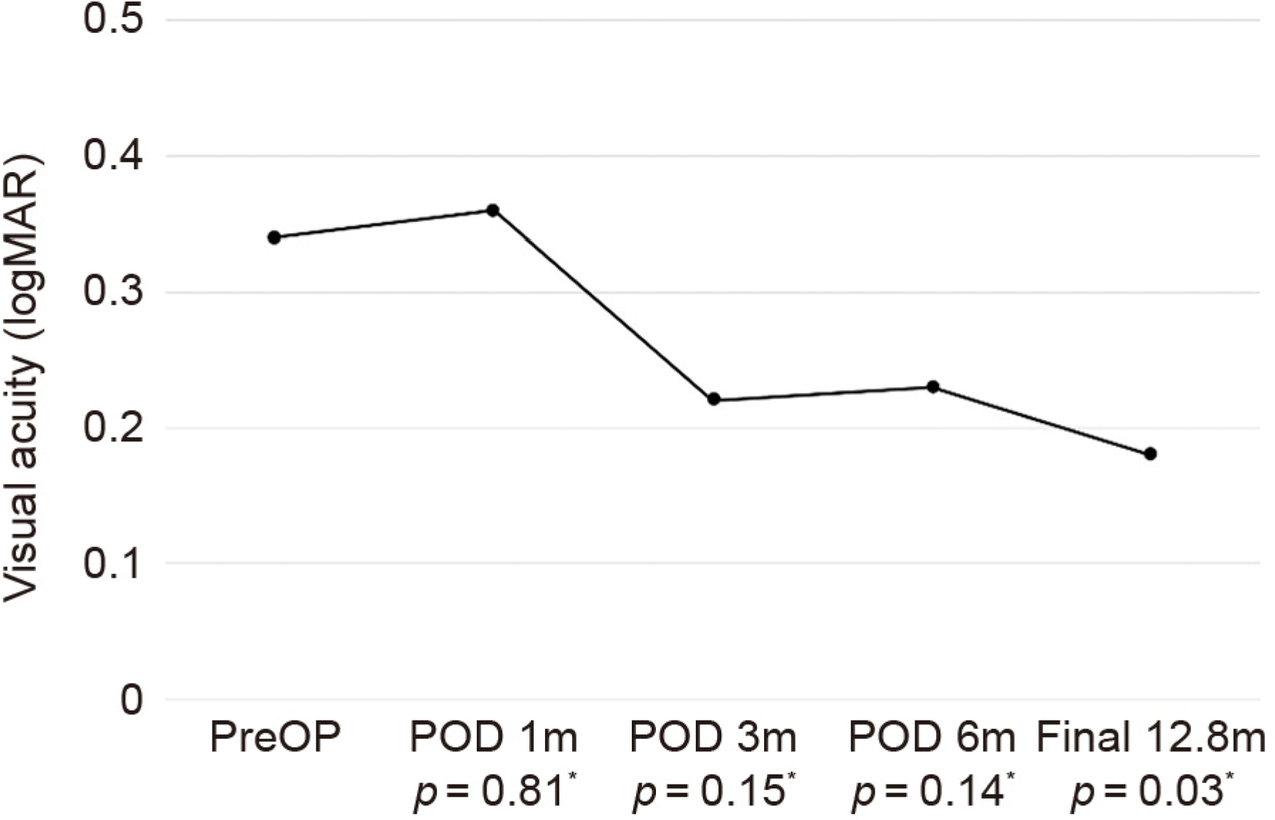
Forty-nine patients (50 eyes) were enrolled in the study. The mean follow-up period was 12.8 ± 6.6 months. A best-cor-rected VA of 6/12 or better was achieved in 43 eyes (86%). The mean VA significantly improved from 0.32 logarithm of the mini-mum angle of resolution (logMAR) at baseline to 0.18 logMAR at last follow-up (p = 0.03). The refractive status after intrascleral fixation of the PC IOL revealed a mean hyperopic shift of +1.09 ± 1.28 diopters from the predicted spherical equivalent. Postoperative vitreous hemorrhages occurred in six cases and were cleared without visual compromise. Cystoid macular edema was well-controlled by topical nonsteroidal anti-inflammatory drugs (NSAID) medications in two cases. In two cases, IOL dislocation recurred and required re-operation. There were no serious adverse events of suture-related complications, retinal detachment, corneal compromise, or endophthalmitis in any of the patients.
Go to : 
References
1. Gross JG, Kokame GT, Weinberg DV; Dislocated In-The-Bag Intraocular Lens Study Group. In-the-bag intraocular lens dislocation. Am J Ophthalmol. 2004; 137:630–5.
2. Krė pštė L, Kuzmienė L, Miliauskas A, Janulevič ienė I. Possible predisposing factors for late intraocular lens dislocation after abdominal cataract surgery. Medicina (Kaunas). 2013; 49:229–34.
3. Fernández-Buenaga R, Alio JL, Pérez-Ardoy AL, et al. Late in-the-bag intraocular lens dislocation requiring explantation: risk factors and outcomes. Eye (Lond). 2013; 27:795–801. quiz 802.

4. Davis D, Brubaker J, Espandar L, et al. Late in-the-bag abdominal intraocular lens dislocation: evaluation of 86 consecutive cases. Ophthalmology. 2009; 116:664–70.
5. Zeh WG, Price FW Jr. Iris fixation of posterior chamber intraocular lenses. J Cataract Refract Surg. 2000; 26:1028–34.

6. Por YM, Lavin MJ. Techniques of intraocular lens suspension in the absence of capsular/zonular support. Surv Ophthalmol. 2005; 50:429–62.

7. Azar DT, Wiley WF. Double-knot transscleral suture fixation abdominal for displaced intraocular lenses. Am J Ophthalmol. 1999; 128:644–6.
8. Bloom SM, Wyszynski RE, Brucker AJ. Scleral fixation suture for dislocated posterior chamber intraocular lens. Ophthalmic Surg. 1990; 21:851–4.

9. Chan CK. An improved technique for management of dislocated posterior chamber implants. Ophthalmology. 1992; 99:51–7.

10. Chang S, Coll GE. Surgical techniques for repositioning a abdominal intraocular lens, repair of iridodialysis, and secondary abdominal lens implantation using innovative 25-gauge forceps. Am J Ophthalmol. 1995; 119:165–74.
11. Friedberg MA, Pilkerton AR. A new technique for repositioning and fixating a dislocated intraocular lens. Arch Ophthalmol. 1992; 110:413–5.

12. Kokame GT, Yamamoto I, Mandel H. Scleral fixation of dislocated posterior chamber intraocular lenses: temporary haptic external-ization through a clear corneal incision. J Cataract Refract Surg. 2004; 30:1049–56.
13. Maguire AM, Blumenkranz MS, Ward TG, Winkelman JZ. Scleral loop fixation for posteriorly dislocated intraocular lenses. Operative technique and long-term results. Arch Ophthalmol. 1991; 109:1754–8.
14. Schneiderman TE, Johnson MW, Smiddy WE, et al. Surgical abdominal of posteriorly dislocated silicone plate haptic intraocular lenses. Am J Ophthalmol. 1997; 123:629–35.
15. Koh HJ, Kim CY, Lim SJ, Kwon OW. Scleral fixation technique using 2 corneal tunnels for a dislocated intraocular lens. J Cataract Refract Surg. 2000; 26:1439–41.

17. Drolsum L. abdominal follow-up of secondary flexible, open loop, anterior chamber intraocular lenses. J Cataract Refract Surg. 2003; 29:498–503.
18. Biro Z. Results and complications of secondary intraocular lens implantation. J Cataract Refract Surg. 1993; 19:64–7.

19. Downing JE. Ten-year follow up comparing anterior and posterior chamber intraocular lens implants. Ophthalmic Surg. 1992; 23:308–15.

20. Evereklioglu C, Er H, Bekir NA, et al. Comparison of secondary implantation of flexible open-loop anterior chamber and scler-al-fixated posterior chamber intraocular lenses. J Cataract Refract Surg. 2003; 29:301–8.

21. Vote BJ, Tranos P, Bunce C, et al. abdominal outcome of combined pars plana vitrectomy and scleral fixated sutured posterior chamber intraocular lens implantation. Am J Ophthalmol. 2006; 141:308–12.
22. Gabor SG, Pavlidis MM. Sutureless intrascleral posterior chamber intraocular lens fixation. J Cataract Refract Surg. 2007; 33:1851–4.

23. Agarwal A, Kumar DA, Jacob S, et al. Fibrin glue-assisted abdominal posterior chamber intraocular lens implantation in eyes with abdominal posterior capsules. J Cataract Refract Surg. 2008; 34:1433–8.
24. Yamane S, Inoue M, Arakawa A, Kadonosono K. Sutureless 27-gauge needle-guided intrascleral intraocular lens implantation with lamellar scleral dissection. Ophthalmology. 2014; 121:61–6.

25. Totan Y, Karadag R. Trocar-assisted sutureless intrascleral abdominal chamber foldable abdominal lens fixation. Eye (Lond). 2012; 26:788–91.
26. Ohta T, Toshida H, Murakami A. Simplified and safe method of abdominalless intrascleral posterior chamber intraocular lens fixation: Y-fixation technique. J Cataract Refract Surg. 2014; 40:2–7.
27. Takayama K, Akimoto M, Taguchi H, et al. Transconjunctival abdominalless intrascleral intraocular lens fixation using intrascleral abdominal guided with catheter and 30-gauge needles. Br J Ophthalmol. 2015; 99:1457–9.
28. Kristianslund O, Råen M, Østern A, Drolsum L. Late in-the-bag abdominal lens dislocation: a randomized clinical trial comparing lens repositioning and lens exchange. Ophthalmology. 2017; 124:151–9.
29. Güell JL, Barrera A, Manero F. A review of suturing techniques for posterior chamber lenses. Curr Opin Ophthalmol. 2004; 15:44–50.
30. Heilskov T, Joondeph BC, Olsen KR, Blankenship GW. Late abdominal after transscleral fixation of a posterior chamber abdominal lens. Arch Ophthalmol. 1989; 107:1427.
31. Scharioth GB, Prasad S, Georgalas I, et al. Intermediate results of sutureless intrascleral posterior chamber intraocular lens fixation. J Cataract Refract Surg. 2010; 36:254–9.

32. Hayashi K, Hayashi H, Nakao F, Hayashi F. Intraocular lens tilt and decentration, anterior chamber depth, and refractive error after transscleral suture fixation surgery. Ophthalmology. 1999; 106:878–82.
33. Durak A, Oner HF, Koçak N, Kaynak S. Tilt and decentration after primary and secondary transsclerally sutured posterior chamber abdominal lens implantation. J Cataract Refract Surg. 2001; 27:227–32.
34. Sinha R, Bansal M, Sharma N, et al. Transscleral suture-fixated versus intrascleral haptic-fixated intraocular lens: a comparative study. Eye Contact Lens. 2017; 43:389–93.

35. Flynn HW Jr. Pars plana vitrectomy in the management of sub-luxed and posteriorly dislocated intraocular lenses. Graefes Arch Clin Exp Ophthalmol. 1987; 225:169–72.

36. Smiddy WE. Dislocated posterior chamber intraocular lens. A new technique of management. Arch Ophthalmol. 1989; 107:1678–80.
37. Campo RV, Chung KD, Oyakawa RT. Pars plana vitrectomy in the management of dislocated posterior chamber lenses. Am J Ophthalmol. 1989; 108:529–34.

38. Mittra RA, Connor TB, Han DP, et al. Removal of dislocated abdominal lenses using pars plana vitrectomy with placement of an open-loop, flexible anterior chamber lens. Ophthalmology. 1998; 105:1011–4.
39. Althaus C, Sundmacher R. Endoscopically controlled optimization of abdominal suture fixation of posterior chamber lenses in the ciliary sulcus. Ophthalmology. 1993; 90:317–24.
40. Bading G, Hillenkamp J, Sachs HG, et al. abdominal safety and functional outcome of combined pars plana vitrectomy and scleral-fixated sutured posterior chamber lens implantation. Am J Ophthalmol. 2007; 144:371–7.
41. Yang CS, Chao YJ. abdominal outcome of combined vitrectomy and transscleral suture fixation of posterior chamber intraocular lenses in the management of posteriorly dislocated lenses. J Chin Med Assoc. 2016; 79:450–5.
42. Donaldson KE, Gorscak JJ, Budenz DL, et al. Anterior chamber and sutured posterior chamber intraocular lenses in eyes with poor capsular support. J Cataract Refract Surg. 2005; 31:903–9.

43. Suto C, Hori S, Fukuyama E, Akura J. Adjusting intraocular lens power for sulcus fixation. J Cataract Refract Surg. 2003; 29:1913–7.

44. Ma DJ, Choi HJ, Kim MK, Wee WR. Clinical comparison of abdominal sulcus and pars plana locations for posterior chamber abdominal lens transscleral fixation. J Cataract Refract Surg. 2011; 37:1439–46.
45. Kumar DA, Agarwal A, Packiyalakshmi S, et al. Complications and visual outcomes after glued foldable intraocular lens implantation in eyes with inadequate capsules. J Cataract Refract Surg. 2013; 39:1211–8.

46. Yamane S, Sato S, Maruyama-Inoue M, Kadonosono K. Flanged intrascleral intraocular lens fixation with double-needle technique. Ophthalmology. 2017; 124:1136–42.

47. Khan MA, Gupta OP, Smith RG, et al. Scleral fixation of abdominal lenses using Gore-Tex suture: clinical outcomes and safety profile. Br J Ophthalmol. 2016; 100:638–43.
48. Koushan K, Mikhail M, Beattie A, et al. Corneal endothelial cell loss after pars plana vitrectomy and combined phacoemulsification –vitrectomy surgeries. Can J Ophthalmol. 2017; 52:4–8.
49. Numa A, Nakamura J, Takashima M, Kani K. abdominal corneal endothelial changes after intraocular lens implantation. Anterior vs posterior chamber lenses. Jpn J Ophthalmol. 1993; 37:78–87.
Go to : 
 | Figure 1.Technique for the intrascleral fixation of intraocular lens (IOL). (A) Scleral tunnel created 1.5 mm from the limbus using a paracentesis incision diamond knife. Detailed view (inset) of the diamond knife. (B) Transscleral incision created perpendicular to the scleral tunnel. 3-dimensional view (inset) of the scleral tunnel and transscleral incision. (C) The leading haptic of the secondary IOL is grasped with 23-gauge intraocular forceps. (D) The leading haptic is pulled through the transscleral incision, and left externalized. (E) The McPherson forceps grasps the externalized tip, inserts the IOL haptic into the tunnel. (F) The tailing haptic is then inserted into the second scleral tunnel, and the two tunnels were closed in a conventional manner. |
 | Figure 2.Mean best corrected visual acuity (BCVA) changes after surgery. The mean BCVA before surgery was 0.34 ± 0.47 (logMAR) and the BCVA improved to 0.18 ± 0.32 at final follow-up. Note the temporary deterioration of BCVA at the first month after surgery. PreOP = preoperative; POD = postoperative day; m = month(s). *Paired t-test. |
Table 1.
Preoperative clinical characteristics
Table 2.
Changes of BCVA, IOP, ECD and refraction after surgery
| Baseline | Final | p-value* | |
|---|---|---|---|
| BCVA (logMAR) | 0.34 ± 0.47 | 0.18 ± 0.32 | 0.03 |
| IOP (mmHg) | 16.0 ± 5.1 | 14.4 ± 3.3 | 0.01 |
| ECD (cells/mm2) | 2,316.1 ± 541.5 | 2,206.5 ± 536.0 | 0.00 |
| Refraction (SE, diopter) | +8.3 ± 4.4 | −0.2 ± 1.8 | 0.00 |


 PDF
PDF ePub
ePub Citation
 Print
Print


 XML Download