

 PDF
PDF ePub
ePub Citation
 Print
Print


Abstract
Purpose
To report the effects of combined low dose bevacizumab and low dose triamcinolone intravitreal injection compared with single bevacizumab intravitreal injection in patients with macular edema secondary to branch retinal vein occlusion.
Methods
Thirty eyes of 30 patients diagnosed with branch retinal vein occlusion were evaluated. The combined injection group (15 eyes of 15 patients) was treated with intravitreal injection of combined low dose bevacizumab (0.625 mg/0.025 mL) and low dose triamcinolone (1 mg/0.025 mL). The single injection group (15 eyes of 15 patients) was treated with intravitreal injection of bevacizumab (1.25 mg/0.05 mL). The best-corrected visual acuity (BCVA), central macular thickness (CMT), and intraocular pressure (IOP) of treated eyes were measured before injection and at 1 month, 2 months, and 3 months after injection.
Results
In the combined injection group, the BCVA increased significantly at 1 month, 2 months, and 3 months after injection (p < 0.05). However, in the single injection group, the BCVA increased significantly only at 3 months after injection (p < 0.05). In both groups, the CMT decreased significantly at 1 month, 2 months, and 3 months after injection (p < 0.05). The IOP showed no significant change at 3 months after injection (p > 0.05) in both groups. The BCVA, CMT, and IOP after injection showed no significant differences between the combined injection group and the single injection group (p > 0.05).
Figures and Tables
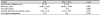
Table 2
Visual acuity, central macular thickness, and intraocular pressure results in patients with branch retinal vein occlusion
Values are presented as mean ± SD unless otherwise indicated.
BCVA = best corrected visual acuity; IVB = intravitreal bevacizumab injection; IVTA = intravitreal triamcinolone injection; CMT = central macular thickness; IOP = intraocular pressure.
*Mann-Whitney test; †p-value compared with baseline, Wilcoxon signed rank test.
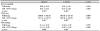
Table 3
Comparison of visual acuity between intravitreal bevacizumab injection and intravitreal bevacizumab-triamcinolone injection

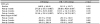
Table 4
Comparison of central macular thickness between intravitreal bevacizumab injection and intravitreal bevacizumab-triamcinolone injection
Notes
References
1. Rogers S, McIntosh RL, Cheung N, et al. The prevalence of retinal vein occlusion: pooled data from population studies from the United States, Europe, Asia, and Australia. Ophthalmology. 2010; 117:313–319.e1.

2. Cugati S, Wang JJ, Rochtchina E, Mitchell P. Ten-year incidence ofretinal vein occlusion in an older population: the Blue Mountains Eye Study. Arch Ophthalmol. 2006; 124:726–732.
3. Argon laser photocoagulation for macular edema in branch vein occlusion. The Branch Vein Occlusion Study Group. Am J Ophthalmol. 1984; 98:271–282.
4. Chang MA, Fine HF, Bass E, et al. Patients' preferences in choosing therapy for retinal vein occlusions. Retina. 2007; 27:789–797.
5. McIntosh RL, Mohamed Q, Saw SM, Wong TY. Interventions for branch retinal vein occlusion: an evidence-based systematic review. Ophthalmology. 2007; 114:835–854.
6. Zhou JQ, Xu L, Wang S, et al. The 10-year incidence and risk factors of retinal vein occlusion: The Beijing Eye Study. Ophthalmology. 2013; 120:803–808.
7. Scott IU, Ip MS, VanVeldhuisen PC, et al. A randomized trial comparing the efficacy and safety of intravitreal triamcinolone with standard care to treat vision loss associated with macular Edema secondary to branch retinal vein occlusion: the Standard Care vs Corticosteroid for Retinal Vein Occlusion (SCORE) study report 6. Arch Ophthalmol. 2009; 127:1115–1128.
8. Pielen A, Feltgen N, Isserstedt C, et al. Efficacy and safety of intravitreal therapy in macular edema due to branch and central retinal vein occlusion: a systematic review. PLoS One. 2013; 8:e78538.
9. Li j, Paulus YM, Shuai Y, et al. New developments in the classification, pathogenesis, risk factors, natural history, and treatment of branch retinal vein occlusion. J Ophthalmol. 2017; 2017:4936924.
10. Lee K, Jung H, Sohn J. Comparison of injection of intravitreal drugs with standard care in macular edema secondary to branch retinal vein occlusion. Korean J Ophthalmol. 2014; 28:19–25.
11. Ali RI, Kapoor KG, Khan AN, Gibran SK. Efficacy of combined intravitreal bevacizumab and triamcinolone for branch retinal vein occlusion. Indian J Ophthalmol. 2014; 62:396–399.
12. Noma H, Funatsu H, Yamasaki M, et al. Aqueous humour levels of cytokines are correlated to vitreous levels and severity of macular oedema in branch retinal vein occlusion. Eye (Lond). 2008; 22:42–48.
13. Aiello LP, Avery RL, Arrigg PG, et al. Vascular endothelial growth factor in ocular fluid of patients with diabetic retinopathy and other retinal disorders. N Engl J Med. 1994; 331:1480–1487.
14. Noma H, Funatsu H, Mimura T, et al. Role of soluble vascular endothelial growth factor receptor-2 in macular oedema with central retinal vein occlusion. Br J Ophthalmol. 2011; 95:788–792.
15. Noma H, Mimura T. Aqueous soluble vascular endothelial growth factor receptor-2 in macular edema with branch retinal vein occlusion. Curr Eye Res. 2013; 38:1288–1290.
16. Noma H, Mimura T, Yasuda K, Shimura M. Cytokine kinetics after monthly intravitreal bevacizumab for retinal vein occlusion associated with macular oedema. Ophthalmic Res. 2016; 56:207–214.
17. Yamada R, Nishida A, Shimozono M, et al. Predictive factors for recurrence of macular edema after successful intravitreal bevacizumab therapy in branch retinal vein occlusion. Jpn J Ophthalmol. 2015; 59:389–393.
18. Yasuda S, Kondo M, Kachi S, et al. Rebound of macular edema after intravitreal bevacizumab therapy in eyes with macular edema secondary to branch retinal vein occlusion. Retina. 2011; 31:1075–1082.
19. Rezar S, Eibenberger K, Bühl W, et al. Anti-VEGF treatment in branch retinal vein occlusion: a real-world experience over 4 years. Acta Ophthalmol. 2015; 93:719–725.
20. Yumusak E, Buyuktortop N, Ornek K. Early results of dexamethasone implant, ranibizumab, and triamcinolone in macular edema due to branch retinal vein occlusion. Eur J Ophthalmol. 2016; 26:54–59.
21. Sun Y, Qu Y. Comparison of intravitreal bevacizumab with intravitreal triamcinolone acetonide for treatment of cystoid macular edema secondary to retinal vein occlusion: a meta-analysis. Int J Ophthalmol. 2015; 8:1234–1239.
22. Thompson JT. Cataract formation and other complications of intravitreal triamcinolone for macular edema. Am J Ophthalmol. 2006; 141:629–637.
23. Kiddee W, Trope GE, Sheng L, et al. Intraocular pressure monitoring post intravitreal steroids: a systematic review. Surv Ophthalmol. 2013; 58:291–310.
24. Jonas JB, Degenring R, Kreissig I, Akkoyun I. Safety of intravitreal high-dose reinjections of triamcinolone acetonide. Am J Ophthalmol. 2004; 138:1054–1055.
25. Marticorena J, Gomez-Ulla F, Romano MR, Luna I. Repeated pseudoendophthalmitis after combined photodynamic therapy and intravitreal triamcinolone. Graefes Arch Clin Exp Ophthalmol. 2007; 245:1403–1404.
26. Demir M, Oba E, Guven D, et al. Results of intravitreal triamcinolone acetonide in patients with macular edema secondary to branch retinal vein occlusion. Int J Clin Pharm. 2014; 36:438–442.
27. Ehrlich R, Ciulla TA, Moss AM, Harris A. Combined treatment of intravitreal bevacizumab and intravitreal triamcinolone in patients with retinal vein occlusion: 6 months of follow-up. Graefes Arch Clin Exp Ophthalmol. 2010; 248:375–380.
 XML Download