

 PDF
PDF ePub
ePub Citation
 Print
Print


Abstract
Purpose
To compare axial length applanation ultrasonography (A-scan) (CineScan B-Scan; Quantel Medical, Bozeman, MT, USA) and low-coherence reflectometry (Lenstar LS900®; Haag-Streit, Bern, Switzerland), the accuracy of the predictive post-operative refraction of both instruments, and the intraocular lens (IOL) calculators.
Methods
A total of 250 eyes of 191 patients who received cataract surgery were included in the study. The axial length was measured by the A-scan and Lenstar LS900®. The SRK-2, SRK/T, and Olsen formulas were used to calculate the IOL power, and the difference between the predictive and actual postoperative refractions after 6 weeks and the probability that they were within 0.25 diopters (D) and 0.5 D were compared.
Results
The mean axial lengths measured by the A-scan and Lenstar LS900® were 23.42 ± 0.94 mm and 23.55 ± 0.95 mm, respectively, which showed a statistically significant difference (paired t-test, p = 0.000). When comparing the difference between the predictive and actual postoperative refractions, the results of the A-scan using the SRK-2 and SRK/T formulas were significant toward the hyperopia, and the results of the Lenstar LS900® using the SRK-2, SRK/T, and Olsen formulas were significant toward the myopia (paired t-test, p = 0.001 and p < 0.001, respectively). When comparing the mean absolute difference between the two refractions and the probability that they were within 0.25 D and 0.5 D, the Lenstar LS900® using the Olsen formula significantly showed the highest accuracy (McNemar test, p = 0.045 and p = 0.002; p = 0.010 and p = 0.002, respectively).
Conclusions
The A-scan using the SRK-2 and SRK/T formulas showed that the actual postoperative refraction was more hyperopic than the predicted refraction, whereas the Lenstar LS900® resulted in more myopic findings. The accuracy of predictive postoperative refraction was highest with the Lenstar LS900® using the Olsen formula.
Go to : 
References
1. Giers U, Epple C. Comparison of A-scan device accuracy. J Cataract Refract Surg. 1990; 16:235–42.

2. Tehrani M, Krummenauer F, Blom E, Dick HB. Evaluation of the practicality of optical biometry and applanation ultrasound in 253 eyes. J Cataract Refract Surg. 2003; 29:741–6.
3. Findl O, Derxler W, Menapace R, et al. Improved prediction of abdominal lens power using partial coherence interferometry. J Catacact Refract Surg. 2001; 27:861–7.
4. Holzer MP, Mamusa M, Auffarth GU. Accuracy of a new partial coherence interferometry analyser for biometric measurements. Br J Ophthalmol. 2009; 93:807–10.
5. Cruysberg LP, Doors M, Verbakel F, et al. Evaluation of the Lenstar LS 900 non-contact biometer. Br J Ophthalmol. 2010; 94:106–10.
6. Hsieh YT, Wang IJ. Intraocular lens power measured by partial abdominal interferometry. Optom Vis Sci. 2012; 89:1697–701.
7. Cooke DL, Cooke TL. Comparison of 9 intraocular lens power abdominal formulas. J Cataract Refract Surg. 2016; 42:1157–64.
8. Melles RB, Holladay JT, Chang WJ. Accuracy of Intraocular lens calculation formulas. Ophthalmology. 2018; 125:169–78.
9. Bjeloš Ronč ević M, Bušić M, Cima I, et al. Comparison of optical low-coherence reflectometry and applanation ultrasound biometry on intraocular lens power calculation. Graefes Arch Clin Exp Ophthalmol. 2011; 249:67–75.
10. Haigis W, Lege B, Miller N, Schneider B. Comparison of abdominal ultrasound biometry and partial coherence interferometry for intraocular lens calculation according to Haigis. Graefes Arch Clin Exp Ophthalmol. 2000; 238:765–73.
11. Ademola-Popoola DS, Nzeh DA, Saka SE, et al. Comparison of ocular biometry measurements by applanation and immersion A-scan techniques. J Curr Ophthalmol. 2015; 27:110–4.
12. Rohrer K, Frueh BE, Wälti R, et al. Comparison and evaluation of ocular biometry using a new noncontact optical low-coherence reflectometer. Ophthalmology. 2009; 116:2087–92.
13. Lin HY, Chen HY, Fam HB, et al. Comparison of corneal power abdominal from VERION image-guided surgery system and four other devices. Clin Ophthalmol. 2017; 11:1291–9.
14. Kim JW, Lee H, Jung JW, et al. Comparison of ocular biometry using low-coherence reflectometry with other devices for intraocular lens power calculation. J Korean Ophthalmol Soc. 2015; 56:1558–65.
15. Salouti R, Nowroozzadeh MH, Zamani M, et al. Comparison of the ultrasonographic method with 2 partial coherence interferometry methods for intraocular lens power calculation. Optometry. 2011; 82:140–7.
16. Moon JS, Shin JA, Bae GH, Chung SK. Comparison of biometric measurements and refractive results between applanation abdominal and three different interferometries. J Korean Ophthalmol Soc. 2015; 56:1720–7.
17. Shin JW, Seong M, Kang MH, et al. Comparison of ocular abdominal and postoperative refraction in cataract patients between Lenstar(R) and IOL Master(R). J Korean Ophthalmol Soc. 2012; 53:833–8.
18. Kongsap P. Comparison of a new optical biometer and a standard biometer in cataract patients. Eye Vis (Lond). 2016; 3:27. eCollection 2016.
19. Savini G, Hoffer KJ, Shammas HJ, et al. Accuracy of a new swept-source optical coherence tomography biometer for IOL power calculation and comparison to IOLMaster. J Refract Surg. 2017; 33:690–5.
20. Lake D, Fong K, Wilson R. Early refractive stabilization after abdominal phacoemulsification: What is the optimum time for spectacle prescription? J Cataract Refract Surg. 2005; 31:1845.
21. Özcura F, Aktaş S, Sağ dı k HM, Tetikoğ lu M. Comparison of the biometric formulas used for applanation A-scan ultrasound biometry. Int Ophthalmol. 2016; 36:707–12.
22. Kijima T, Kozawa T, Kora Y, et al. Accuracy of intraocular power calculation formulas. Nippon Ganka Gakkai Zasshi. 1999; 103:470–6.
23. Yalvaç IS, Nurözler A, Unlü N, et al. Calculation of intraocular lens power with the SRK II formula for axial high myopia. Eur J Ophthalmol. 1996; 6:375–8.
Go to :
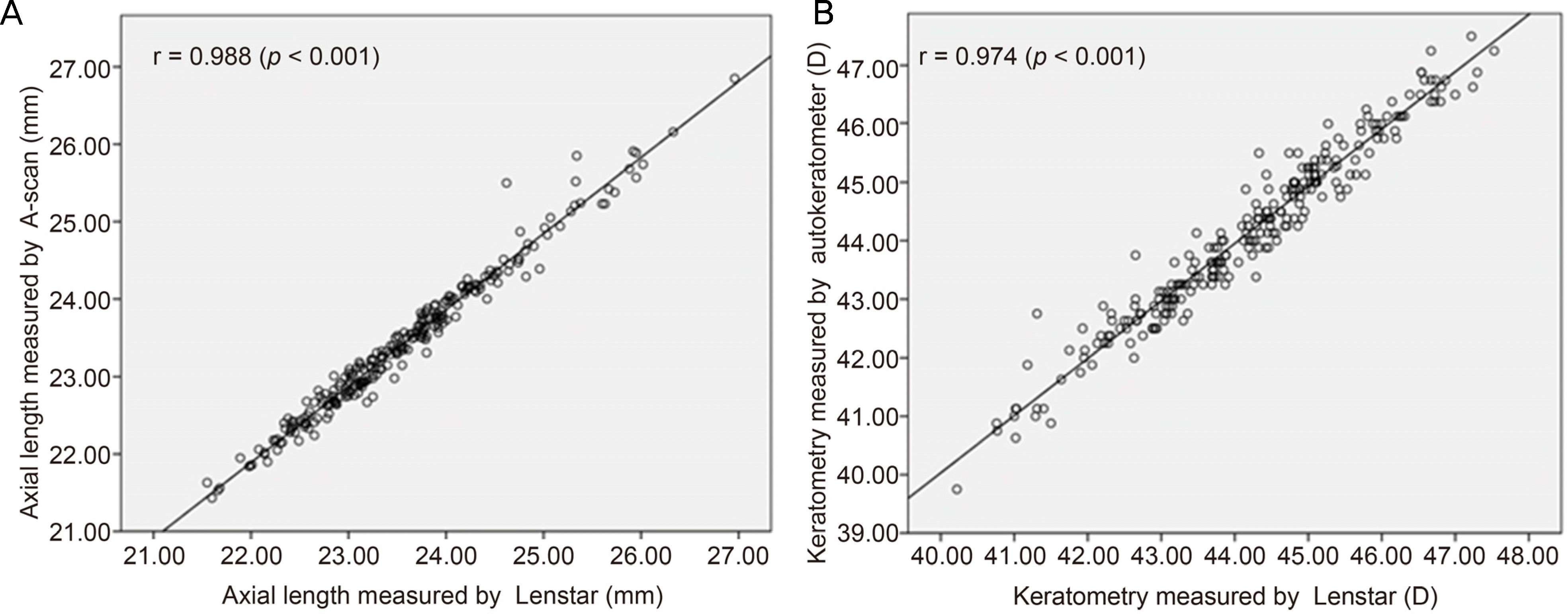 | Figure 1.Correlation between biometric data (axial length, keratometry) measured by Lenstar LS900® and other devices (Pearson correlation analysis). Axial length measured by Lenstar LS900® and A-scan (A). Keratometry measured by Lenstar LS900® and autokeratometer (B). r = Pearson correlation coefficient. |
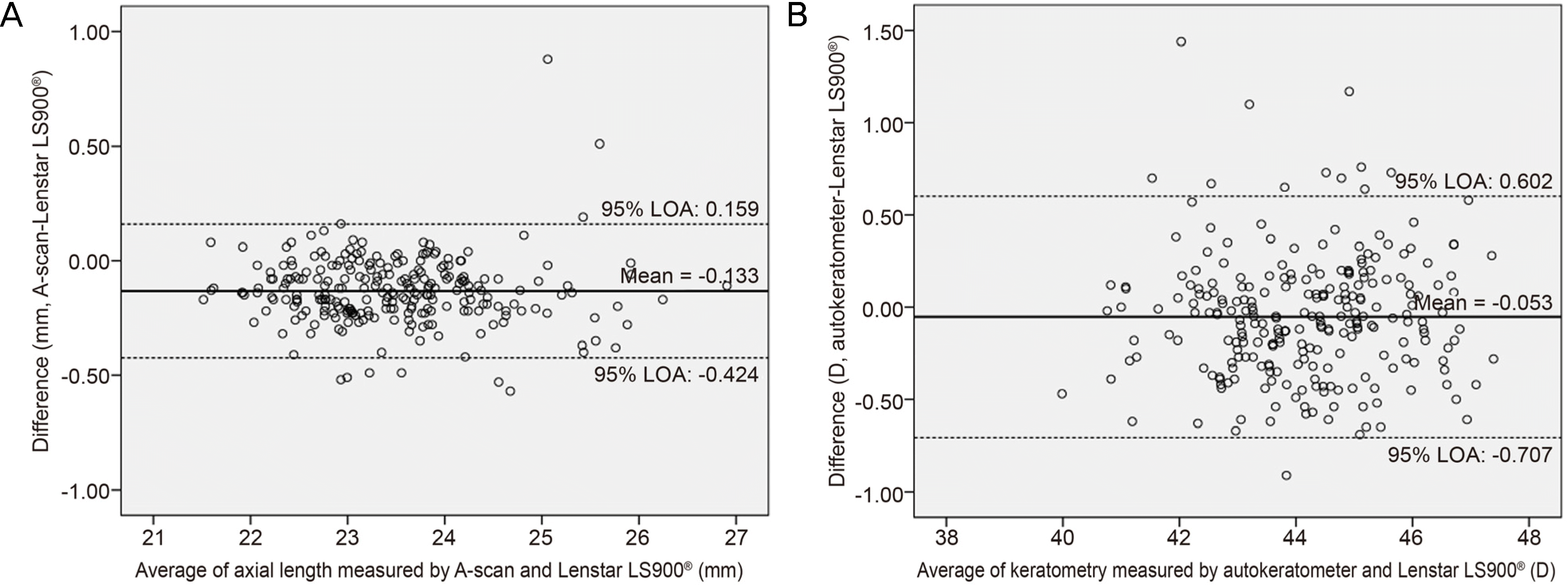 | Figure 2.Bland-Altman plot of axial length and keratometry. Bland-Altman plot of axial length between A-scan and Lenstar LS900® (A), keratometry between autokeratometer and Lenstar LS900® (B). LOA = limit of agreement. |
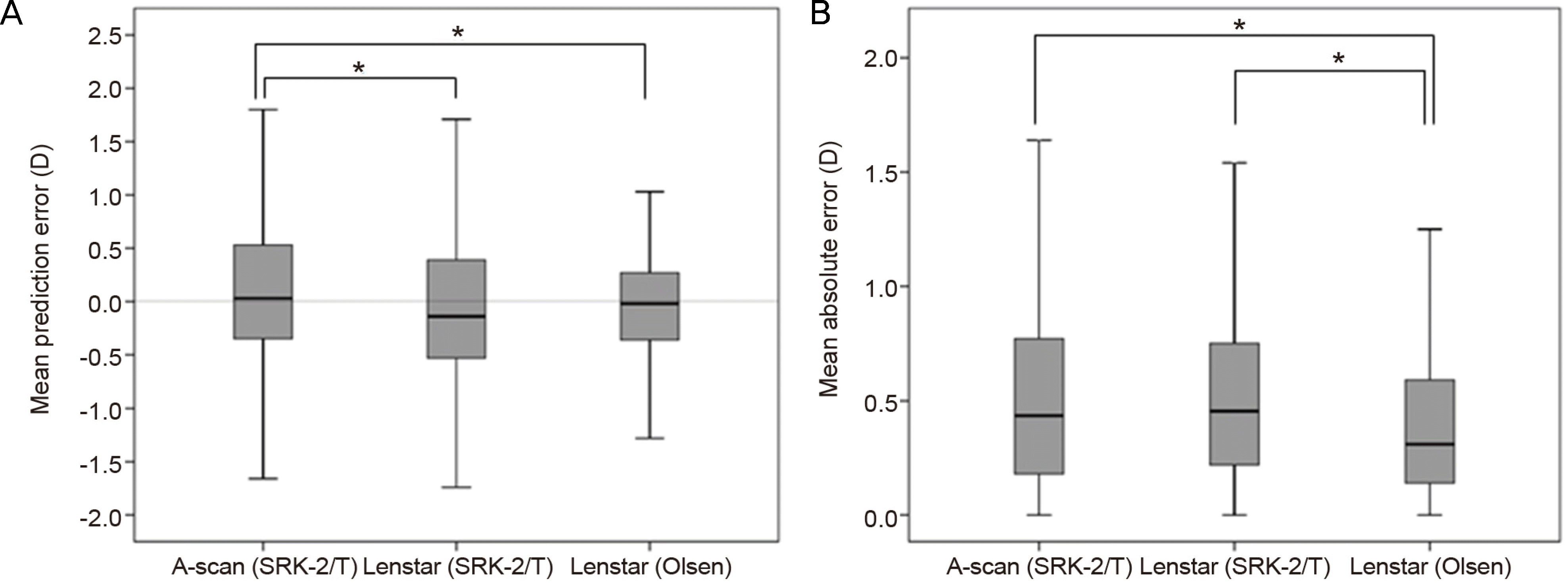 | Figure 3.Comparisons of accuracy of predictive postoperative refraction by mean prediction error (A), mean absolute error (B). ‘A-scan (SRK-2/T)’ is ‘measurement of A-scan using SRK-2 and SRK/T formulas’, ‘Lenstar (SRK-2/T)’ is ‘measurement of Lenstar LS900® using SRK-2 and SRK/T formulas' and ‘Lenstar (Olsen)’ is ‘measurement of Lenstar LS900® using Olsen formula’. Bold line in gray box means ‘median value’, bottom line of gray box means ‘first quartile’, top line of gray box means ‘third quartile’, lower end of vertical line means ‘minimum value’, and upper end of vertical line means ‘maximum value’. * p < 0.05, Statistically significant (Paired t-test). |
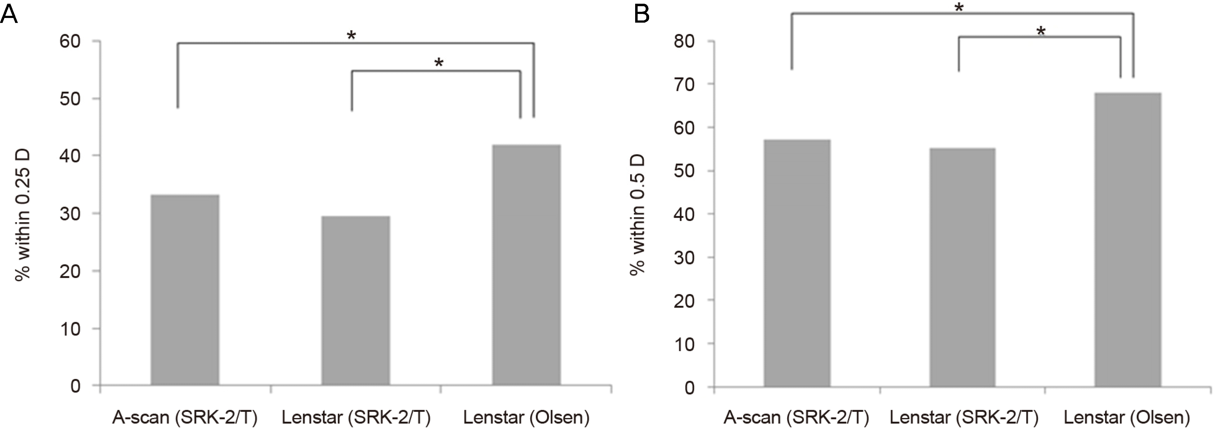 | Figure 4.Comparisons of accuracy using probability within 0.25 D and 0.5 D. Comparisons of probability that the difference between the predictive and actual postoperative refraction is within 0.25 D (A) and within 0.5 D (B). * p < 0.05, Statistically significant (McNemar test). |
Table 1.
Demographic data of enrolled patients
Table 2.
Comparison of biometric data (axial length, mean keratometry, central corneal thickness, anterior chamber depth, lens thickness) by Lenstar LS900®, A-scan and autokeratometer
|
A-scan and Autokeratometer |
Lenstar LS900® |
p-value* | |||
|---|---|---|---|---|---|
| Mean (SD) | Range | Mean (SD) | Range | ||
| AL (mm) | 23.42 (0.94) | 21.43 to 26.85 | 23.55 (0.95) | 21.55 to 26.96 | <0.001 |
| Mean keratometry (D) | 44.13 (1.47) | 40.22 to 47.53 | 44.17 (1.47) | 40.22 to 47.53 | 0.089 |
Table 3.
Values of mean absolute error and probability that it is of within 0.25 D and 0.50 D according to axial length (IOL formula) and IOL model
| Mean absolute error (D) |
Probability of mean absolute error within 0.25 D and 0.50 D (%) |
||||||||||
|---|---|---|---|---|---|---|---|---|---|---|---|
| Within 0.25 D | Within 0.50 D | ||||||||||
| n | A-scan (SRK) | Lenstar LS900® (SRK) | Lenstar LS900® (Olsen) | A-scan (SRK) | Lenstar LS900® (SRK) | Lenstar LS900® (Olsen) | A-scan (SRK) | Lenstar LS900® (SRK) | Lenstar LS900® (Olsen) | ||
| AL (IOL formula) | <25 mm | 232 | 0.513 | 0.517 | 0.398 | 34.9 | 30.6 | 43.1 | 58.2 | 56 | 66.8 |
| (SRK-2) | (<0.001*) | (<0.001*) | (0.079‡) | (0.003‡) | (0.050‡) | (0.011‡) | |||||
| ≥25 mm | 18 | 0.754 | 0.595 | 0.410 | 11.1 | 16.7 | 27.8 | 44.4 | 44.4 | 83.3 | |
| (SRK/T) | (0.010†) | (0.008†) | (0.375‡) | (0.500‡) | (0.039‡) | (0.016‡) | |||||
| IOL model | ZCB00 | 142 | 0.536 | 0.519 | 0.407 | 35.9 | 25.4 | 40.1 | 54.9 | 54.9 | 67.6 |
| (0.002*) | (<0.001*) | (0.519‡) | (0.004‡) | (0.023‡) | (0.021‡) | ||||||
| SN60WF | 108 | 0.524(0.003*) | 0.527(<0.001*) | 0.388 | 29.6(0.034‡) | 35.2(0.175‡) | 44.4 | 60.2(0.243‡) | 55.6(0.045‡) | 68.5 | |
 XML Download