

 PDF
PDF ePub
ePub Citation
 Print
Print


INTRODUCTION
Acute phlegmonous esophagitis, a rare and life-threatening disorder, is characterized by bacterial infection of the submucosal and muscularis layers of the esophagus. It causes necrosis with serious complications, which include esophageal stenosis or perforation, mediastinitis, and empyema (1). Old age, diabetes mellitus, alcoholism, malnutrition, and immunosuppression are well known predisposing factors (2). In diagnosing acute phlegmonous esophagitis, computed tomography (CT) typically reveals an intramural, circumferential, low-attenuation area of the esophagus surrounded by an enhanced peripheral rim (1). To the best of our knowledge, only a few reports on phlegmonous esophagitis have appeared in the literature (123456789). Herein, we present a rare case of acute phlegmonous esophagitis complicated with mediastinitis in a patient with diabetes mellitus and alcoholic liver cirrhosis. We also report the patient's clinical course and CT findings.
CASE REPORT
A 56-year-old man was admitted to the emergency room with a 2-month history of fever, as well as pain in the pharynx, chest, and abdomen. He had a 7-year history of type 2 diabetes mellitus. He also had alcoholic liver cirrhosis and a history of hepatitis C virus infection. The patient complained of pain and tenderness in the pharynx, chest, and upper abdomen. Physical examination revealed a weight loss of 4 kg over 2 months. His body temperature was 38.8℃, and his heart rate was 138 beats/min. Both his blood pressure and respiratory rate were within the normal range. Laboratory examinations revealed leukopenia (leukocyte count: 2200 cells/μL) and an increased C-reactive protein level (3.5 mg/dL). Other examinations revealed hyperglycemia (230 mg/dL), an aspartate aminotransferase/alanine aminotransferase level of 270/77 U/L, and a total bilirubin level of 3.7 mg/dL.
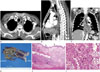
CT images revealed diffuse esophageal wall thickening with an intramural, circumferential, low-attenuation area surrounded by an enhanced peripheral rim involving the proximal portion of the thoracic inlet to the gastroesophageal junction (Fig. 1A–C). CT images revealed increased mediastinal fat attenuation, localized mediastinal fluid collection, and minimal pleural effusion in both hemithoraces, which were suggestive of acute mediastinitis.
The patient had no history of acid or base ingestion. In light of the clinical features and CT findings, phlegmonous esophagitis, corrosive esophagitis, or reflux esophagitis were considered as differential diagnoses, and a diagnosis of acute phlegmonous esophagitis with mediastinitis was eventually arrived at; however, an initial upper gastrointestinal endoscopy was not performed.
One hour after conducting the CT scan, the surgeons decided to perform an emergency surgery. A right posterolateral thoracotomy, esophageal dissection with multifocal esophageal myotomy from the thoracic inlet to the gastroesophageal junction, and drainage were sequentially performed. During the procedure, pus was observed in the mediastinum and between the submucosal and muscularis layers of the esophagus. The friable muscularis layer of the esophagus was also observed. A drainage tube was inserted, and empiric antibiotic treatment with intravenous clindamycin and flomoxef was initiated. The patient required postoperative admission to the intensive care unit. Surgery for postoperative bleeding and gastrostomy for enteral nutrition were performed the following day.
A right-sided pleural effusion culture revealed the presence of Klebsiella pneumoniae (K. pneumoniae); therefore, the patient's antibiotic therapy was changed to piperacillin/tazobactam and ciprofloxacin.
On the sixth day of hospitalization, fever, an elevated C-reactive protein level, and leukocytosis persisted, and air was leaking from the chest tube in the right hemithorax. A follow-up chest CT scan revealed a phlegmon extending to the paravertebral area, as well as poor delineation of the outer wall of the mid-to-lower esophagus. Surgeons suspected a new complication of esophageal perforation, although it was hard to delineate the perforation site. On the 10th day of hospitalization, a segmental esophagectomy from the upper thoracic esophagus to the gastroesophageal junction was performed, as well as a cervical esophagostomy, which revealed an esophageal perforation 2 cm in diameter at the level of the left atrium. A gross esophagectomy specimen revealed an esophageal perforation in the mid portion of the specimen, loss of mucosal folds, and dark-brown discoloration of the mucosa (Fig. 1D). Microscopic examination revealed abundant inflammatory cells, including numerous lymphocytes, neutrophils, and necrotic inflammatory cell debris in the damaged muscularis layer of the esophagus, suggesting phlegmonous esophagitis (Fig. 1E, F).
On the 18th day of hospitalization, there was sanguineous drainage through the percutaneous endoscopic gastrostomy (PEG) tube. One day later, primary repair of the PEG site and an additional feeding jejunostomy for enteral nutrition were performed. On the 20th day of hospitalization, the patient's C-reactive protein levels gradually decreased; the patient was extubated and transferred to the general ward. On the 22nd day of hospitalization, the chest tube was removed, and chest radiography revealed an improvement of the atelectasis and pleural effusion. On the 33rd day of hospitalization, the patient was transferred to another hospital near his home. He is currently scheduled to undergo esophageal reconstruction and the closure of jejunostomy in our hospital.
DISCUSSION
We report a rare case of acute phlegmonous esophagitis in a patient with underlying diabetes mellitus and alcoholic liver cirrhosis. A complication of an esophageal perforation was demonstrated by an esophagectomy.
Phlegmonous enteritis is reported to be a rare, life-threatening disorder characterized by bacterial infection of the submucosal and muscularis layers of the digestive tract (1). The stomach is the most commonly involved site, with more than 100 cases reported in the literature. The mortality rate due to phlegmonous enteritis is 42% (7). Acute phlegmonous esophagitis is even more rare than phlegmonous gastritis, and a few cases of phlegmonous esophagitis with or without stomach involvement have been reported (12345689). There are several predisposing factors such as alcoholism, old age, malnutrition with a low albumin level, and uncontrolled diabetes, for acute phlegmonous esophagitis (1). In this case, all these predisposing factors were present, creating the potential for a combined effect. There are several causative organisms implicated in cases of phlegmonous enteritis. K. pneumoniae is considered to be the most common of these (1). Pathogenesis involves damage to the intestinal tract, resulting in susceptibility to bacterial infections (3). In our patient, K. pneumoniae was identified in the pleural effusion culture.
Radiological CT findings demonstrating an intramural, circumferential, low-attenuation area of the esophagus surrounded by an enhanced peripheral rim are essential for the diagnosis of acute phlegmonous esophagitis. The histopathological features include thickening of the submucosa and infiltration of neutrophils and plasma cells with intramural hemorrhage, necrosis, thrombosis of the submucosal blood vessels, and an abscess in the submucosal and muscularis layers of the esophagus (45). We think that all of these known findings might be similar and can be correlated with those in our case.
The differential diagnoses of diffuse esophageal wall thickening on CT could be a dissecting intramural hematoma, corrosive esophagitis, reflux esophagitis, or a diffuse esophageal spasm. However, a diffuse intramural hematoma and a diffuse esophageal spasm are not likely to involve infection or inflammation. In the case of a diffuse intramural hematoma, precontrast CT images showed high-attenuation wall thickening of the entire esophagus. A history of acid or base ingestion is crucial for diagnosing corrosive esophagitis. Reflux esophagitis is often accompanied by a sliding esophageal hernia and mainly involves the mid-to-lower esophagus without peripheral rim enhancement on the CT. A diffuse esophageal spasm is usually diagnosed by barium esophagogram, and a CT does not typically reveal prominent enhancement of the mucosa or peripheral rim enhancement of the esophagus.
The treatment strategy for this rare condition includes managing the infection by systemic antibiotic administration, preventing contamination progression, providing nutritional support, preserving digestive tract continuity, and providing timely surgical intervention if required (10). Surgical resection is required when there is extensive phlegmonous esophagogastritis, since this involves esophageal necrosis, esophageal stricture progression, gastric mucosa atrophy, and acute peritonitis (2). As a CT scan is the most useful diagnostic modality for acute phlegmonous esophagitis, rapid access to a CT scan is important for making an accurate diagnosis and deciding the requirement of a surgical intervention. Radiologists must evaluate the extent and severity of the condition and any comorbidity using CT images. Particularly in cases of patients with predisposing factors, such as alcoholism or uncontrolled diabetes, radiologists must consider this condition.
In summary, we presented a rare case of acute phlegmonous esophagitis with mediastinitis complicated by an esophageal perforation in a patient with diabetes mellitus and alcoholic liver cirrhosis. The patient was treated with antibiotic therapy and surgical intervention that included drainage and esophagectomy.
 XML Download