

 PDF
PDF ePub
ePub Citation
 Print
Print


INTRODUCTION
Diverticular disease is a common colonic disease. Diverticulitis occurs in about 10–25% of patients with diverticulosis and is sometimes combined with perforation, abscess, or fistula formation (1). Although rare, colon cancer can develop from the mucosa of a colonic diverticulum. This rarity, together with sometimes non-specific imaging findings, complicates differential diagnosis from common diverticular disease. In this report, we describe a rare case of mucinous adenocarcinoma arising in a colonic diverticulum without diverticulitis or abscess formation, mimicking a perforated diverticular abscess.
CASE REPORT
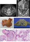
An 82-year-old female was admitted to our emergency department with acute abdominal pain localized to the right lower quadrant. Her pain started one day before, was persistent, and rated as moderate (Numeric Rating Scale 6). She did not have other gastrointestinal symptoms such as vomiting or diarrhea. Physical examination on admission showed tenderness and rebound tenderness at the right lower quadrant and a fever of 38.3℃. On laboratory examination, her white blood cell count was 18700/μL (normal, 4000–10000/μL) with neutrophilia of 78.12%. The C-reactive protein was elevated to 5.75 mg/dL (normal, 0.0–0.5 mg/dL). The patient had no specific medical history except for hypertension and hyperlipidemia. She denied a previous history of acute colonic diverticulitis. Contrast-enhanced abdominal computed tomography (CT) revealed a 4.8 × 3.9 × 3.8 cm peripheral-enhancing, low-attenuated, exophytic mass like lesion at the medial wall of the proximal ascending colon. The lesion connected to the lumen of the proximal ascending colon via an out-pouching sac, indicating a colonic diverticulum. The outer margin of the diverticulum was not defined, but contained a small amount of air (Fig. 1A, B). There was no calcification within the mass like lesion. Mild soft tissue stranding was noted in surrounding fat. An involved segment of colon showed mural thickening with preservation of the layering pattern. There were multiple enlarged lymph nodes at the right lower quadrant, the largest of which was 9 × 17 mm in diameter. Lymph nodes were round or oval with regular borders and homogeneous enhancement (mean, 45 HU on pre-contrast images; mean, 150 HU on contrast-enhanced images). No intraperitoneal free air was seen. The appendix and other pelvic organs were unremarkable. Based on abdominal CT findings of an out-pouching diverticulum, low-attenuated mass like lesion surrounding a diverticulum, and mural thickening of involved colonic segment with preservation of the mural layer and clinical and laboratory features of acute abdominal pain, fever, and leukocytosis, the differential diagnosis was perforated colonic diverticulitis with abscess formation. However, the possibility of colon cancer was also considered because of the exophytic mass, which showed a relatively mild degree of soft tissue stranding in peri-lesional fat. The patient underwent ileocecectomy with lymph node dissection. Intraoperatively, an 8 cm mass was identified at the ascending colon without abscess formation.
Macroscopically, there was a large diverticular opening at the proximal ascending colon, but the surrounding mucosa was otherwise grossly unremarkable (Fig. 1C). Cut sections revealed an irregular mass in the pericolic soft tissue, involving the colonic wall (Fig. 1D). Microscopically, the diverticular opening was lined by mucosa invaginating into the muscularis propria. Tumor cell nests with an abundant mucin pool occupied the diverticular space and extended through the diverticular wall to the pericolic soft tissue with involvement of a pericolic lymph node (Fig. 1E, F). Immunohistochemical findings suggested a neoplasm of intestinal origin rather than a metastatic malignancy (CDX-2+, CK7-, CK20-, GCDFP-15-, TTF-1-, PAX-8-, ER-). Based on the pathologic findings, a moderately differentiated mucinous adenocarcinoma arising in a colonic diverticulum was confirmed. After surgery, the patient received adjuvant chemotherapy.
DISCUSSION
Colonic diverticular disease is common, but colon cancer arising in a colonic diverticulum is rare. Since colon cancer associated with colonic diverticular abscess was first described by Tolley in 1967 (2), only a few cases of colon cancer associated with a diverticulum have been reported (345). Several investigators have tried to show an increased incidence of colonic carcinoma in patients with diverticulitis, suggesting that chronic inflammation leads to metaplasia and neoplastic changes (46). However, other investigators have argued that there is no specific relationship between diverticulitis and carcinoma based on the following evidence: 1) the rarity of adenocarcinomas arising within diverticular mucosa, 2) the absence of occasional dysplasia in diverticular mucosa, and 3) the often documented occurrence of adenocarcinomas in areas with no diverticula (7). In our case, histopathological examination of the colonic specimen showed carcinoma within a diverticulum without involvement of the colonic mucosa. There was no histopathological evidence of chronic inflammation or abscess.
Mucinous adenocarcinoma is a histopathological subtype of colorectal cancer characterized by abundant extracellular mucin pools (8). In previous studies that compared imaging findings of mucinous colorectal carcinoma with non-mucinous carcinoma, mucinous carcinomas frequently showed bowel wall thickening greater than 2 cm, eccentric bowel wall thickening, heterogeneous contrast enhancement of the tumor, poor contrast enhancement of the solid tumor component, a large area of low attenuation within the tumor, and intratumoral calcification on contrast-enhanced CT. They also showed high signal intensity on T2-weighted images, with a peripheral contrast enhancement pattern on MRI, in contrast to non-mucinous carcinomas (79). Consistent with previous findings, the mass in our case showed low attenuation with poor contrast enhancement on CT. The mass had no intratumoral calcification. These findings mimic an abscess cavity, complicating the diagnosis. However, we considered the possibility of colon cancer due to the weak soft tissue stranding in peri-lesional fat.
Two cases of a mucinous adenocarcinoma developing in a diverticulum have been reported (35). Kwon et al. (3) reported a rare case of mucinous adenocarcinoma arising from a rectal diverticulum. In that case, radiologic and endoscopic findings suggested primary rectal cancer, and the rectal diverticulum was not visible on radiologic and endoscopic examination. In the second case (5), a mucinous adenocarcinoma was incidentally identified in perforated colonic diverticulitis with abscess formation. To the best of our knowledge, the present case is the first report of a mucinous adenocarcinoma arising in a colonic diverticulum without diverticulitis or abscess formation. Due to the rarity of carcinoma development within a diverticulum, it is easily missed. Furthermore, difficulties in differentiating between colon cancer and diverticulitis with or without abscess formation are well recognized (10). Meanwhile, malignant tumors arising within a diverticulum can easily penetrate the serosa due to lack of a muscular layer (4). In our case, the tumor penetrated through the diverticular wall into pericolic soft tissue and a pericolic lymph node. It is important to consider the possibility of malignancy when diagnosing diverticular disease, especially when unusual imaging features are observed.
In summary, we describe a rare case of mucinous adenocarcinoma arising in the colonic diverticulum, mimicking a perforated diverticular abscess. Despite its rarity, the possibility of a malignant tumor masquerading as diverticular disease should be considered to improve patient management and prognosis.
 XML Download