

 PDF
PDF ePub
ePub Citation
 Print
Print


INTRODUCTION
The diagnosis of the spinal meningiomas is not very difficult based on radiologic findings and typical locations. However, diagnostic difficulties occur when the meningioma has atypical imaging characteristics or is in an unfamiliar location. Spinal meningiomas, which accounts for approximately 12% of all meningioma, are mostly intradural extramedullary in location (1). Some cases of intradural spinal meningiomas are associated with extradural extensions; however, it is uncommon to have pure epidural location. Extradural spinal meningiomas account for only 2.5% to 3.5% of all spinal meningioma (2). We report a case of pathologically confirmed spinal epidural meningioma. Additionally, we review the literature and the radiographic features of meningioma in unusual locations.
CASE REPORT
A 58-year-old woman presented with progressive weakness and paraesthesia of both lower legs for four months. The woman showed paraparesis of motor Grade IV on both legs and had difficulty maintaining her balance due to weakness. She had hyperesthesia and dysesthesia on light touch, pain, and temperature sense below the T9 dermatome level. The position and vibration senses were intact. She did not have symptoms related to sphincter dysfunction.
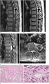
A spine MRI demonstrated a well-defined mass approximately 3.0 × 1.0 cm in diameter in the posterior epidural space of the seventh thoracic vertebral level on sagittal T1-weighted (Fig. 1A) and T2-weighted images (Fig. 1B). The mass caused compression and signal change of the spinal cord, suggesting compressive myelopathy. However, adjacent bone marrow was intact with no evidence of osseous infiltration. T1-weighted images with contrast enhancement showed strong enhancement of the mass with a left neural foraminal extension of the T7 vertebrae (Fig. 1C, D). Preoperatively, the radiologic differential diagnosis was lymphoma or metastatic tumor, but no primary lesion was found in the quick preoperative radiologic evaluation.
The patient received excision with partial laminectomy of T7. Microscopic examination of the epidural space mass revealed an atypical meningioma. Histologically, the tumor was dominated by multiple vessels interspersed with small meningothelial tumor cells. Focal necrosis was also seen (Fig. 1E). The tumor cells showed increased mitoses up to 10 per 10 high-power fields despite bland cytological features (Fig. 1F). Tumor cells were also positive for epithelial membrane antigen (not shown). The final histological diagnosis was an atypical (WHO Grade II) meningioma.
After the surgery, the patient did not undergo further treatment such as post-operative radiotherapy. Nonetheless, the neurologic symptoms were relieved, and no evidence of tumor recurrence was shown on three month follow up CT.
DISCUSSION
Spinal meningiomas represent approximately 12% of all meningioma (1). Most meningiomas are intradural tumors that arise at the thoracic level. In the spine, meningiomas are thought to arise from the denticulate ligaments and more than 95% are classified as WHO Grade I (3). Extradural component is thought to be extended from an intradural mass. It is uncommon for a spinal meningioma to have an epidural location; in fact, exclusively extradural spinal meningiomas are infrequent and account for only 2.5% to 3.5% of all spinal meningioma (2).
Spinal extradural meningiomas show the same histology, peak incidence in the fifth and sixth decades, the same frequent location in the thoracic spine and the same well-known sex preponderance of female patients compared with intradural meningiomas (2).
The clinical findings of epidural meningiomas are not significantly different from those of intradural located ones. Patients may present with back pain, sensory and motor changes, and finally sphincter disturbances in late phases (2).
MRI is the best imaging modality to diagnose spinal meningiomas. It delineates the location and extent of the tumor, guiding the plan of the surgery. However, extradural spinal meningiomas are uncommon, and they can be mistaken as others. It affects the extent of the surgery and patient's outcome, so the diagnosis of the extradural spinal mass needs to be carefully done.
Spinal meningiomas have typical features on MRI. On T1-weighted images, the mass often has signal intensity similar to that of the spinal cord and exhibits mildly increased signal intensity on T2-weighted images (1). In addition, the pattern of contrast enhancement is strong and homogeneous. Foraminal extension does not favor diagnosis of a meningioma over diagnosis of a schwannoma or neurofibroma (1). The latter two lesions usually demonstrate high signal intensity on T2-weighted images with cystic change and inhomogeneous enhancement (1). Therefore, the two tumors are easy to exclude. In addition, lymphoma, a malignant epidural mass, is the other misdiagnosis of meningioma due to homogeneous and strong enhancement and low signal intensity on T2-weighted images due to its dense cellularity (235). Moreover, hyperostosis is observed less frequently in patients with epidural spinal meningiomas than in patients with cranial meningiomas (9). Hyperostosis was not clear in our case.
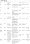
We have reviewed other 12 cases of spinal extradural meningiomas previously reported since 2002 in Table 1. They were most commonly located in the thoracic spine followed by the cervical spine, and patients showed predominance of female. On imaging studies, cases appeared as intraspinal extradural masses with mostly homogeneous enhancement. In most cases, the mass showed extradural en plaque lesion suggesting the dural based origin of the mass, also known as dural tail sign, making it dumbbell shape appearance. Nevertheless, our case did not present any signs that clarified that the mass originated from the dura. Compared to other reported cases, our case showed more well-delineated margin. These findings made it more difficult to make the accurate diagnosis and it could easily be mistaken as other extradural tumor such as lymphoma.
Pathologically, spinal meningiomas tend to be well-defined discrete lesions with a dural attachment. Microscopically, the common patterns are meningothelial, fibroblastic, transitional, and psammomatous and most case reports describe features of a miningothelial or psammomatous meningioma (27). The WHO classification of meningiomas are divided into three grades. Among many subtypes, meningiothelial, fibrous and transitional are the most common, and belong to Grade I. Our patient's pathology report suggested atypical meningioma (WHO Grade II) with increased mitotic activity, focal necrosis and frequent vascular tumor emboli. Among reviewed 12 cases in Table 1, there is no case confirmed as atypical meningioma. Apart from one case of choroid meningioma (WHO Grade II) occurred in a 9-year-old girl, all of the other cases were Grade I meningiomas.
In literature review of major spinal meningioma series, including both intra- and extradural subtypes, recurrence rates of surgical excision are reported as 3% to 7% (2). Although, there was no case which showed recurrence of the tumor among the reviewed 12 cases, the recurrence rate after surgery for extradural meningioma is four times higher than that of intradural meningioma (410). Our patient yet did not show any tumor recurrence for three months, but careful observation and follow up is still necessary.
In conclusion, we describe here a rare case of spinal extradural meningioma in the thoracic level with atypical one (WHO Grade II). However, it must be not forgotten that meningiomas may occur in rare locations and with variable imaging characteristics.
 XML Download