Abstract
Purpose
This study aimed to determine which factors are related to perforated appendicitis. We also conducted a survey to identify the causative organism.
Methods
From January 2011 to December 2014, 569 pediatric patients (322 male) younger than 19 years old who underwent an appendectomy due to acute appendicitis at Hallym University Sacred Heart Hospital were enrolled. Patients' medical records were reviewed retrospectively to determine their clinical manifestations, laboratory and imaging results, and pathogens.
Results
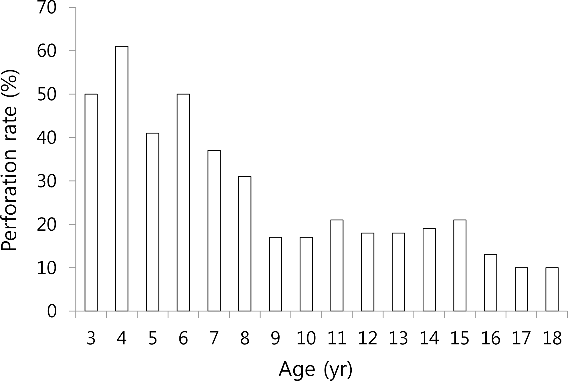
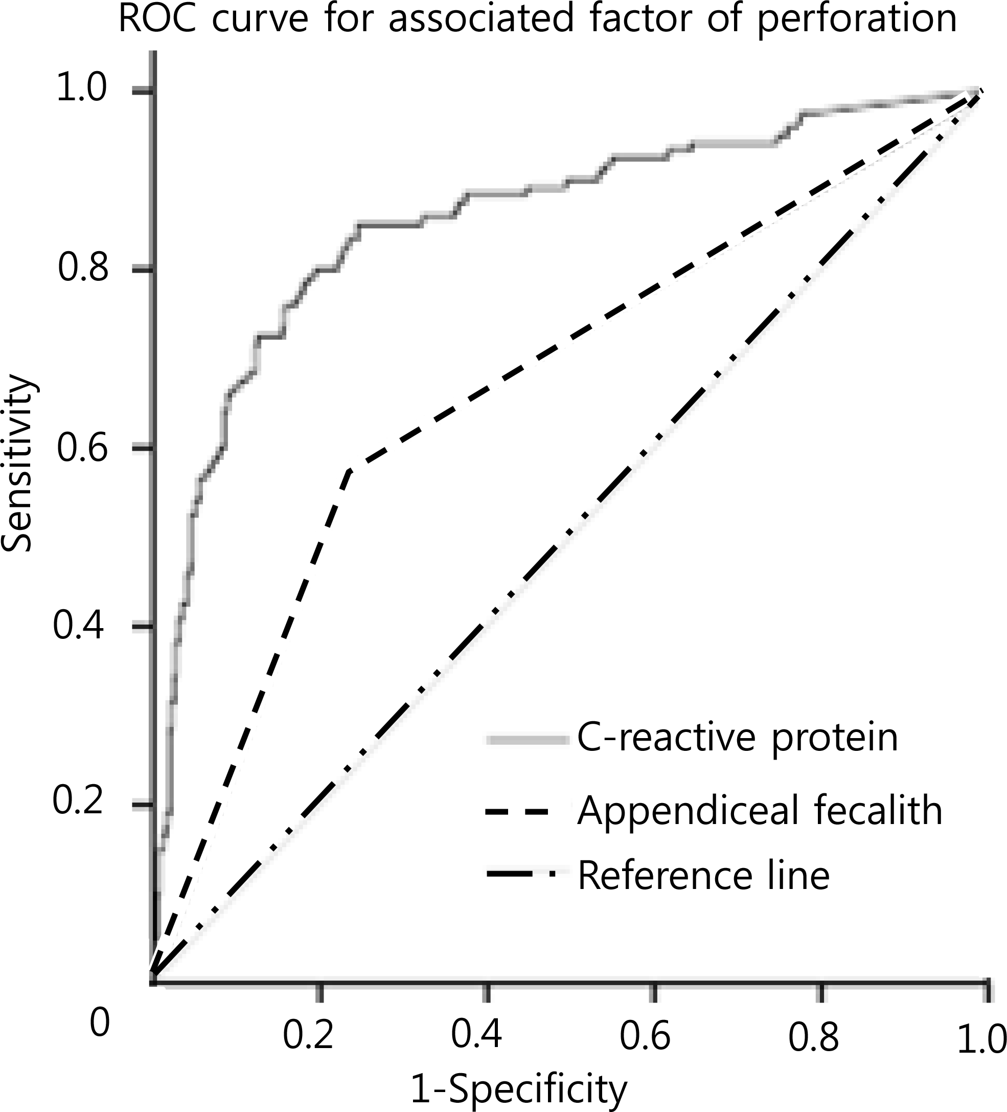
About 127 patients (22%) had perforated appendicitis. The rate of perforated appendicitis in preschool, late childhood, and adolescent ages were 50%, 27%, and 16.8%, respectively. The risk factors of perforation were high C-reactive protein levels and the presence of appendiceal fecalith (P<0.001). Of the 24 samples of peritoneal fluid and periappendiceal pus that were collected intraoperatively, 16 were culture positive. The most common pathogen was Escherichia coli (n=10), and others were Pseudomonas aeruginosa, Streptococcus spp., and Staphylococcus spp.
Conclusions
The perforation rate of appendicitis among patients younger than 5 years old was 50%, and this decreased in proportion with age. Clinicians should be aware of the possibility of perforation when patients with appendicitis have high C-reactive protein levels or the presence of appendiceal fecalith on imaging.
Go to : 
References
1. Cherry JD, Harrison GJ, Kaplan SL, Steinbach WJ, Hotez PJ. Appendicitis and pelvic abscess. Cherry JD, editor. Feigin & Cherry's textbook of pediatric infectious diseases. 7th Ed.Philadelphia: Saunders Co;2013. p. 679–89.
2. Abou Merhi B, Khalil M, Daoud N. Comparison of Alvarado score evaluation and clinical judgment in acute appendicitis. Med Arch. 2014; 68:10–3.
3. Aiken JJ, Oldham KT. Acute appendicitis. Kliegman RM, Nelson WE, editors. editors.Nelson textbook of pediatrics. 20th Ed.Philadelphia: Elsevier;2016. p. 1887–93.

4. Mc Cabe K, Babl FE, Dalton S. Paediatric Research in Emergency Departments International Collaborative (PREDICT). Management of children with possible appendicitis: a survey of emergency physicians in Australia and New Zealand. Emerg Med Australas. 2014; 26:481–6.

5. Chen CY, Chen YC, Pu HN, Tsai CH, Chen WT, Lin CH. Bacteriology of acute appendicitis and its implication for the use of prophylactic antibiotics. Surg Infect (Larchmt). 2012; 13:383–90.

6. Adams DH, Fine C, Brooks DC. High-resolution real-time ultrasonography. A new tool in the diagnosis of acute appendicitis. Am J Surg. 1988; 155:93–7.
8. Stefanutti G, Ghirardo V, Gamba P. Inflammatory markers for acute appendicitis in children: are they helpful? J Pediatr Surg. 2007; 42:773–6.

9. Naiditch JA, Lautz TB, Daley S, Pierce MC, Reynolds M. The implications of missed opportunities to diagnose appendicitis in children. Acad Emerg Med. 2013; 20:592–6.

10. Choi JY, Ryoo E, Jo JH, Hann T, Kim SM. Risk factors of delayed diagnosis of acute appendicitis in children: for early detection of acute appendicitis. Korean J Pediatr. 2016; 59:368–73.

11. Horwitz JR, Gursoy M, Jaksic T, Lally KP. Importance of diarrhea as a presenting symptom of appendicitis in very young children. Am J Surg. 1997; 173:80–2.

12. Bansal S, Banever GT, Karrer FM, Partrick DA. Appendicitis in children less than 5 years old: influence of age on presentation and outcome. Am J Surg. 2012; 204:1031–5.

13. Peng YS, Lee HC, Yeung CY, Sheu JC, Wang NL, Tsai YH. Clinical criteria for diagnosing perforated appendix in pediatric patients. Pediatr Emerg Care. 2006; 22:475–9.

14. Ngim CF, Quek KF, Dhanoa A, Khoo JJ, Vellusamy M, Ng CS. Pediatric appendicitis in a developing country: what are the clinical predictors and outcome of perforation? J Trop Pediatr. 2014; 60:409–14.

15. Mathews EK, Griffin RL, Mortellaro V, Beierle EA, Harmon CM, Chen MK, et al. Utility of immature granulocyte percentage in pediatric appendicitis. J Surg Res. 2014; 190:230–4.

16. Bonadio W, Peloquin P, Brazg J, Scheinbach I, Saunders J, Okpalaji C, et al. Appendicitis in preschool aged children: regression analysis of factors associated with perforation outcome. J Pediatr Surg. 2015; 50:1569–73.

17. Hung MH, Lin LH, Chen DF. Clinical manifestations in children with ruptured appendicitis. Pediatr Emerg Care. 2012; 28:433–5.

18. Gladman MA, Knowles CH, Gladman LJ, Payne JG. Intraoperative culture in appendicitis: traditional practice challenged. Ann R Coll Surg Engl. 2004; 86:196–201.

19. Foo FJ, Beckingham IJ, Ahmed I. Intraoperative culture swabs in acute appendicitis: a waste of resources. Surgeon. 2008; 6:278–81.

20. Park KW. Appendicitis in children. Korean J Pediatr. 1993; 36:1044–6.
21. Chan KW, Lee KH, Mou JW, Cheung ST, Sihoe JD, Tam YH. Evidence-based adjustment of antibiotic in pediatric complicated appendicitis in the era of antibiotic resistance. Pediatr Surg Int. 2010; 26:157–60.

22. Boueil A, Guegan H, Colot J, D'Ortenzio E, Guerrier G. Peritoneal fluid culture and antibiotic treatment in patients with perforated appendicitis in a Pacific Island. Asian J Surg. 2015; 38:242–6.

Go to : 
 | Fig. 1.The perforation rate of acute appendicitis according to age are shown. Approximately the perforation rate was higher in younger age. The peak of perforation rate was found in 4 years age group. |
 | Fig. 2.Receiver operating characteristic (ROC) curves of C-reactive protein and appendiceal fecalith. |
Table 1.
Clinical Characteristic and Laboratory Results of Subjects
| At admission | Perforation (n=442) | No perforation (n=127) | P-value |
|---|---|---|---|
| Age (yr) | 12.3±3.7 | 10±4.1 | <0.001 |
| Nausea or vomiting | 296 (67.0) | 101 (79.5) | 0.007 |
| Diarrhea | 50 (11.7) | 29 (23.8) | <0.001 |
| Duration of symptom∗ (day) | 1.3 | 1.9 | 0.026 |
| Abdominal tenderness | 422 (99.1) | 120 (98.4) | 0.512 |
| Abdominal rebound tenderness | 357 (83.8) | 112 (91.8) | 0.027 |
| White cell count (/µL) | 13,363±4,685 | 17,575±5,172 | <0.001 |
| Absolute neutrophil count (/µL) | 11,312±13,714 | 15,074±4,777 | <0.001 |
| Neutrophil (%) | 77.5±11.9 | 85.3±5.9 | 0.006 |
| CRP (mg/L) | 21.1±35.1 | 91.3±64.5 | <0.001 |
| ESR (mm/hr) | 13.8±18.5 | 35.3±19.6 | <0.001 |
| Appendiceal fecalith | 101 (23) | 71 (56) | <0.001 |
Table 2.
Associated Factors of Perforation of Acute Appendicitis


 PDF
PDF ePub
ePub Citation
 Print
Print


 XML Download