Abstract
Purpose
To evaluate interpretation errors involving spine MRIs by residents in their second to fourth year of training, classified as minor, intermediate and major discrepancies, as well as the types of commonly discordant lesions with or without clinical significance.
Materials and Methods
A staff radiologist evaluated both preliminary and final reports of 582 spine MRIs performed in the emergency room from March 2011 to February 2013, involving (1) the incidence of report discrepancy, classified as minor if there was sufficient description of the main MR findings without ancillary or incidental lesions not influencing the main diagnosis, treatment, or patients’ clinical course; intermediate if the correct diagnosis was made with insufficient or inadequate explanation, potentially influencing treatment or clinical course; and major if the discrepancy affected the main diagnosis; and (2) the common causes of discrepancy. We analyzed the differences in the incidence of discrepancy with respect to the training years of residents, age and sex of patients.
Results
Interpretation discrepancy occurred in 229 of the 582 cases (229/582, 39.3%), including 146 minor (146/582, 25.1%), 40 intermediate (40/582, 6.9%), and 43 major cases (43/582, 7.4%). The common causes of major discrepancy were: over-diagnosis of fracture (n = 10), missed cord lesion (n = 9), missed signal abnormalities associated with diffuse marrow (n = 5), and failure to provide differential diagnosis of focal abnormal marrow signal intensity (n = 5). No significant difference was found in the incidence of minor, intermediate, and major discrepancies according to the levels of residency, patients’ age or sex.
Conclusion
A 7.4% rate of major discrepancies was found in preliminary reporting of emergency MRIs of spine interpreted by radiology residents, probably related to a relative lack of clinical experience, indicating the need for additional training, especially involving spine trauma, spinal cord and bone marrow lesions.
Go to : 
References
1. Yablon CM, Wu JS, Newman LR, Downie BK, Hochman MG, Eisenberg RL. A needs assessment of musculoskeletal fellowship training: a survey of practicing musculoskeletal radiologists. AJR Am J Roentgenol. 2013; 200:732–740.

2. Walls J, Hunter N, Brasher PM, Ho SG. The DePICTORS Study: discrepancies in preliminary interpretation of CT scans between on-call residents and staff. Emerg Radiol. 2009; 16:303–308.

3. Chung JH, Strigel RM, Chew AR, Albrecht E, Gunn ML. Overnight resident interpretation of torso CT at a level 1 trauma center an analysis and review of the literature. Acad Radiol. 2009; 16:1155–1160.
4. Miyakoshi A, Nguyen QT, Cohen WA, Talner LB, Anzai Y. Accuracy of preliminary interpretation of neurologic CT examinations by on-call radiology residents and assessment of patient outcomes at a level I trauma center. J Am Coll Radiol. 2009; 6:864–870.

5. Hochberg AR, Rojas R, Thomas AJ, Reddy AS, Bhadelia RA. Accuracy of on-call resident interpretation of CT angiography for intracranial aneurysm in subarachnoid hemorrhage. AJR Am J Roentgenol. 2011; 197:1436–1441.

6. Sistrom C, Deitte L. Factors affecting attending agreement with resident early readings of computed tomography and magnetic resonance imaging of the head, neck, and spine. Acad Radiol. 2008; 15:934–941.

7. Bruni SG, Bartlett E, Yu E. Factors involved in discrepant preliminary radiology resident interpretations of neuroradiological imaging studies: a retrospective analysis. AJR Am J Roentgenol. 2012; 198:1367–1374.

8. Filippi CG, Schneider B, Burbank HN, Alsofrom GF, Linnell G, Ratkovits B. Discrepancy rates of radiology resident interpretations of on-call neuroradiology MR imaging studies. Radiology. 2008; 249:972–979.

Go to : 
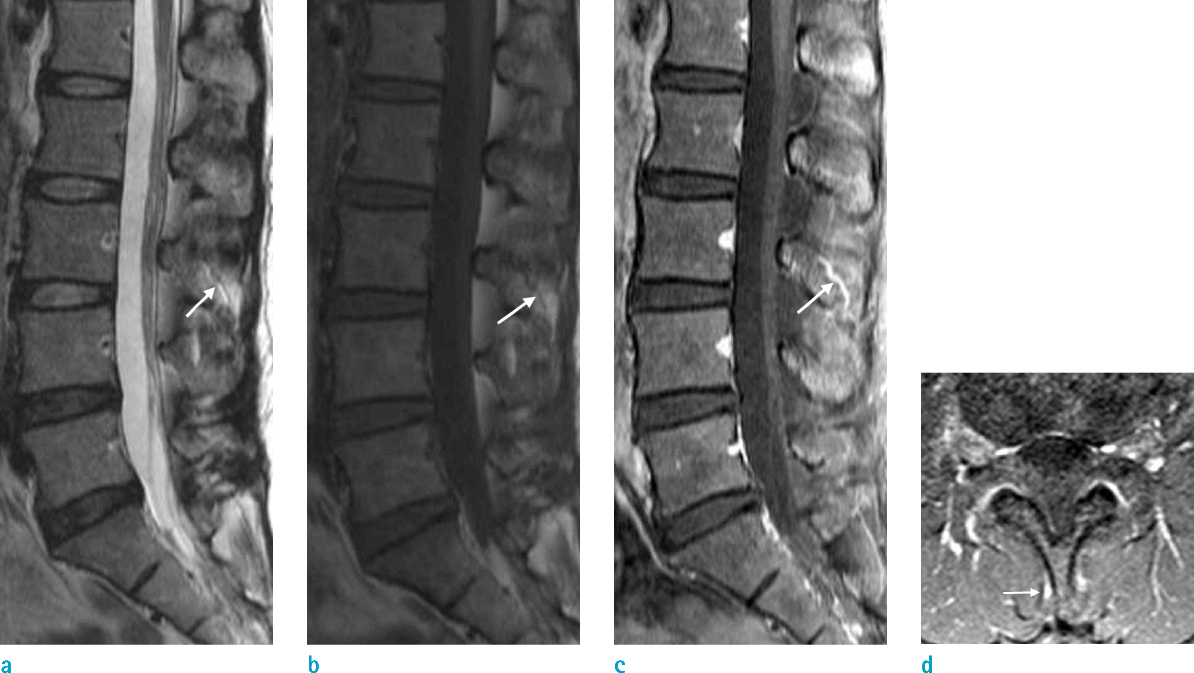
 | Fig. 1.Lumbar spine MRI of a 39-year-old woman with back pain. Linear lesion at the spinous process of L3 presenting high signal intensity on T2-weighted mid-sagittal image (a, arrow) and low signal intensity on T1-weighted mid-sagittal image (b, arrow), which enhances the vascular structure along the spinous process on T1-weighted enhanced mid-sagittal (c, arrow) and axial scans (d, arrow). This lesion was described as a fracture of spinous process, and determined as a major discrepancy. |
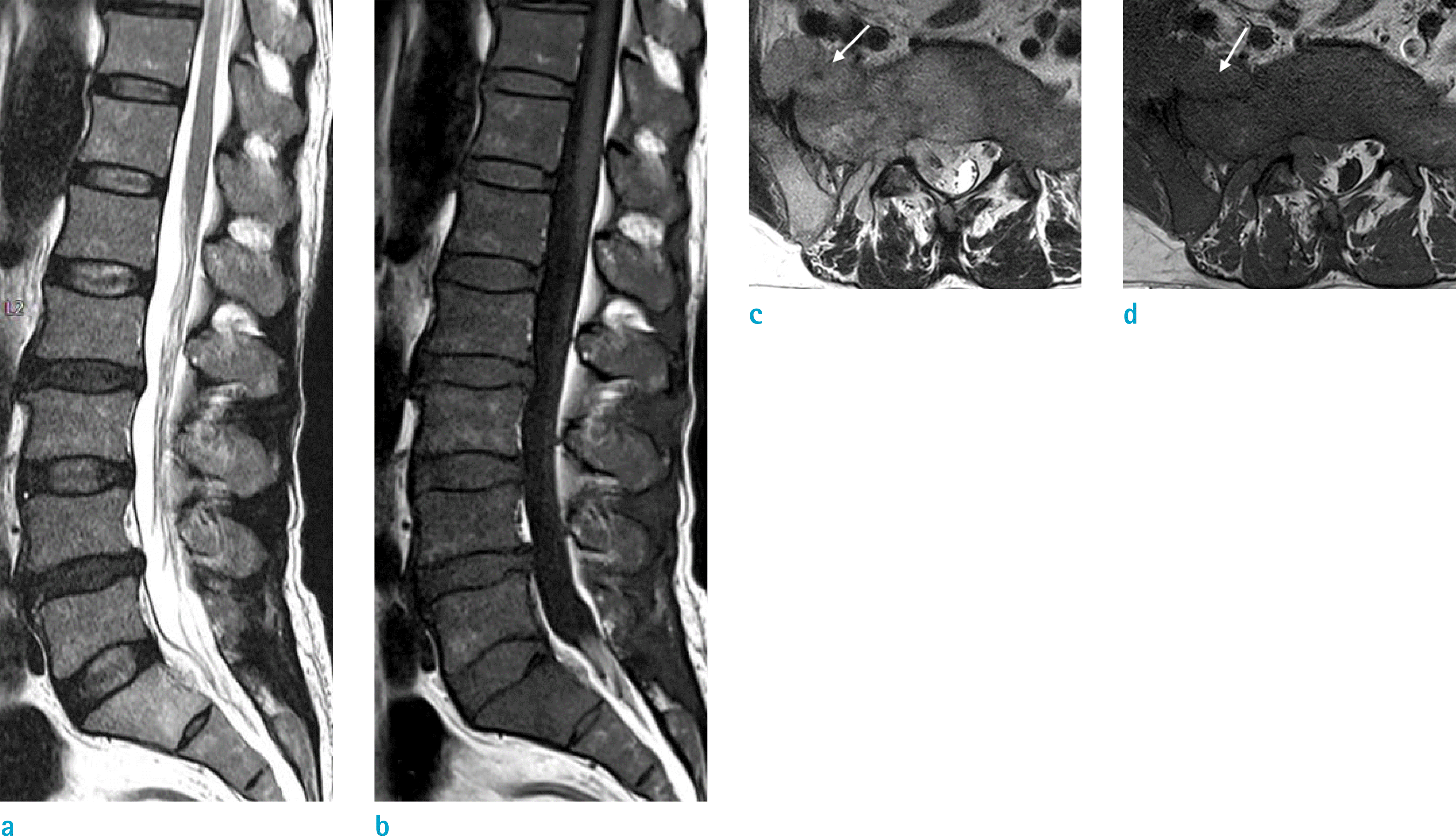
 | Fig. 2.Lumbar spine MRI of a 71-year-old man with leg weakness. Herniated disc is noted at L4–5 level on T2-weighted mid-sagittal scan (a), but diffuse abnormal marrow signal was found on the T1-weighted mid-sagittal image (b). Intermediate signal intensity mass lesion is also seen in the right sacral ala on T2-weighted axial image (c, arrow), which shows a low signal intensity mass on T1-weighted axial scan (d, arrow). These findings were not described in the preliminary report. This patient was diagnosed as acute leukemia, and the case was classified as a major discrepancy. |
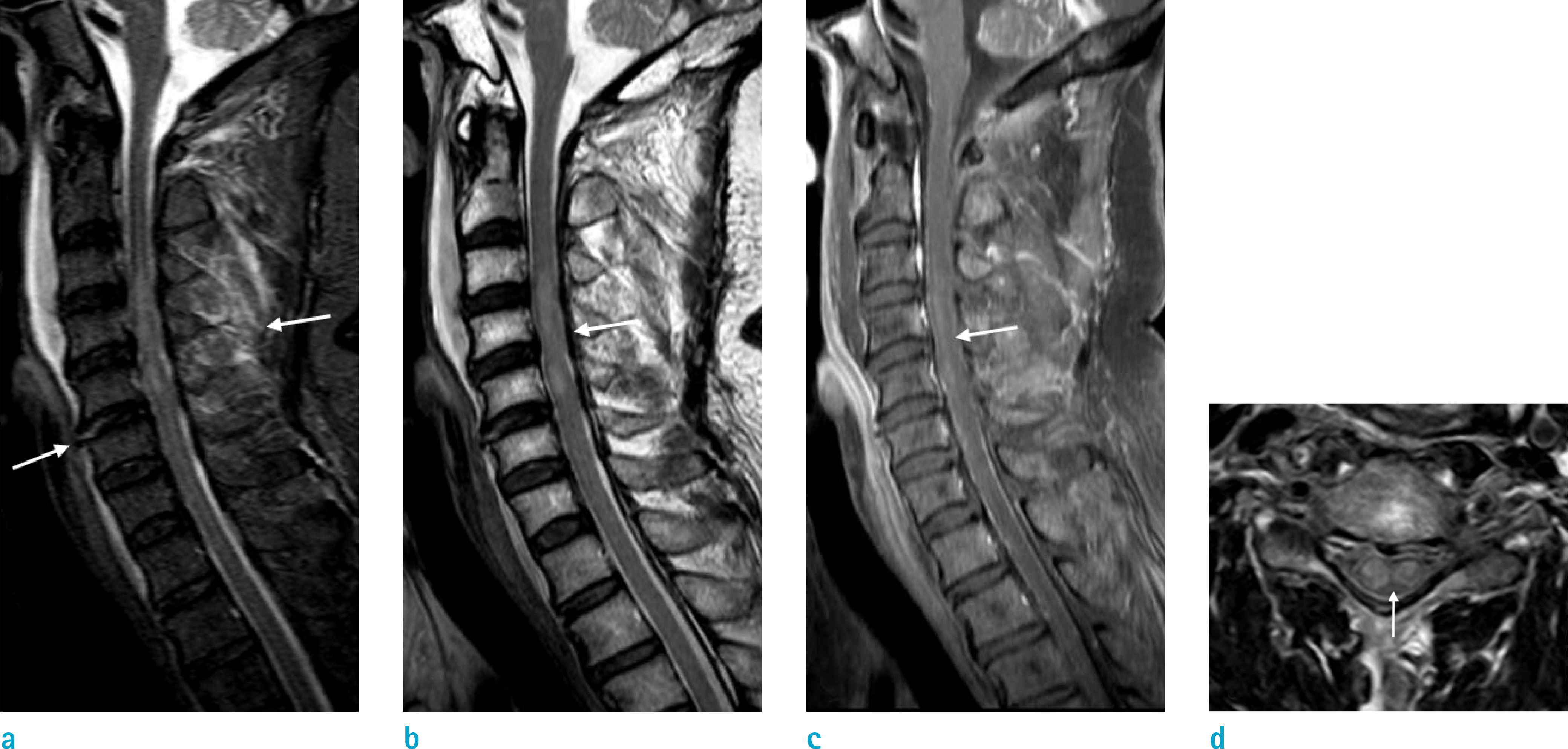
 | Fig. 3.Cervical spine MRI of a 66-year-old man following a traffic accident. Linear intra-discal high signal lesion with prevertebral hemorrhage and high signal intensity of the posterior ligamentous complex suggested traumatic discoligamentous injury on T2-weighted STIR sagittal image (a, arrows). Intramedullar high signal intensity with cord swelling is also observed on both T2-weighted STIR (a) and T2-weighted mid-sagittal scan (b, arrow), with enhancement on the T1-weighted enhanced mid-sagittal image (c, arrow), indicative of cord injury. This finding is also clearly visible on the T2-weighted axial image (d, arrow). The traumatic lesions including the acute cord injury were not mentioned in the preliminary report by the on-call resident, and were classified as a major discrepancy. |
Table 1.
Main Final Diagnosis of the 582 Emergency Spine MRI
Table 2.
Causes of Interpretation Discrepancies


 PDF
PDF ePub
ePub Citation
 Print
Print


 XML Download