

 PDF
PDF ePub
ePub Citation
 Print
Print


Abstract
This work reports the acceptance testing and commissioning experience of the Robotic Intensity-Modulated Radiation Therapy (IMRT) M6 system with a newly released InCiseTM2 Multileaf Collimator (MLC) installed at the Yonsei Cancer Center. Acceptance testing included a mechanical interdigitation test, leaf positional accuracy, leakage check, and End-to-End (E2E) tests. Beam data measurements included tissue-phantom ratios (TPRs), off-center ratios (OCRs), output factors collected at 11 field sizes (the smallest field size was 7.6 mm×7.7 mm and largest field size was 115.0 mm×100.1 mm at 800 mm source-to-axis distance), and open beam profiles. The beam model was verified by checking patient-specific quality assurance (QA) in four fiducial-inserted phantoms, using 10 intracranial and extracranial patient plans. All measurements for acceptance testing satisfied manufacturing specifications. Mean leaf position offsets using the Garden Fence test were found to be 0.01±0.06 mm and 0.07±0.05 mm for X1 and X2 leaf banks, respectively. Maximum and average leaf leakages were 0.20% and 0.18%, respectively. E2E tests for five tracking modes showed 0.26 mm (6D Skull), 0.3 mm (Fiducial), 0.26 mm (Xsight Spine), 0.62 mm (Xsight Lung), and 0.6 mm (Synchrony). TPRs, OCRs, output factors, and open beams measured under various conditions agreed with composite data provided from the manufacturer to within 2%. Patient-specific QA results were evaluated in two ways. Point dose measurements with an ion chamber were all within the 5% absolute-dose agreement, and relative-dose measurements using an array ion chamber detector all satisfied the 3%/3 mm gamma criterion for more than 90% of the measurement points. The Robotic IMRT M6 system equipped with the InCiseTM2 MLC was proven to be accurate and reliable.
References
1. Eshleman JS, Berkenstock KG, Singapuri KP, Fuller C. The CyberKnife® M6TM radiosurgery system. J Lanc Gen Hosp. 2013; 8:44–49.
2. Kim N, Lee H, Kim JS, et al. Clinical outcomes of multileaf collimator-based CyberKnife for spine stereotactic body radiation therapy. The British journal of radiology. 2017; 90:20170523.

3. Barbotteau Y, Rignon A, Crespin M, et al. 22. CyberKnife M6 InCise-2 multileaf collimator setup, modelization and quality insurance program. Physica Medica: European Journal of Medical Physics. 2016; 32:351–352.
4. Kim J, Park K, Yoon J, et al. Feasibility Study of a Custom-made Film for End-to-End Quality Assurance Test of Robotic Intensity Modulated Radiation Therapy System. Progress in Medical Physics. 2016; 27:189–195.
5. Sudahar H, Kurup PG, Murali V, Velmurugan J. Influence of smoothing algorithms in Monte Carlo dose calculations of cyberknife treatment plans: A lung phantom study. Journal of cancer research and therapeutics. 2012; 8:367.
6. O'Connor P, Seshadri V, Charles P. Detecting MLC errors in stereotactic radiotherapy plans with a liquid filled ionization chamber array. Australasian physical & engineering sciences in medicine. 2016; 39:247–252.
7. Hoogeman MS, van de Water S, Levendag PC, van der Holt B, Heijmen BJ, Nuyttens JJ. Clinical introduction of Monte Carlo treatment planning: a different prescription dose for non-small cell lung cancer according to tumor location and size. Radiotherapy and Oncology. 2010; 96:55–60.
8. Sharma S, Ott J, Williams J, Dickow D. Dose calculation accuracy of the Monte Carlo algorithm for CyberKnife compared with other commercially available dose calculation algorithms. Medical Dosimetry. 2011; 36:347–350.
9. Francescon P, Kilby W, Satariano N. Monte Carlo simulated correction factors for output factor measurement with the CyberKnife system–results for new detectors and correction factor dependence on measurement distance and detector orientation. Physics in Medicine & Biology. 2014; 59:N11.
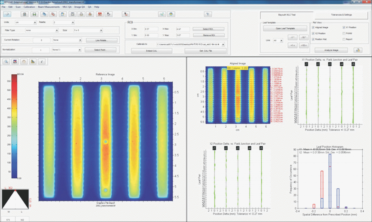
Fig. 2.
Results of leakage test: (a) EBT3 film and image analysis of X1 closed leakage and (b) EBT3 film and image analysis of X2 closed leakage.
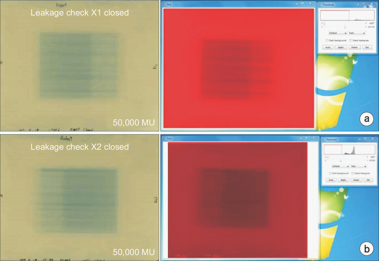
Fig. 3.
Visual comparisons of OCRs between measurement and composite data at a depth of 15 mm. OCRs with (a) X field size of 7.6 mm, 15.4 mm, and 23 mm and (b) Y field size of 7.7 mm, 15.4 mm, and 23.1 mm. OCRs with (c) X field size of 30.8 mm, 46.2 mm, 69.2 mm, and 100 mm and (d) Y field size of 30.8 mm, 46.2 mm, 69.3 mm, and 100.1 mm. OCRs with (e) X field size of 38.4 mm, 53.8 mm, 84.6 mm, and 115 mm and (f) Y field size of 38.5 mm, 53.9 mm, 84.7 mm, and 100.1 mm.
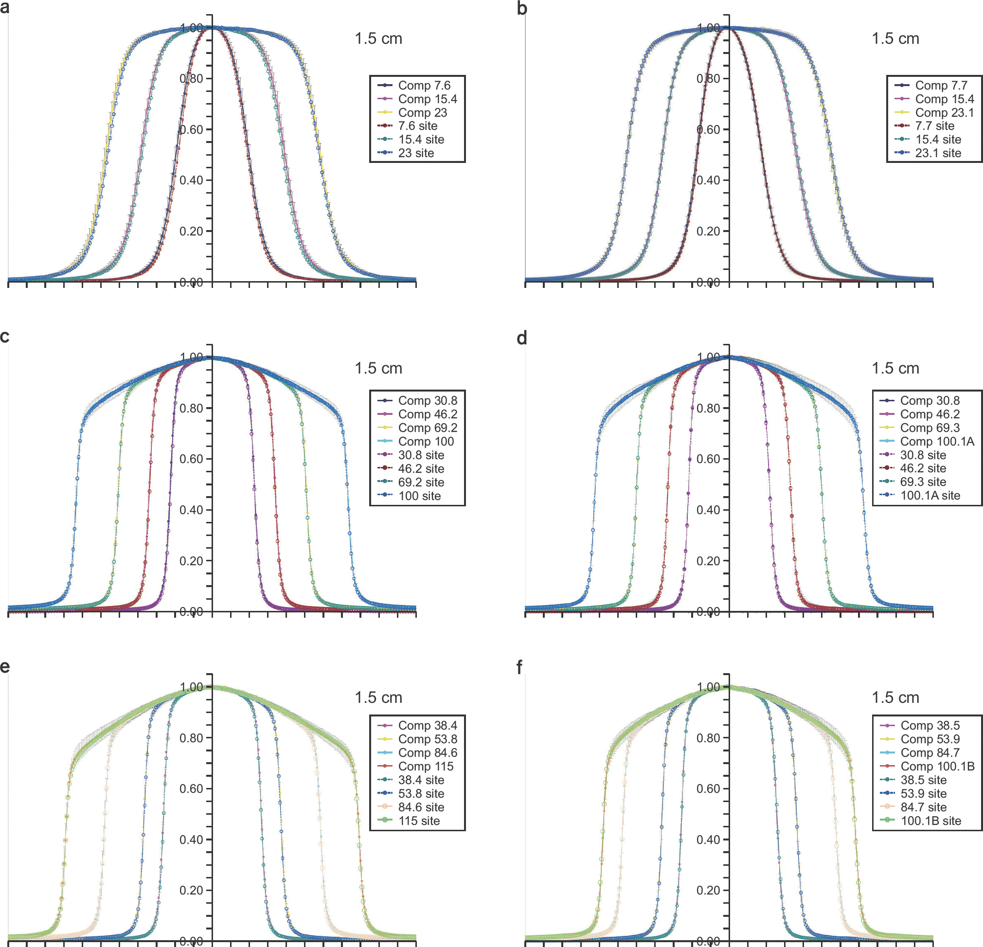
Fig. 4.
Point dose errors of patient-specific QA: (a) stereotactic dose verification phantom, (b) pelvis phantom, and (c) thorax phantom.
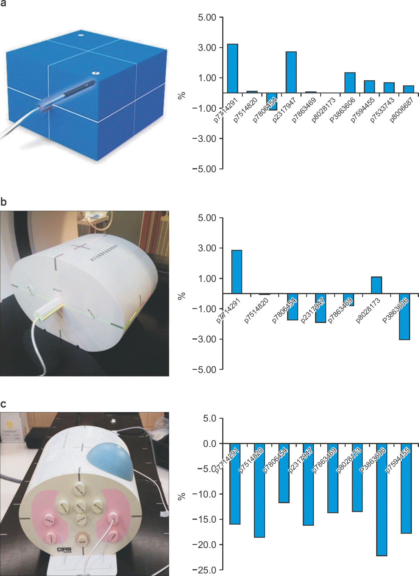
Table 1.
Percent differences of TPRs between measurement and composite data.
Table 2.
Percent differences of OFs between measurement and composite data.
 XML Download