Abstract
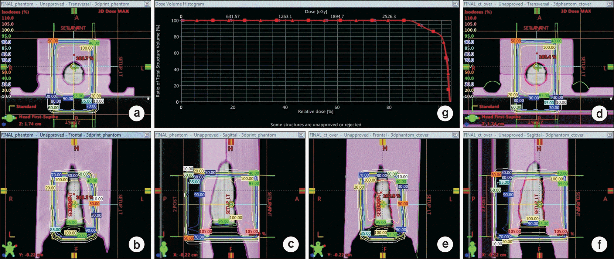
Creating individualized build-up material for superficial photon beam radiation therapy at irregular surface is complex with rice or commonly used flat shape bolus. In this study, we implemented a workflow using 3D printed patient specific bolus and describe our clinical experience. To provide better fitted build-up to irregular surface, the 3D printing technique was used. The PolyLactic Acid (PLA) which processed with nontoxic plant component was used for 3D printer filament material for clinical usage. The 3D printed bolus was designed using virtual bolus structure delineated on patient CT images. Dose distributions were generated from treatment plan for bolus assigned uniform relative electron density and bolus using relative electron density from CT image and compared to evaluate the inhomogeneity effect of bolus material. Pretreatment QA is performed to verify the relative electron density applied to bolus structure by gamma analysis. As an in-vivo dosimetry, Optically Stimulated Luminescent Dosimeters (OSLD) are used to measure the skin dose. The plan comparison result shows that discrepancies between the virtual bolus plan and printed bolus plan are negligible. (0.3% maximum dose difference and 0.2% mean dose difference). The dose distribution is evaluated with gamma method (2%, 2 mm) at the center of GTV and the passing rate was 99.6%. The OSLD measurement shows 0.3% to 2.1% higher than expected dose at patient treatment lesion. In this study, we treated Mycosis fungoides patient with patient specific bolus using 3D printing technique. The accuracy of treatment plan was verified by pretreatment QA and in-vivo dosimetry. The QA results and 4 month follow up result shows the radiation treatment using 3D printing bolus is feasible to treat irregular patient skin.
Go to : 
REFERENCES
1. HOPPE. Richard T. Mycosis fungoides: radiation therapy. Dermatologic therapy. 2003; 16:347–354.
2. SCHOLTZ W. Ueber den Einfluss der Röntgenstrahlen auf die Haut in gesundem und krankem Zustande. Archiv für Dermatologie und Syphilis. 1902; 59:421–446.

3. MICAILY Bizhan, et al. Radiotherapy for unilesional mycosis fungoides. International Journal of Radiation Oncology Biology Physics. 1998; 42:361–364.

4. HOPPE RT, et al. Electron-beam therapy for mycosis fungoides: the Stanford University experience. Cancer treatment reports. 1979; 63:691–700.
5. MAJITHIA Lonika, et al. Treating Cutaneous T-Cell Lymphoma with Highly Irregular Surfaces with Photon Irradiation Using Rice as Tissue Compensator. Frontiers in oncology. 2015; 5.

6. Yoon Kyoungjun, et al. Development of new 4D phantom model in respiratory gated volumetric modulated arc therapy for lung SBRT. Progress in Medical Physics. 2014; 25:100–109.

7. Ju Sang Gyu, et al. New technique for developing a proton range compensator with use of a 3-dimensional printer. International Journal of Radiation Oncology Biology Physics. 2014; 88:453–458.

8. Jin-Suk HA, et al. Customized 3D Printed Bolus for Breast Reconstruction for Modified Radical Mastectomy (MRM). Progress in Medical Physics. 2016; 27:196–202.
9. Holtzer NA, Galis J, Paalman MI, Heukelom S. 3D printing of tissue equivalent boluses and molds for external beam radiotherapy. In: Estro 33, Vienna.
10. BORCA. Casanova Valeria, et al. Dosimetric characterization and use of GAFCHROMIC EBT3 film for IMRT dose verification. Journal of applied clinical medical physics. 2013; 14:158–171.
11. KUTCHER. Gerald J, et al. Comprehensive QA for radiation oncology: report of AAPM radiation therapy committee task group 40. Medical physics. 1994; 21:581–618.
12. YUSOF. Hanum Fasihah, et al. On the use of optically stimulated luminescent dosimeter for surface dose measurement during radiotherapy. PloS one. 2015; 10:e0128544.
Go to : 
 | Fig. 1.Design procedure of the 3D virtual bolus in TPS. (a) The contour is delineated with 2 mm expansion from the body structure, (b) the virtual bolus is designed as a box shape which cover the treatment lesion and exclude the contour drawn previously and divided by two parts to reduce the production time. |
 | Fig. 2.The isodose lines difference of the treatment plan in (a) axial, (b) coronal and (c) sagittal view for actual treatment plan without applying HU value to bolus structure. The isodose lines difference of the treatment plan in (d) axial, (e) coronal and (f) sagittal view for actual treatment plan with applying HU value to bolus structure. (g) DVH difference between two plans. Box is represent the without HU assign plan and triangle is represent the HU assigned plan. |
 | Fig. 3.(a) A series of clinical photographs of patient before treatment setup, (b) after treatment setup, (c) patient picture before treatment and 4 months after treatment. |
Table 1.
3D printing conditions and physical properties of the PLA filament.
Table 2.
The in-vivo dosimetry result with the OSLD measurement.


 PDF
PDF ePub
ePub Citation
 Print
Print


 XML Download