

 PDF
PDF ePub
ePub Citation
 Print
Print


Abstract
For the ViewRay® system (ViewRay Inc., Cleveland, OH, USA) which is representative of magnetic resonance (MR) guided radiotherapy machine, it is important to evaluate effectiveness of AAPM's TG-51 protocol and the effect of the magnetic field on absolute dosimetry. In order to measure the absolute dose, MR-compatible chamber and water phantom system manufactured in this study were used. The materials of the water phantom system were plastic of polymethyl methacrylate (PMMA) and non-ferrous materials. Due to the inherent feature of the ViewRay®, all Co-60 sources are not located at gantry angle of 0° while being located at gantry angle of 90°. For this reason, absolute dosimetry was performed based on the measurements in solid water phantom (SWP) and water which determine the SWP to water correction factor. For evaluation of output constancy with gantry angle, measurements were made with ionization chamber inserted in cylindrical water-equivalent phantom. For measured doses in water, the values of dose deviation according to a reference dose of 200 cGy for Head 1, Head 2 and Head 3 were -0.27%, -0.45% and -0.22%, respectively. For measured doses in SWP, the values of dose deviation according to a reference dose of 200 cGy for Head 1, Head 2 and Head 3 were -1.91%, -2.07% and -1.84%, respectively. All values of dose measured in SWP tended to be less than those measured in water by -1.63%. With the reference gantry angles of 0° and 90°, the maximum values of deviation for Head 1, Head 2 and Head 3 were 0.48%, 1.06% and 0.40%, respectively. The measurement agreement is within the range of results obtainable for conventional treatment machines. The low strength of the magnetic field does not affect dose measurements. Using the SWP to water correction factor, absolute doses for ViewRay® system can be measured.
REFERENCES
1.Park JM., Park SY., Wu HG., Kim JI. Commissioning Experience of Tri-Cobalt-60 MRI-guided Radiation Therapy System. Prog Med Phys. 2015. 26:193–200.

2.Stam MK, et al. Kidney motion during free breathing and breath hold for MR-guided radiotherapy. Phys Med Biol. 2013. 58:2235–45.
3.Seierstad T, et al. MR-guided simultaneous integrated boost in preoperative radiotherapy of locally advanced rectal cancer following neoadjuvant chemotherapy. Radiother Oncol. 2009. 93:279–84.
4.Dawson LA., Jaffray DA. Advances in Image-Guided Radiation Therapy. J Clin Oncol. 2007. 25:938–46.
5.Court L., Rosen I., Mohan R., Dong L. Evaluation of mechanical precision and alignment uncertainties for an integrated CT/LINAC system. Med Phys. 2003. 30:1198–210.
6.Oldham M, et al. Cone-beam-CT guided radiation therapy A model for on-line application. Radiother Oncol. 2005. 75:271–8.
7.Hansen EK, et al. Image-guided radiotherapy using megavoltage cone-beam computed tomography for treatment of paraspinous tumors in the presence of orthopedic hardware. Int J Radiat Oncol Biol Phys. 2006. 66:323–6.
8.Mohan DS., Kupelian PA., Willoughby TR. Short-course intensity-modulated radiotherapy for localized prostate cancer with daily transabdominal ultrasound localization of the prostate gland. Int J Radiat Oncol Biol Phys. 2000. 46:575–80.
9.Choi CH, et al. Quality of tri-Co-60 MR-IGRT treatment plans in comparison with VMAT treatment plans for spine SABR. Br J Radiol. 2017. 90:20160652.
10.Ling CC., Yorke E., Fuks Z. From IMRT to IGRT: frontierland or neverland? Radiother Oncol. 2006. 78:119–22.
11.Tejinder Kataria SG. Image Guided Radiation Therapy. J Nucl Med Radiat Ther. 2014. 5.
12.Mutic S., Dempsey JF. The ViewRay system: magnetic resonance-guided and controlled radiotherapy. Semin Radiat Oncol. 2014. 24:196–9.
13.Wooten HO, et al. Benchmark IMRT evaluation of a Co-60 MRI-guided radiation therapy system. Radiother Oncol. 2015. 114:402–5.
14.Kashani R, et al. Commissioning and Clinical Implementation of the First Online Adaptive MR Image Guided Radiation Therapy Program. Int J Radiat Oncol Biol Phys. 2015. 93:S18–9.
15.Park JM., Park SY., Kim JI., Kang HC., Choi CH. A comparison of treatment plan quality between Tri-Co-60 intensity modulated radiation therapy and volumetric modulated arc therapy for cervical cancer. Phys Med. 2017. 40:11–6.
16.Park JM, et al. Treatment plan comparison between Tri-Co-60 magnetic-resonance image-guided radiation therapy and volumetric modulated arc therapy for prostate cancer. Oncotarget. 2017. 8:91174–84.
17.Park JM, et al. A comparative planning study for lung SABR between tri-Co-60 magnetic resonance image guided radiation therapy system and volumetric modulated arc therapy. Radiother Oncol. 2016. 120:279–85.
18.Almond PR, et al. AAPM's TG-51 protocol for clinical reference dosimetry of high-energy photon and electron beams. Med Phys. 1999. 26:1847–70.
19.McEwen M, et al. Addendum to the AAPM's TG-51 protocol for clinical reference dosimetry of high-energy photon beams. Med Phys. 2014. 41:041501–1.
20.Green O., Goddu S., Mutic S. SU-E-T-352: Commissioning and Quality Assurance of the First Commercial Hybrid MRI-IMRT System. Med Phys. 2012. 39:3785.
21.Goddu S., Green O., Mutic S. WE-G-BRB-08: TG-51 Calibration of First Commercial MRI-Guided IMRT System in the Presence of 0.35 Tesla Magnetic Field. Med Phys. 2012. 39:3968.
22.Day M. EGA A Central axis depth dose data for use in radiotherapy. A survey of depth doses and related data measured in water or equivalent media. Br J Radiol Suppl. 1983. 17:1–147.
23.Tello VM., Tailor RC., Hanson WF. How water equivalent are water-equivalent solid materials for output calibration of photon and electron beams? Med Phys. 1995. 22:1177–89.
24.Seuntjens J., Olivares M., Evans M., Podgorsak E. Absorbed dose to water reference dosimetry using solid phantoms in the context of absorbed-dose protocols. Med Phys. 2005. 32:2945–53.
25.Choi CH, et al. External Auditing on Absorbed Dose Using a Solid Water Phantom for Domestic Radiotherapy Facilities. Prog Med Phys. 2010. 28:50–6.
26.Spindeldreier CK, et al. Radiation dosimetry in magnetic fields with Farmer-type ionization chambers: determination of magnetic field correction factors for different magnetic field strengths and field orientations. Phys Med Biol. 2017. 62:6708–28.
27.Raaijmakers AJ., Raaymakers BW., Lagendijk JJ. Integrating a MRI scanner with a 6 MV radiotherapy accelerator: dose increase at tissue-air interfaces in a lateral magnetic field due to returning electrons. Phys Med Biol. 2005. 50:1363–76.
28.O'Brien DJ., Roberts DA., Ibbott GS'., Sawakuchi GO. Reference dosimetry in magnetic fields: formalism and ionization chamber correction factors. Med Phys. 2016. 43:4915.
29.Bouchard H., de Pooter J., Bielajew A., Duane S. Reference dosimetry in the presence of magnetic fields: conditions to validate Monte Carlo simulations. Phys Med Biol. 2015. 60:6639–54.
Fig. 1
Water phantom system without ferrous materials was manufactured. Water phantom system consists of (a) water phantom tank, (b) plastic ruler, and (c) thumbscrew type moving system.
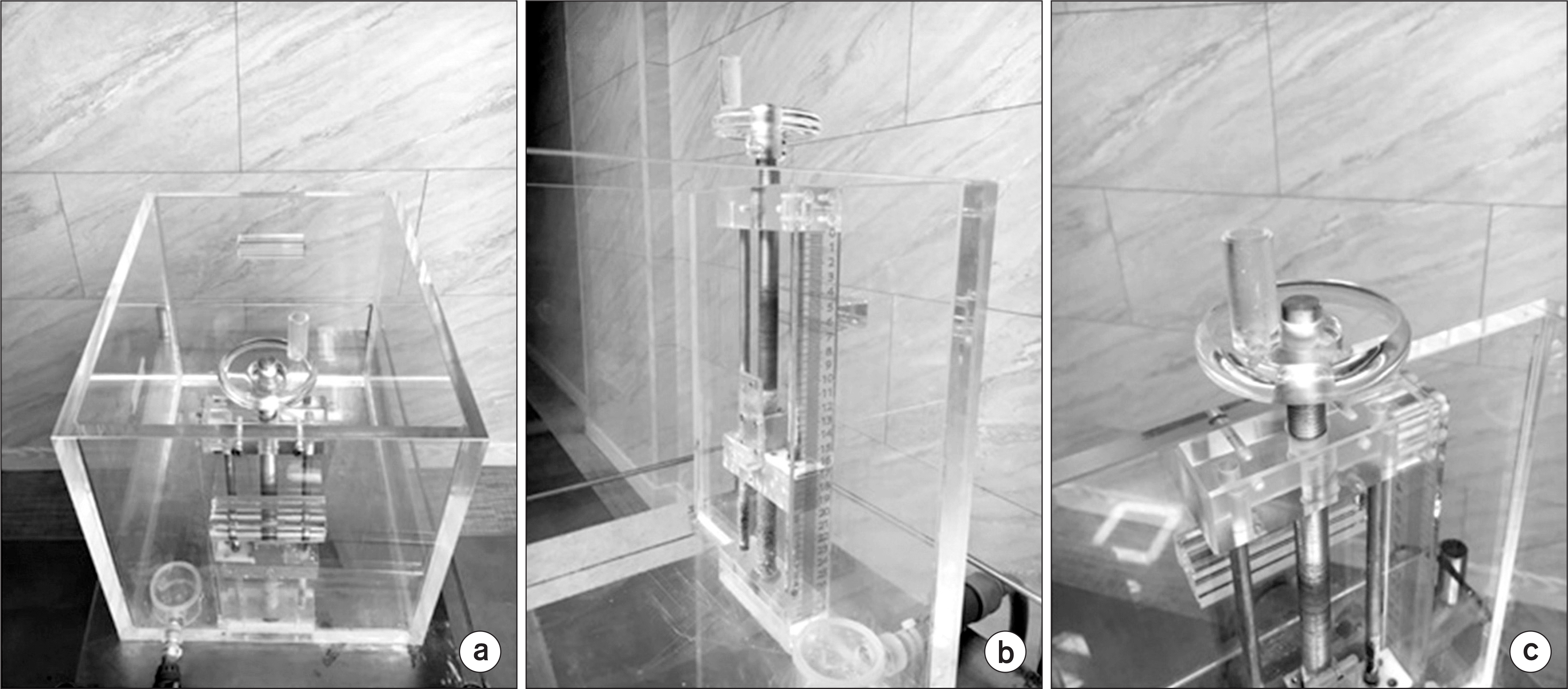
Fig. 2
Dose deviations between measured absolute doses of all Co-60 sources and a reference dose of 100 cGy. After ViewRay® system was commissioned on July 2016, absolute doses were measured for every month.
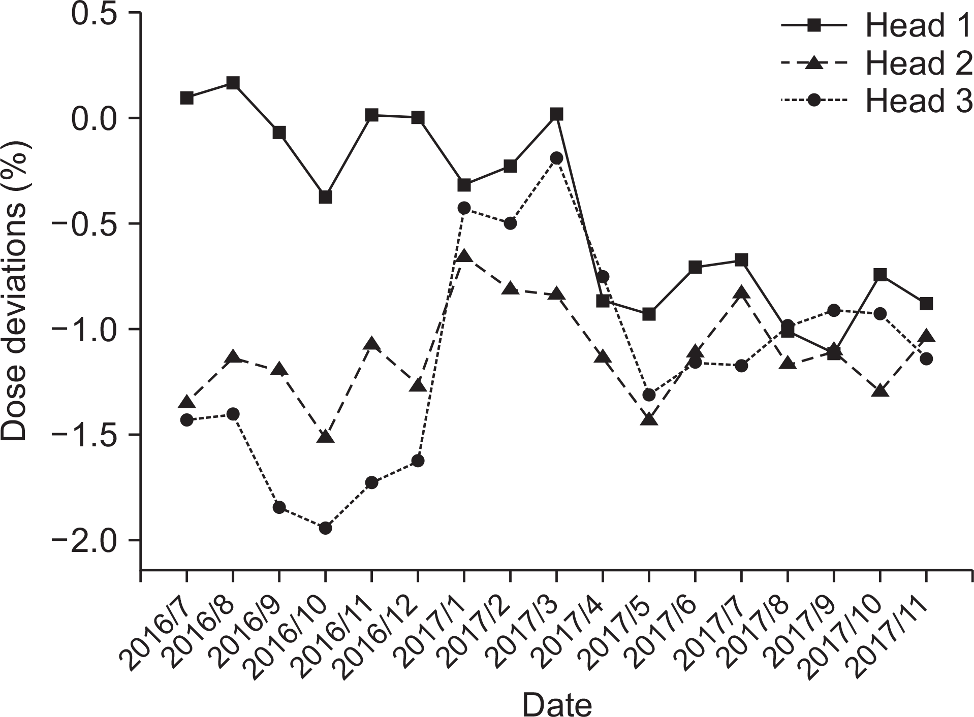
Table 1.
Correction factors of AAPM's TG-51 and absolute dose values in water and solid water phantom (SWP) for Head 1, Head 2, and Head 3. Measurements in water were performed to deliver 200 cGy to reference point when both Head 1 and Head 3 were set to gantry angle of 0°. Measurements in SWP were performed to deliver 200 cGy to reference point when Head 1, Head 2 and Head 3 were set to gantry angle of 90°.
| Correction factor | Output | ||||||
|---|---|---|---|---|---|---|---|
| Pion | Pelec | Ppol | ∗Dw, 0° (cGy) | †Dev200 cGy (%) | ‡DSWP, 90° (cGy) | Dev200 cGy (%) | |
| Head 1 | 1.001 | 1.000 | 0.999 | 199.5 | −0.27 | 196.2 | −1.91 |
| Head 2 | §199.1 | −0.45 | 195.9 | −2.07 | |||
| Head 3 | 199.6 | −0.22 | 196.3 | −1.84 | |||
Table 2.
Output constancy with gantry angle. The reference measurements were gantry angle of 0° for Head 1 and Head 3, and 90° for Head 2.
 XML Download