

 PDF
PDF ePub
ePub Citation
 Print
Print


Abstract
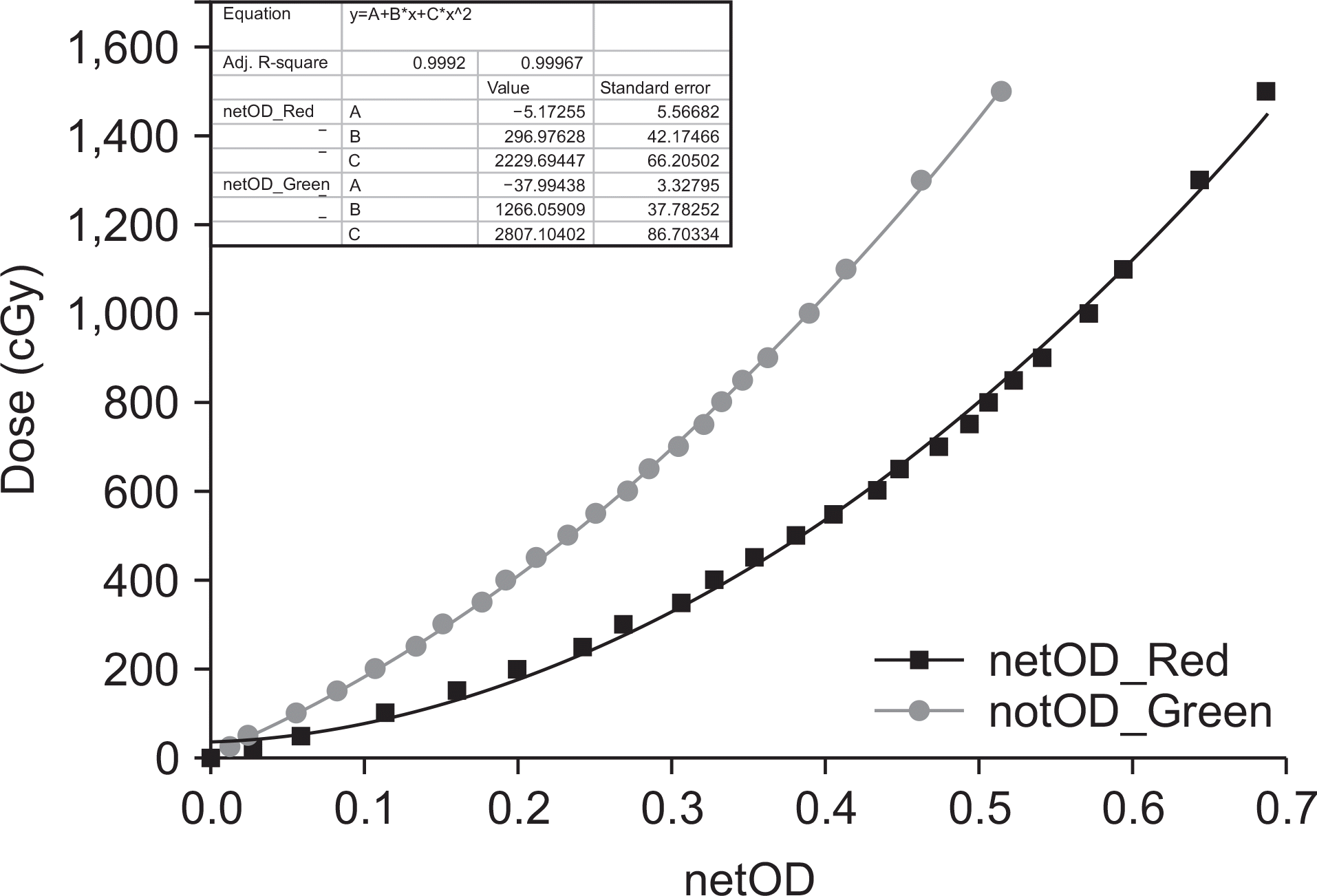
This study assessed the feasibility of a dual-channel (DC) compound method for film dosimetry. The red channel (RC) is usually used to ensure dosimetric quality using a conventional fraction dose because the RC is more accurate at low doses within 3 Gy than is the green channel (GC). However, the RC is prone to rapid degradation of sensitivity at high doses, while degradation of the GC is slow. In this study, the DC compound method combining the RC and GC was explored as a means of providing accurate film dosimetry for high doses. The DC compound method was evaluated at various dose distributions using EBT3 film inserted in a solid-water phantom. Measurements with 10×20 cm2 radiation field and 60° dynamic-wedge were done. Dose distributions acquired using the RC and GC were analyzed with root-mean-squares-error (RMSE) and gamma analyses. The DC compound method was used based on the RC after correcting the GC for high doses in the gamma analysis. The RC and GC produced comparatively more accurate RMSE values for low and high doses, respectively. Gamma passing rates with an acceptance criterion of 3%/3 mm revealed that the RC provided rapid reduction in the high dose region, while the GC displayed a gradual decrease. In the whole dose range, the DC compound method had the highest agreement (93%) compared with single channel method using either the RC (80%) or GC (85%). The findings indicate that the use of DC compound method is more appropriate in dosimetric quality assurance for radiotherapy using high doses.
Go to : 
REFERENCES
1. James MG, Greg B. Quality assurance procedures for stereotactic body radiation therapy. Int J Radiat Oncol Biol Phys. 2008; 71:S122–5.
2. Eric EK, Joseph H, John B, Fang-Fang Y, William S, Sean D, et al. TG Report 142: Quality assurance of medical accelerators. American Association of Physicists in Medicine. Med Phys. 2009; 36:4197–212.
3. Huq MS, Fraass BA, Dunscombe PB, Gibbons JP, Ibbott GS, Medin PM, et al. A method for evaluating quality assurance needs in radiation therapy. Int J Radiat Oncol Biol Phys. 2008; 71:S170–3.

4. Chung JB, Kang SW, Eom KY, Song C, Choi KS, Suh TS. Comparison of dosimetric performance among commercial quality assurance systems for verifying pretreatment plans of stereotactic body radiotherapy using flattening-filter-free beams. J Korean Med Sci. 2016; 31:1742–8.
5. Hayashi N, Watanabe Y, Malmin R, Kato H. Evaluation of triple channel correction acquisition method for radiochromic film dosimetry. J Radiat Res. 2012; 53:930–5.
6. Reinhardt S, Hillbrand M, Wilkens JJ, Assmann W. Comparison of gafchromic EBT2 and EBT3 films for clinical photon and proton beams. Med Phys. 2012; 39:5257–62.
7. Devic S, Seuntjens J, Sham E, Podgorsak EB, Schmidtlein CR, Kirov AS, et al. Precise radiochromic film dosimetry using a flatbed document scanner. Med Phys. 2005; 32:2245–53.
8. Ferreira BC, Lopes MC, Capela M. Evaluation of an Epson flatbed scanner to read gafchromic EBT films for radiation dosimetry. Phys Med Biol. 2009; 54:1073–85.
9. Fukumoto S, Shirato H, Shimzu S, Ogura S, Onimaru R, Kitamura K, et al. Small-volume image-guided radiotherapy using hypofractionated, coplanar, and noncoplanar multiple fields for patients with inoperable Stage I nonsmall cell lung carcinomas. Cancer. 2002; 95:1546–53.
10. Gerszten PC, Burton SA, Welch WC, Brufsky AM, Lembersky BC, Ozhasoglu C, et al. Single-fraction radiosurgery for the treatment of spinal breast metastases. Cancer. 2005; 104:2244–54.
11. Hara R, Itami J, Kondo T, Aruga T, Abe Y, Ito M, et al. Stereotactic single high dose irradiation of lung tumors under respiratory gating. Radiother Oncol. 2002; 63:159–63.
12. Herfarth KK, Debus J, Lohr F, Bahner ML, Rhein B, Fritz P, et al. Stereotactic single-dose radiation therapy of liver tumors: results of a phase I/II trial. J Clin Oncol. 2001; 19:164–70.
13. Borca VC, Pasquino M, Russo G, Grosso P, Cante D, Sciacero P, et al. Dosimetric characterization and use of GAFCHROMIC EBT3 film for IMRT dose verification. J Appl Clinic Med Phys. 2013; 14:158–71.
14. Lewis D, Micke A, Yu X, Chan MF. An efficient protocol for radiochromic film dosimetry combining calibration and measurement in a single scan. Med Phys. 2012; 39:6339–50.
15. Almond PR, Biggs PJ, Coursey BM, Hanson WF, Huq MS, Nath R, et al. AAPM's TG-51 protocol for clinical reference dosimetry of high-energy photon and electron beams. Med Phys. 1999; 26:1847–70.
16. Devic S. Radiochromic film dosimetry: Past, present, and future. Phys Med. 2011; 27:122–34.
17. Moran JM, Radawski J, Fraass BA. A dose-gradient analysis tool for IMRT QA. J Appl Clinic Med Phys. 2005; 6:62–73.
18. Ju T, Simpson T, Deasy JO, Low DA. Geometric interpretation of the g dose distribution comparison technique: Interpolation-free calculation. Med Phys. 2008; 35:879–87.
Go to :
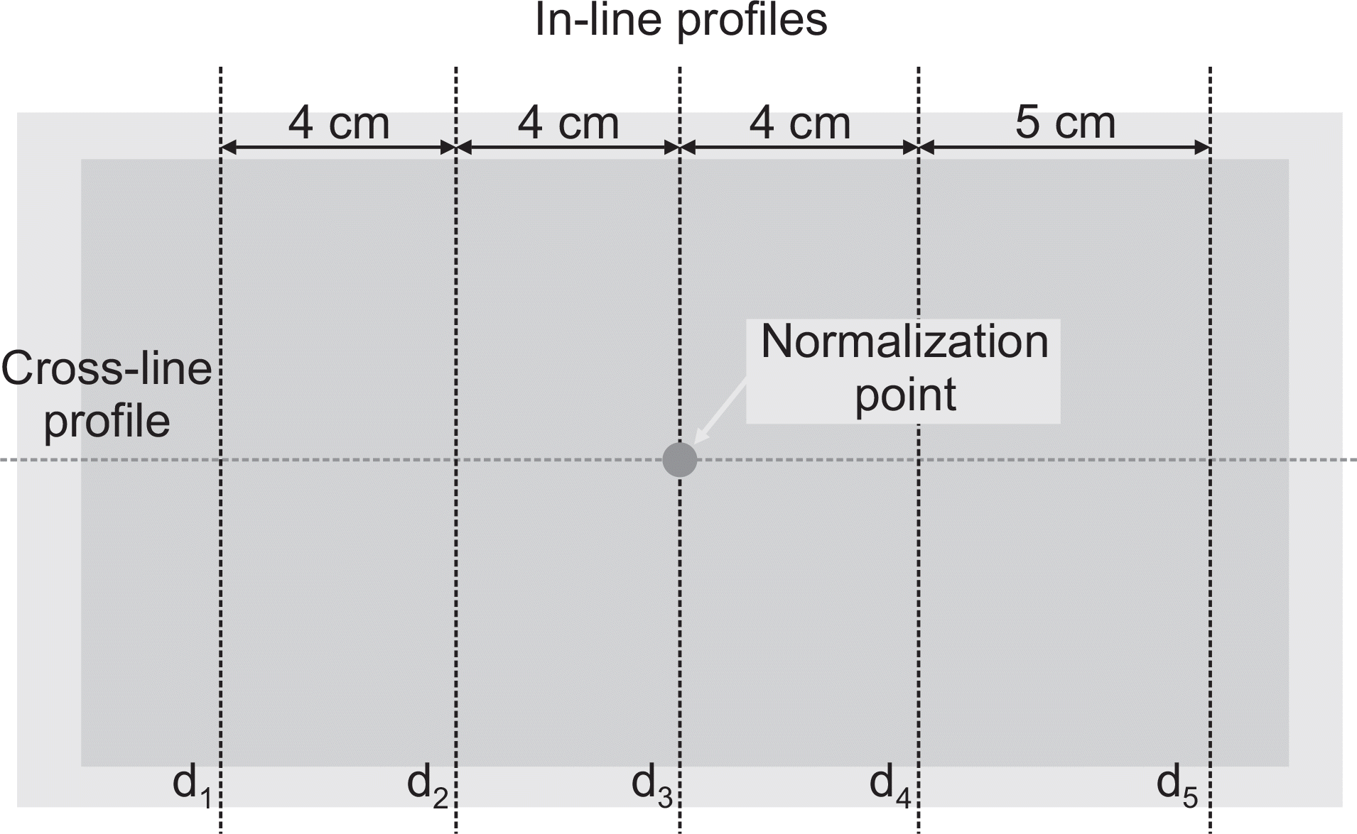 | Fig. 2.EBT film showing a mimic image irradiated radiation for analyzing cross- (red dot line) and in-line (blue dot line) profiles. |
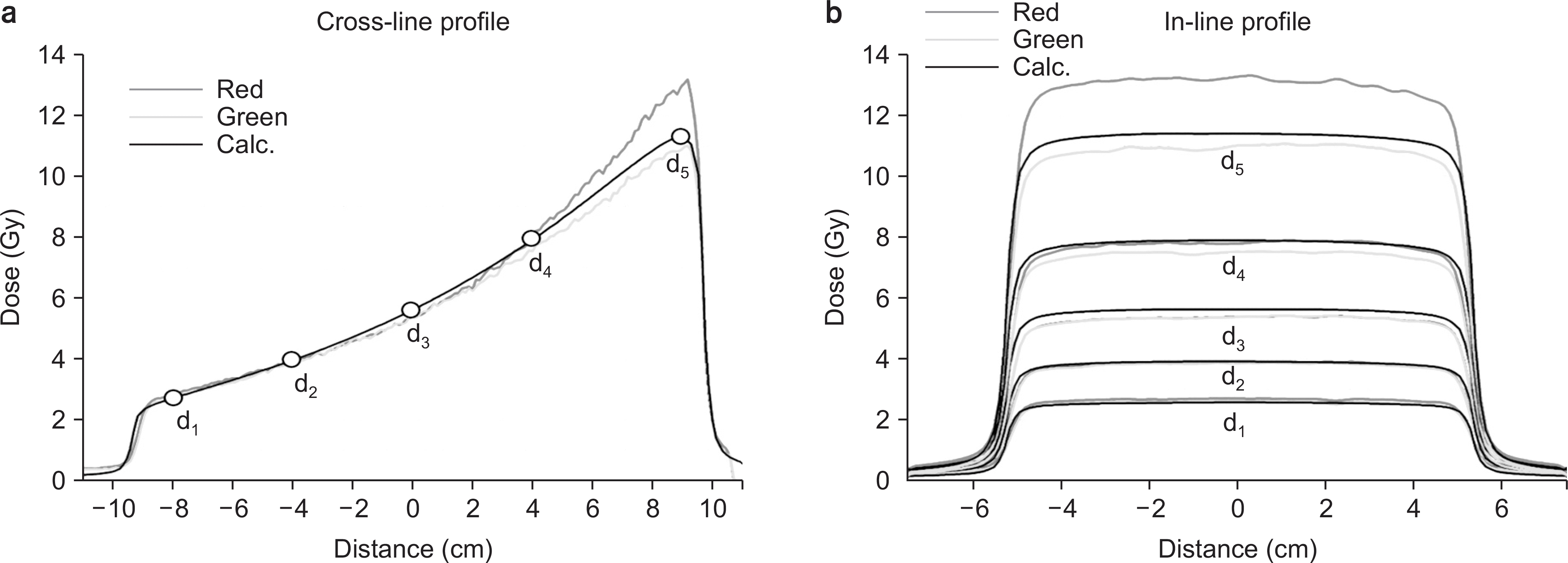 | Fig. 3.The dose profiles ((a) cross-line and (b) in-line) obtained in DRC and DGC compared to DCalc. The cross-line profile was measured in central-axis for dose distribution and the in-line profiles were measured in five positions of d1, d2, d3, d4, and d5, respectively. |
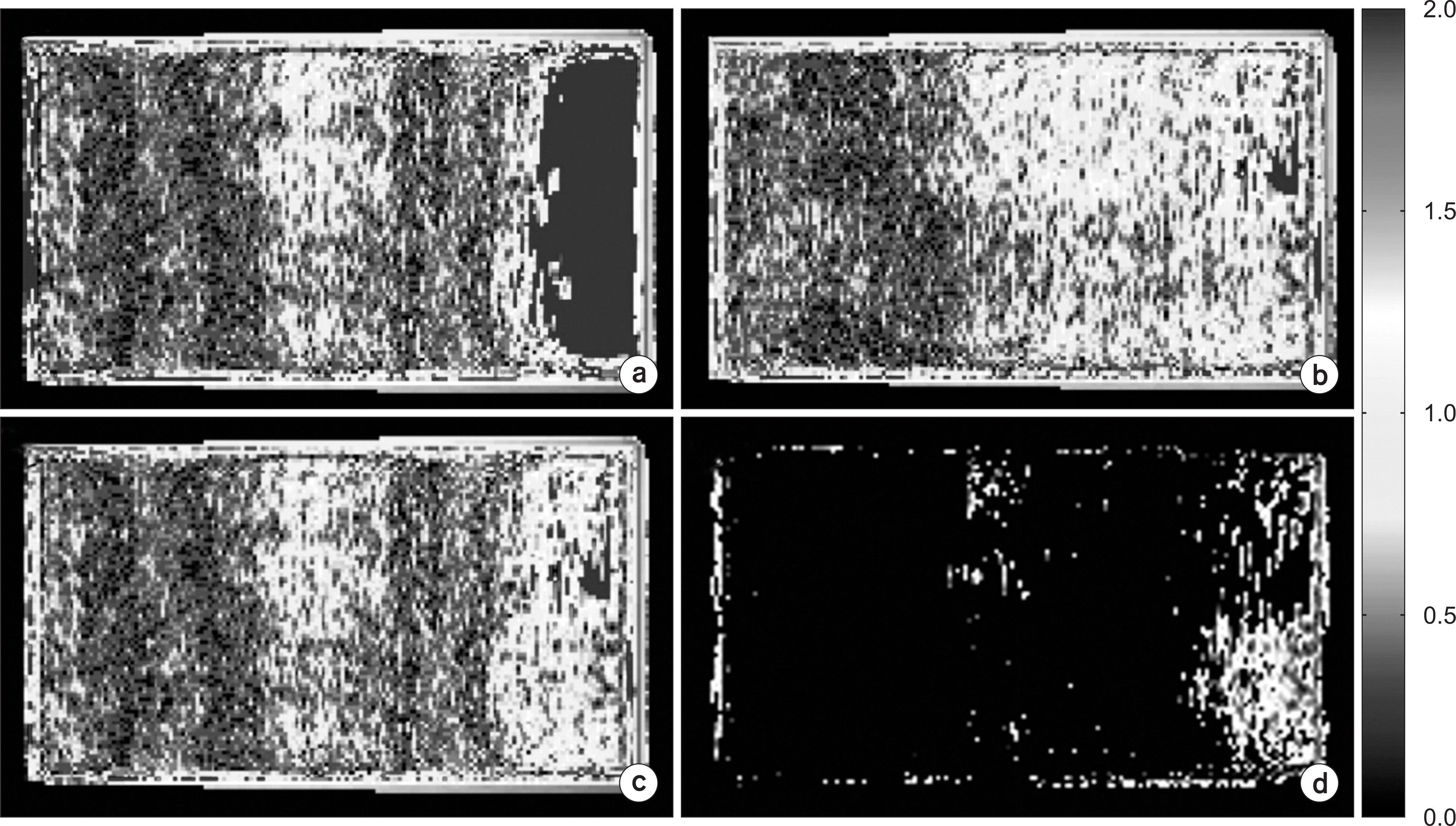 | Fig. 4.Gamma distributions of analysis for (a) red, (b) green channel, (c) dual-channel compound method, and (d) the converted gamma distribution of green channel. |
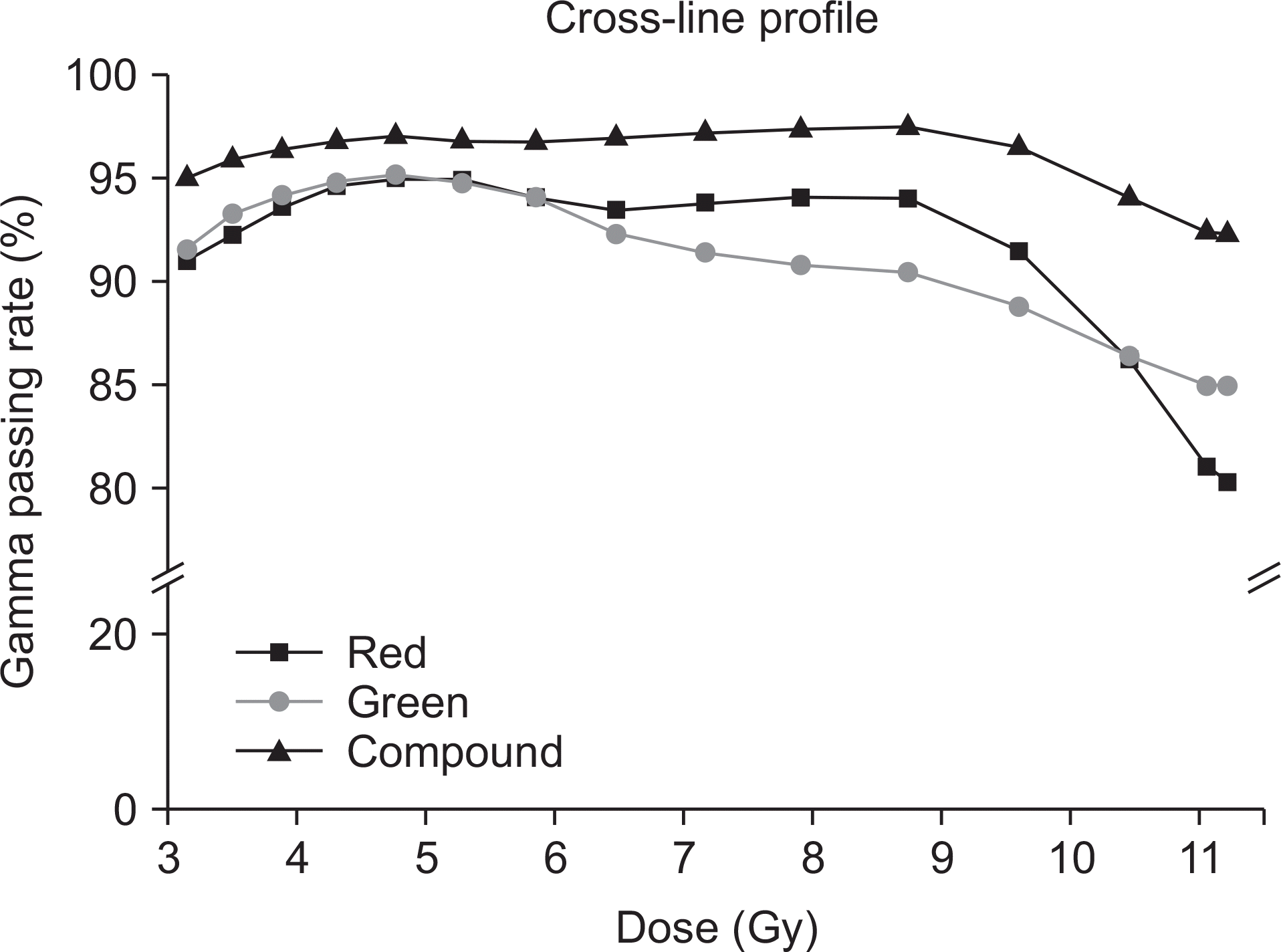 | Fig. 5.Comparison of the gamma passing rates in dose distributions using the red channel, green channel, and dual-channel compound method with increasing doses. |
 XML Download