Abstract
The machine log files recorded by a scanning control unit in proton beam therapy system have been studied to be used as a quality assurance method of scanning beam deliveries. The accuracy of the data in the log files have been evaluated with a standard calibration beam scan pattern. The proton beam scan pattern has been delivered on a gafchromic film located at the isocenter plane of the proton beam treatment nozzle and found to agree within ±1.0 mm. The machine data accumulated for the scanning beam proton therapy of five different cases have been analyzed using a statistical method to estimate any systematic error in the data. The high-precision scanning beam log files in line scanning proton therapy system have been validated to be used for off-line scanning beam monitoring and thus as a patient-specific quality assurance method. The use of the machine log files for patient-specific quality assurance would simplify the quality assurance procedure with accurate scanning beam data.
Go to : 
REFERENCES
1. RR Wilson. Radiological use of fast protons. Radiology. 1946; 47:487–91.
2. Kang JK, MS Kim WI. Jang, et. al. The clinical utilization of radiation therapy in Korea between 2009 and 2013. Radiat Oncol J. 2016; 34:88–95.
3. M Jermann. Particle therapy statistics in 2014. Int J Particle Ther. 2015; 2:50–4.
4. H Li, N Sahoo, F Poenisch et. al. Use of treatment log files in spot scanning proton therapy as part of patient-specific quality assurance. Med Phys. 2013; 40:021703–1. -11.
5. Scandurra D, Albertini F, Meer R, et al. Assessing the quality of proton PBS treatment delivery using machine log files: comprehensive analysis of clinical treatments delivered at PSI Gantry 2. Phys Med Biol. 2016; 61:1171–81.

6. K Chung, Y Han, J Kim et. al. The first private-hospital based proton therapy center in Korea: Status of the Proton Therapy Center at Samsung Medical Center. Radiat Oncol J. 2015; 33:337–43.
7. K Chung, J Kim, DH Kim, S Ahn, Y Han. The proton therapy nozzles at Samsung Medical Center: A Monte Carlo simulation study using TOPAS. J Korean Phys Soc. 2015; 67:170–4.
8. K Chung, Y Han, SH Ahn, JS Kim, H Nonaka. Commissioning and Validation of a Dedicated Scanning Nozzle at Samsung Proton Therapy Center. Prog Med Phys. 2016; 27:267–71.
9. GAFChromicTM EBT3 film specifications. Available at. www.gafchromic.com.
10. G Moliere. Theorie der Streuung schneller geladener Teilchen II. Mehrfach-und Vielfachstreuung. Z Naturforsch A: Phys Sci. 1948; 3a:78–97.
Go to : 
 | Fig. 1.A scanned EBT3 film irradiated with the standard scan pattern composed of 5x5 points (230 MeV). |
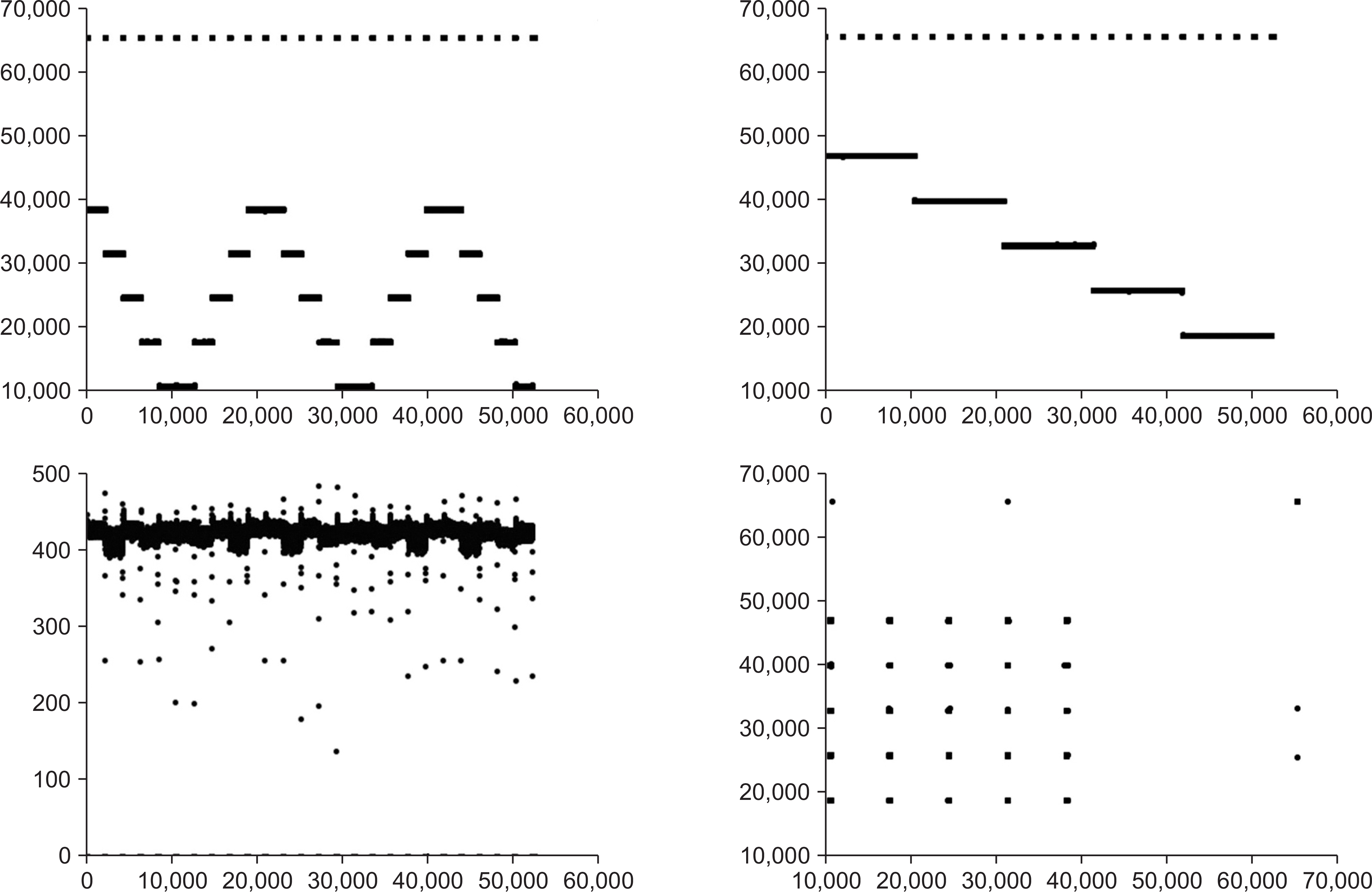
 | Fig. 2.An example of scanning beam information recorded in the scanning control unit (Top left: x-position in time, top right: y-position in time, bottom left: monitor count in time, bottom right: x-position vs. y-position. Raw data without pedestal/noise reduction). |
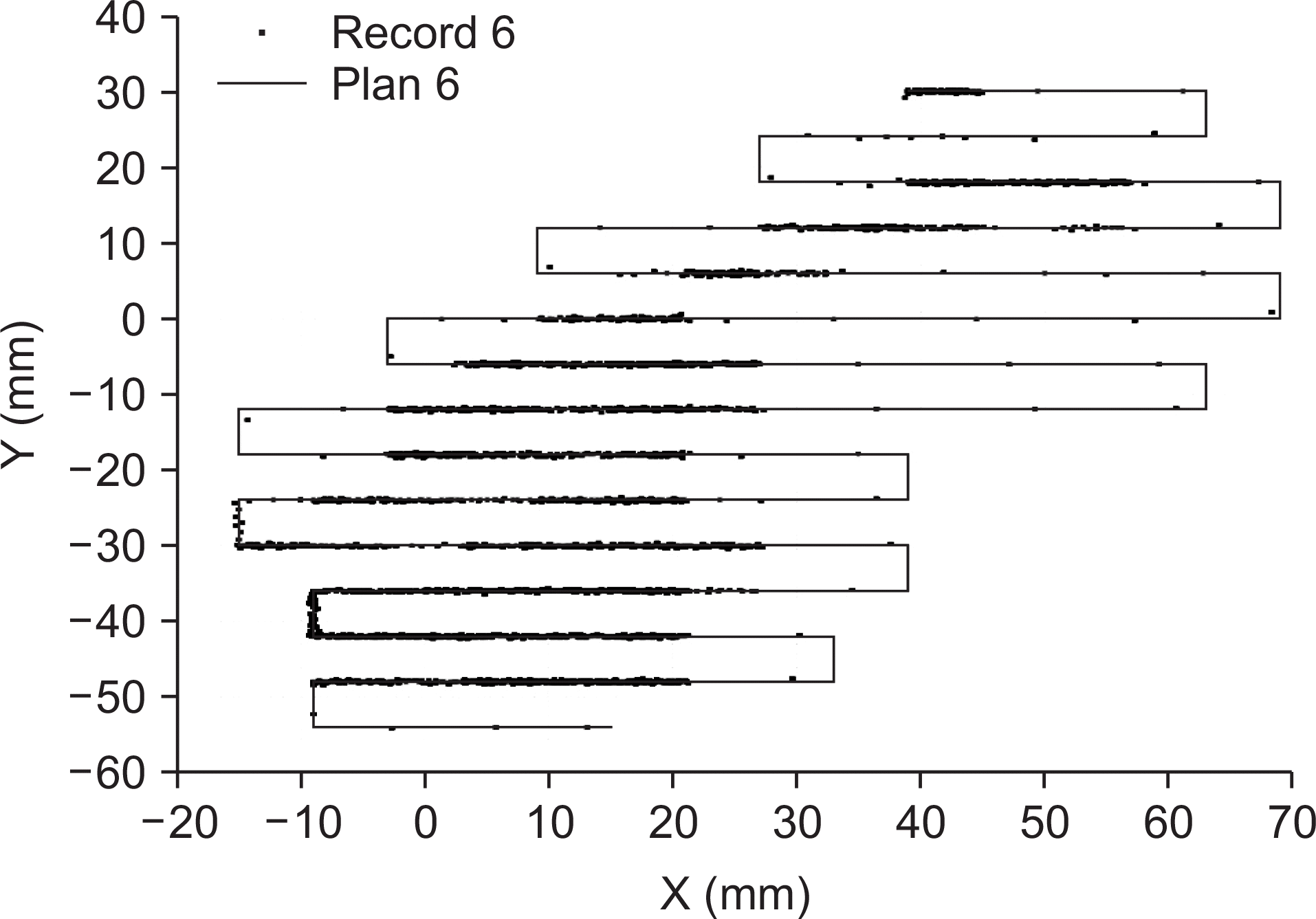
 | Fig. 3.A scanning beam information recorded in the scanning control unit (one sample layer of a patient beam delivery plan). |
Table 1.
The summary of pre-treatment QA log file analysis. The “ds” stands for the displacement of the recorded position from the planned position. For a given field (case), the entire energy layers (number of layers) have been analyzed.
| Cases | Number of layers | Average of ds | Standard deviation |
|---|---|---|---|
| [mm] | of ds [mm] | ||
| Case 1 | 15 | 0.09 | 0.09 |
| Case 2 | 17 | 0.11 | 0.10 |
| Case 3 | 23 | 0.15 | 0.07 |
| Case 4 | 18 | 0.10 | 0.11 |
| Case 5 | 11 | 0.06 | 0.14 |


 PDF
PDF ePub
ePub Citation
 Print
Print


 XML Download