

 PDF
PDF ePub
ePub Citation
 Print
Print


Abstract
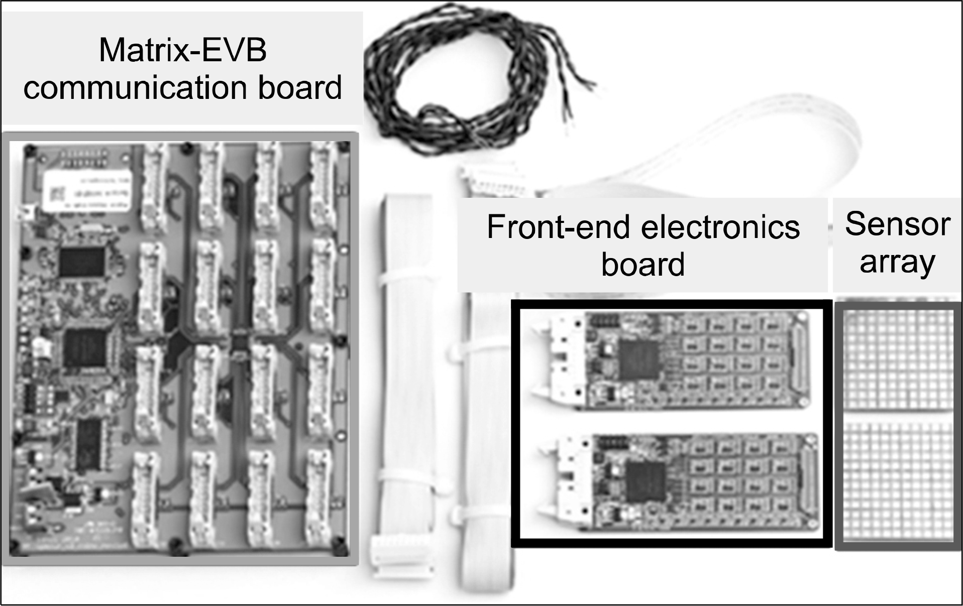
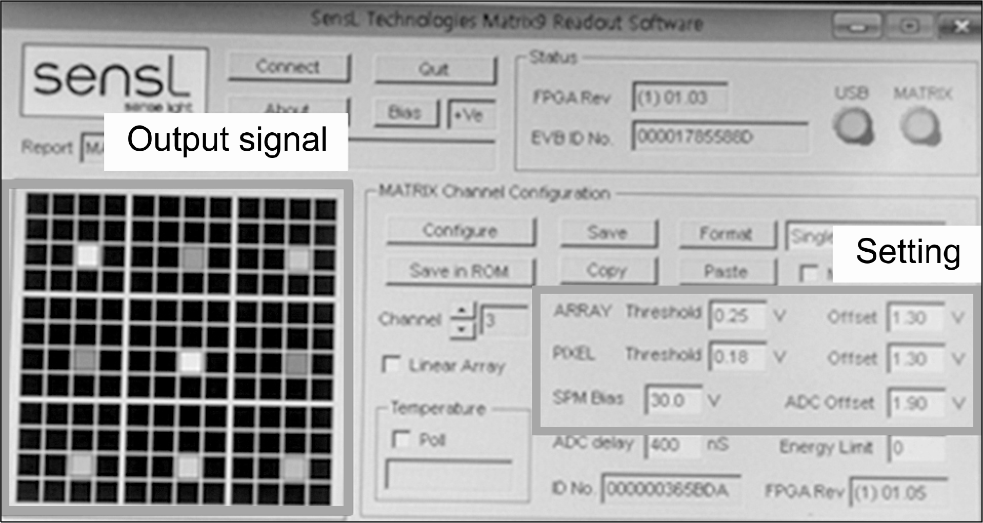
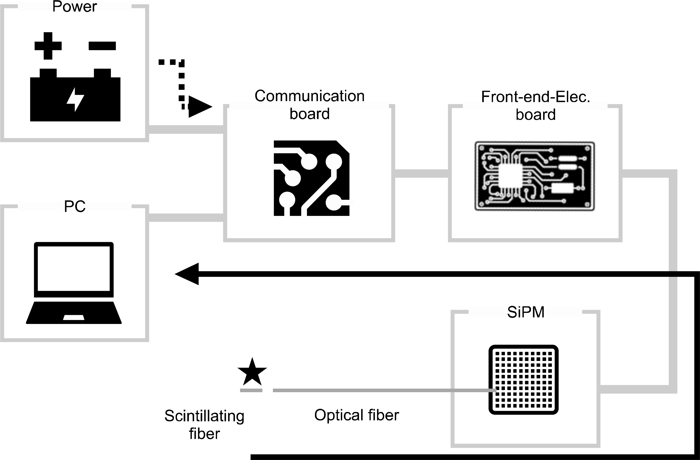
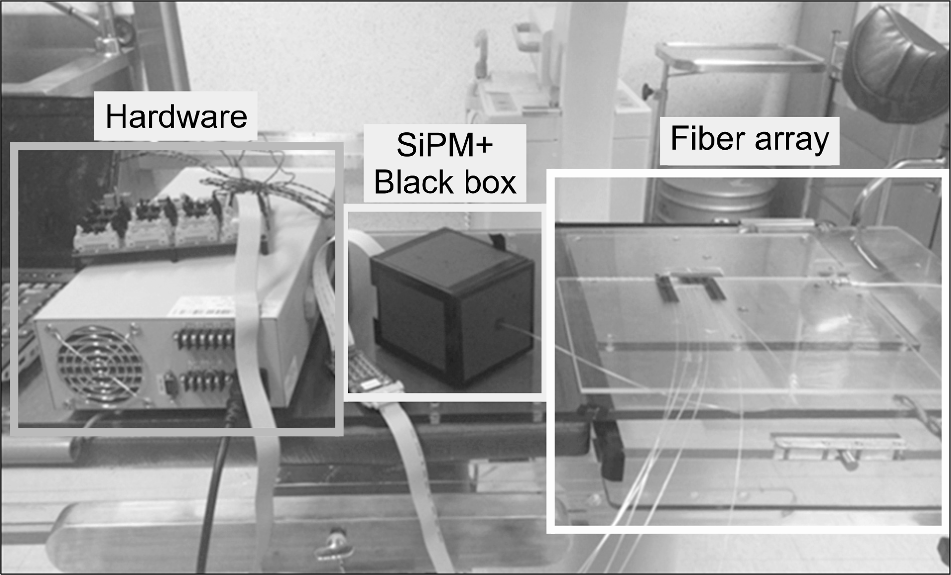
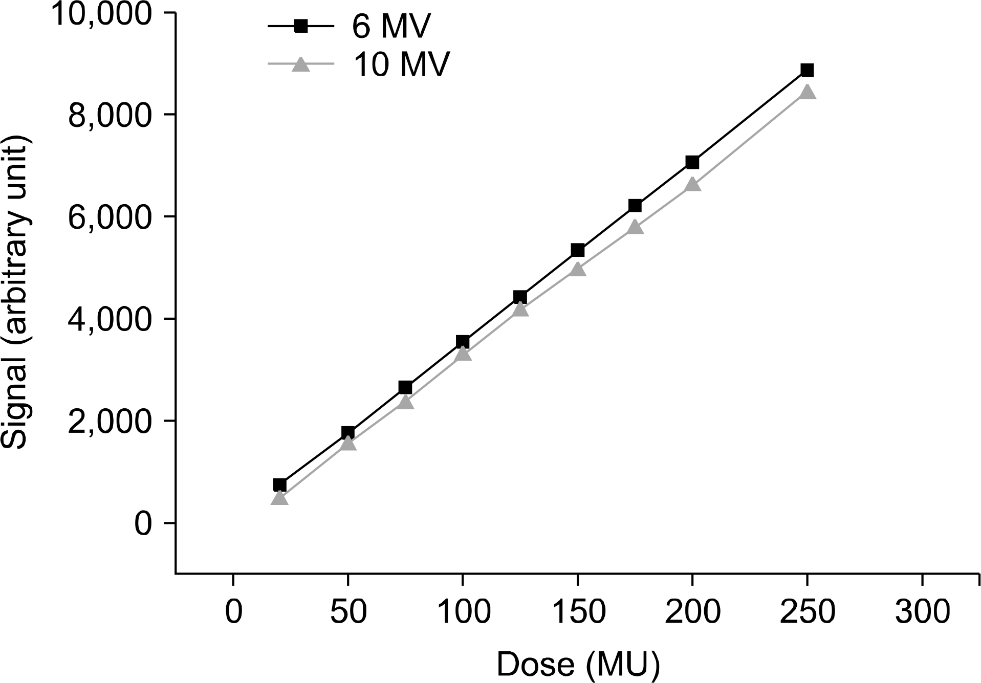
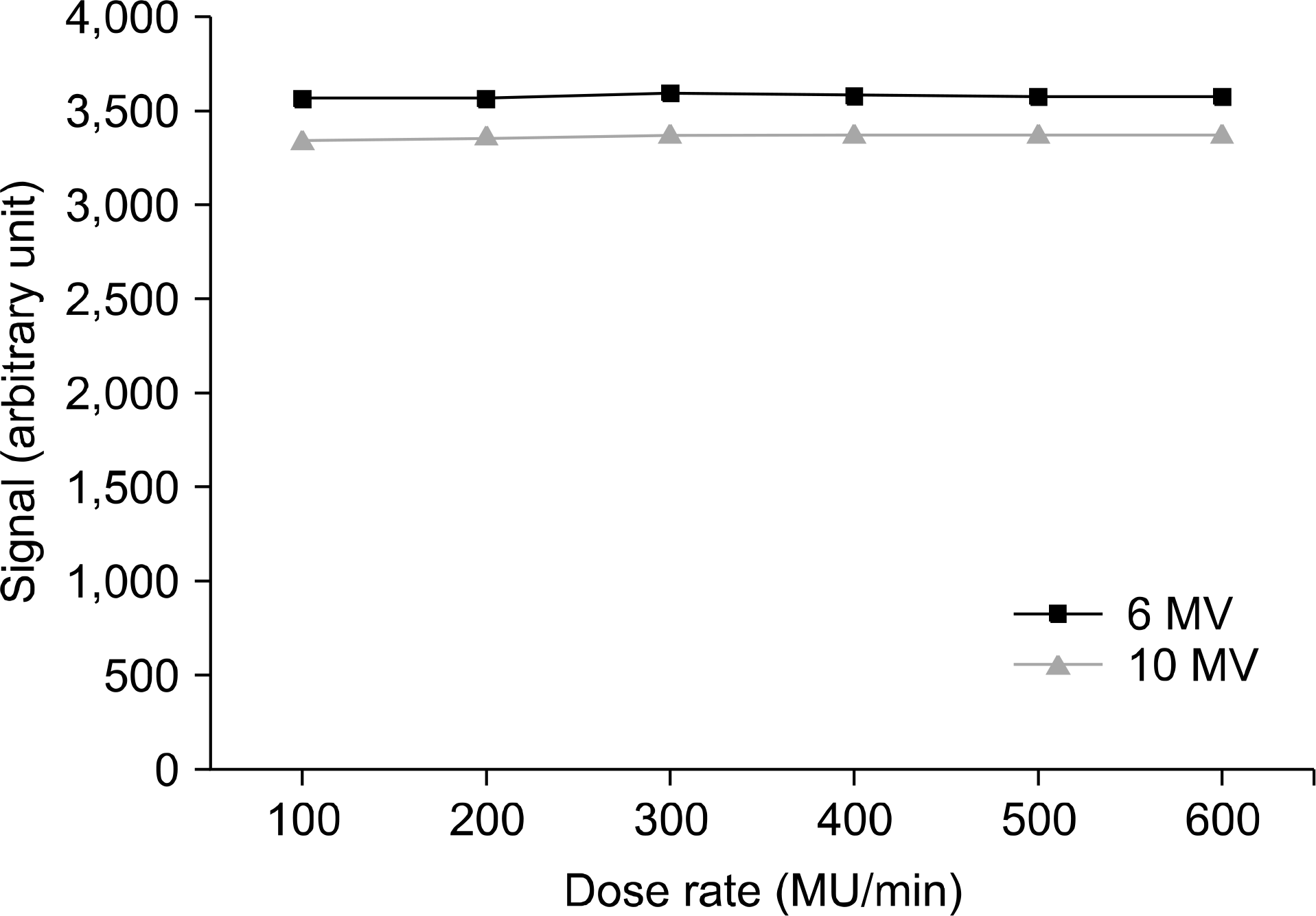
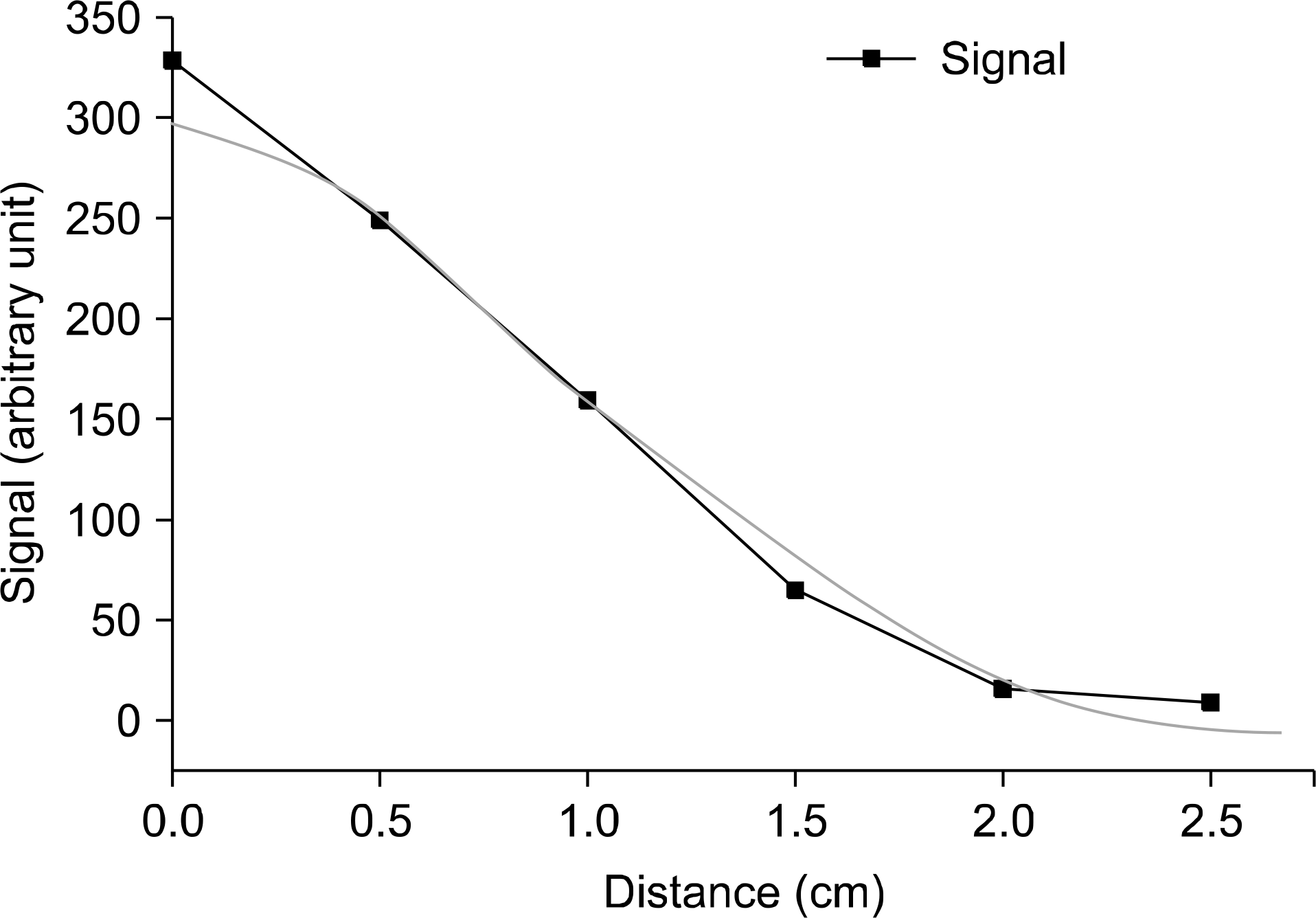
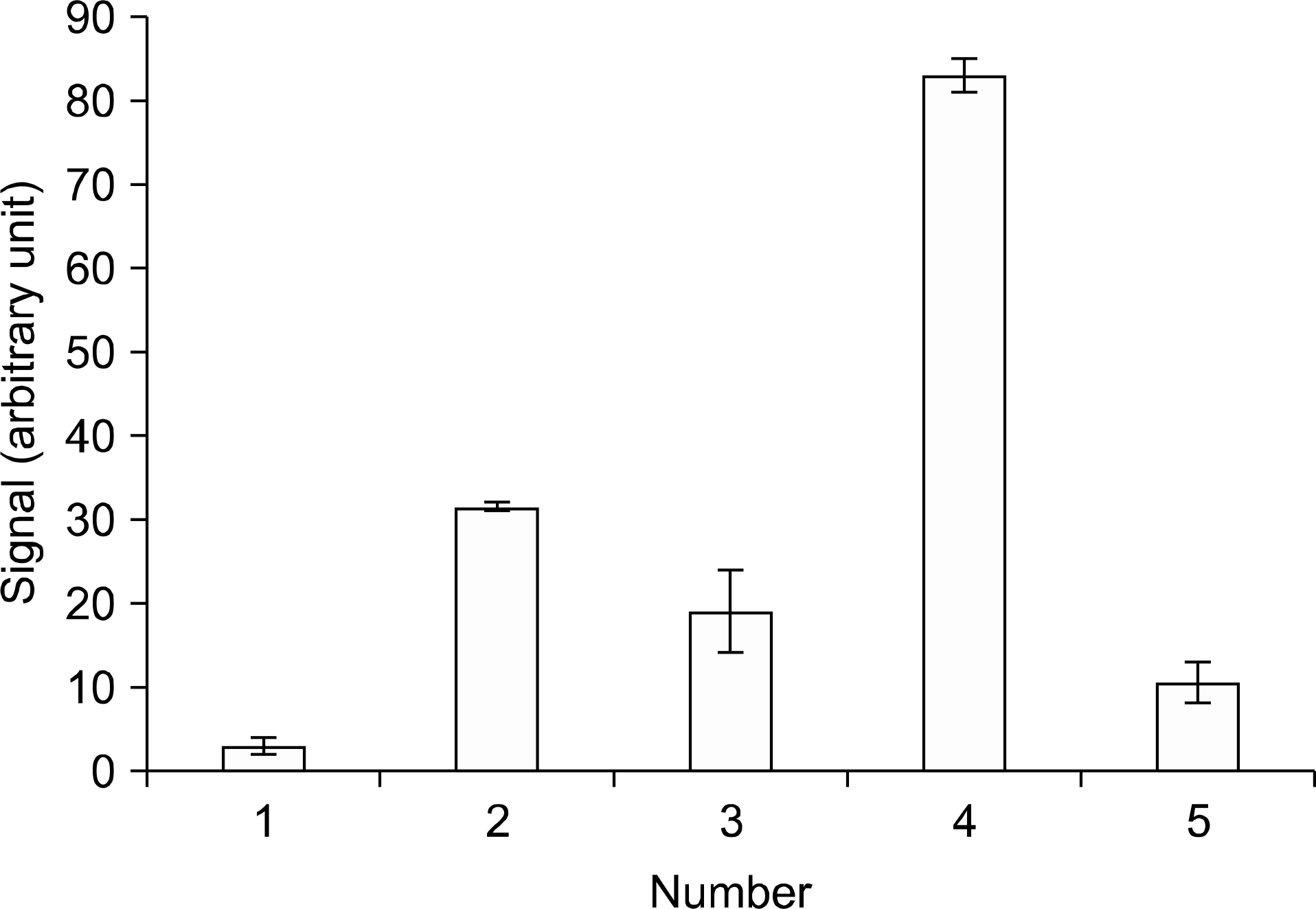
The position verification of the radiation source utilized in brachytherapy forms a critical factor in determining the therapeutic efficiency. Currently, films are used to verify the source position; however, this method is encumbered by the lengthy time interval required from film scanning to analysis, which makes real-time position verification difficult. In general, the source position accuracy is usually tested in a monthly quality assurance check. In this context, this study investigates the feasibility of the real-time position verification of the radiation source in high dose rate (HDR) brachytherapy with the use of scintillating fibers. To this end, we construct a system consisting of scintillating fibers and a silicon photomultiplier (SiPM), optimize the dosimetric software setup and radiation system characteristics to obtain maximum measurement accuracy, and determine the relative ratio of the measured signals dependent upon the position of the scintillating fiber. According to the dosimetric results based on a treatment plan, in which the dwell time is set at 30 and 60 s at two dwell positions, the number of signals is 31.5 and 83, respectively. In other words, the signal rate roughly doubles in proportion to the dwell time. The source position can also be confirmed at the same time. With further improvements in the spatial resolution and scintillating fiber array, the source position can be verified in real-time in clinical settings with the use of a scintillating fiber-based system.
REFERENCES
1. Devlin PM. Brachytherapy: Applications and Techniques. Springer Publishing Company. 2015.
2. Young-Hoon J. Quality Assurance of Brachytherapy System (Physical Aspects). Korean J Med Phys 17-21. 1993.
3. Stewart A, Jones B. Radiobiological concepts for brachytherapy. Devlin PM: Brachytherapy: Applications and Techniques. 1st ed. Baltimore: Lippincott, Williams & Wilkins. 2006.
4. Pötter R, Dimopoulos J, Georg P, et al. Clinical impact of MRI assisted dose volume adaptation and dose escalation in brachytherapy of locally advanced cervix cancer. Radiother Oncol. 83(2):148–155. 2007.

5. Su Jin Lee RNL, Yi BY, Lim SW, Choi JH. Real Time On-line Quality Assurance System for HDR Brachytherapy. Korean J Med Phys. 15(3):156–160. 2004.
6. Beierholm AR, Andersen CE, Lindvold L, Medin J. A comparison of BCF-12 organic scintillators and Al2O3: C crystals for real-time medical dosimetry. Radiat Meas. 43(2):898–903. 2008.
7. Moser SW, Harder WF, Hurlbut CR, Kusner MR. Principles and practice of plastic scintillator design. Radiat Phys Chem 41(1-2): 31-36. 1993.
8. White TO. Scintillating fibres. Nucl. Instr. Meth. Phys. Res. A 273(2-3): 820-825. 1988.
9. Imai S-i. Soramoto S, Mochiki K-i, Iguchi T, Nakazawa M. New radiation detector of plastic scintillation fiber. Rev Sci Instrum. 62(4):1093–1097. 1991.
10. Chichester DL, Watson SM, Johnson JT. Comparison of BCF-10, BCF-12, and BCF-20 scintillating fibers for use in a 1-dimensional linear sensor. IEEE Trans Nucl Sci. 60(5):4015–4021. 2013.
11. Papandreou Z, Leverington BD, Lolos G. Spectral response of scintillating fibers. Nucl. Instr. Meth. Phys. Res. A. 596(3):338–346. 2008.
12. Suzuki K, Bühler P, Fossati S, Marton J, Schafhauser M, Zmeskal J. Development of SciFi/CheFi detector with SiPM readout. Nucl. Instr. Meth. Phys. Res. A. 610(1):75–77. 2009.
13. Bonanno G, Finocchiaro P, Pappalardo A, et al. Precision measurements of photon detection efficiency for SiPM detectors. Nucl. Instr. Meth. Phys. Res. A. 610(1):93–97. 2009.
14. Luxton G, Astrahan MA, Liggett PE, Neblett DL, Cohen DM, Petrovich Z. Dosimetric calculations and measurements of gold plaque ophthalmic irradiators using iridium-192 and iodine-125 seeds. Int J Radiat Oncol. 15(1):167–176. 1988.
15. Lee SH, Kim JR, Cho SJ, et al. Analysis and investigation for the status of radiation therapy QA in Korea. Korean J Med Phys. 21(2):223–231. 2010.
 XML Download