Abstract
The aim of this study is to investigate the delivery accuracy of intensity-modulated radiation therapy (IMRT) plans in the two-headed mode of the ViewRayTM system in comparison with that of the normal operation treatment plan of the machine. For this study, a total of eight IMRT plans and corresponding verification plans were generated (four head and neck, two liver, and two prostate IMRT plans). The delivered dose distributions were measured using ArcCHECKTM with the insertion of an ionization chamber. We measured the delivered dose distributions in three-headed mode (normal operation of the machine), two-headed mode with head 1 disabled, two-headed mode with head 2 disabled, and two-headed mode with head 3 disabled. Therefore, a total of four measurements were performed for each IMRT plan. The global gamma passing rates (3%/3 mm) in three-headed mode, head 1 disabled, head 2 disabled, and head 3 disabled were 99.9±0.1%, 99.8±0.3%, 99.6±0.7%, and 99.7±0.4%, respectively. The difference in the gamma passing rates of the three- and two-headed modes was insignificant. With 2%/2 mm, the rates were 96.6±3.6%, 97.2±3.5%, 95.7±6.2%, and 95.5±4.3%, respectively. Between three-headed mode and head 3 disabled, a statistically significant difference was observed with a p-value of 0.02; however, the difference was minimal (1.1%). The chamber readings showed differences of approximately 1% between three- and two-headed modes, which were minimal. Therefore, the treatment plan delivery in the two-headed mode of the ViewRayTM system seems accurate and robust.
Go to : 
REFERENCES
1.Mera Iglesias M., Aramburu Nunez D., Del Olmo Claudio JL, et al. Multimodality functional imaging in radiation therapy planning: relationships between dynamic contrast-enhanced MRI, diffusion-weighted MRI, and 18F-FDG PET. Comput Math Methods Med 2015: 103843. 2015.

2.Ghose S., Mitra J., Rivest-Henault D, et al. MRI-alone radiation therapy planning for prostate cancer: Automatic fiducial marker detection. Med Phys. 43(5):2218. (2016).

3.Sun J., Dowling J., Pichler P, et al. MRI simulation: end-to- end testing for prostate radiation therapy using geometric pelvic MRI phantoms. Phys Med Biol. 60(8):3097–109. 2015.
4.Ireland RH., Woodhouse N., Hoggard N, et al. An image acquisition and registration strategy for the fusion of hyperpolarized helium-3 MRI and x-ray CT images of the lung. Phys Med Biol. 53(21):6055–63. 2008.

5.Sarkar A., Santiago RJ., Smith R, et al. Comparison of manual vs. automated multimodality (CT-MRI) image registration for brain tumors. Med Dosim. 30(1):20–4. 2005.
6.Mutic S., Dempsey JF. The ViewRay system: magnetic resonance-guided and controlled radiotherapy. Semin Radiat Oncol. 24(3):196–9. 2014.

7.Park JM., Park S., Wu H, et al. Commissioning experience of tri-cobalt-60 MRI-guided radiation therapy system. Prog Med Phys. 26(4):193–200. 2015.

8.Wooten HO., Green O., Yang M, et al. Quality of Intensity Modulated Radiation Therapy Treatment Plans Using a 60Co Magnetic Resonance Image Guidance Radiation Therapy System. Int J Radiat Oncol Biol Phys. 92(4):771–8. 2015.
9.Wooten HO., Rodriguez V., Green O, et al. Benchmark IMRT evaluation of a Co-60 MRI-guided radiation therapy system. Radiother Oncol. 114(3):402–5. 2015.

10.Park JM., Park SY., Kim HJ, et al. A comparative planning study for lung SABR between tri-Co-60 magnetic resonance image guided radiation therapy system and volumetric modulated arc therapy. Radiother Oncol. 120(2):279–85. 2016.

11.Low DA., Harms WB., Mutic S, et al. A technique for the quantitative evaluation of dose distributions. Med Phys. 25(5):656–61. 1998.

Go to : 
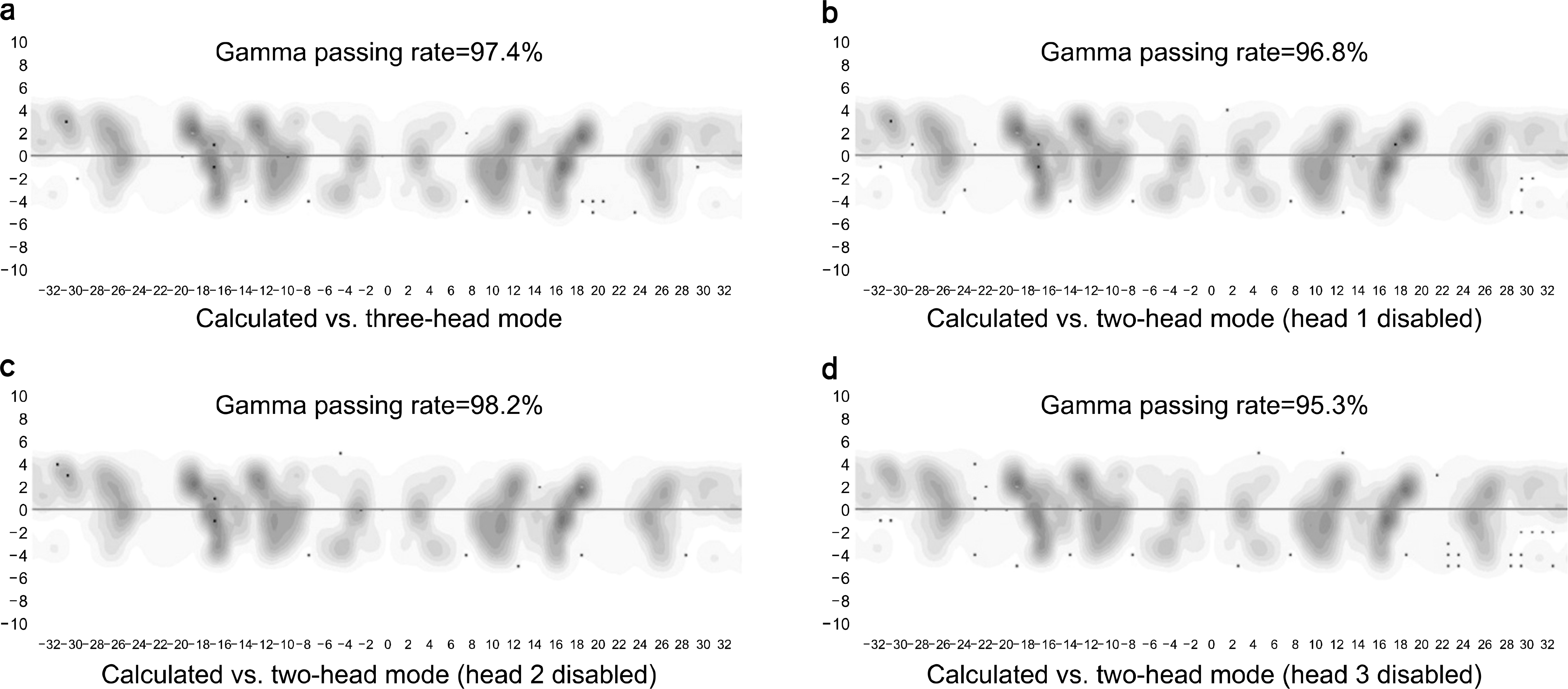
 | Fig. 1Measured dose distributions with the ArcCHECKTM of the intensity modulated radiation therapy (IMRT) plan for the treatment of head and neck cancer. The failed points by the gamma-index method with a 2%/2 mm gamma criterion are shown in red (measured value>calculated value) and blue (measured value<calculated value) colors. The distributions of the three-headed mode (a), two-headed mode with head 1 disabled (b), two-headed mode with head 2 disabled (c) and two-headed mode with head 3 disabled (d) are shown. |
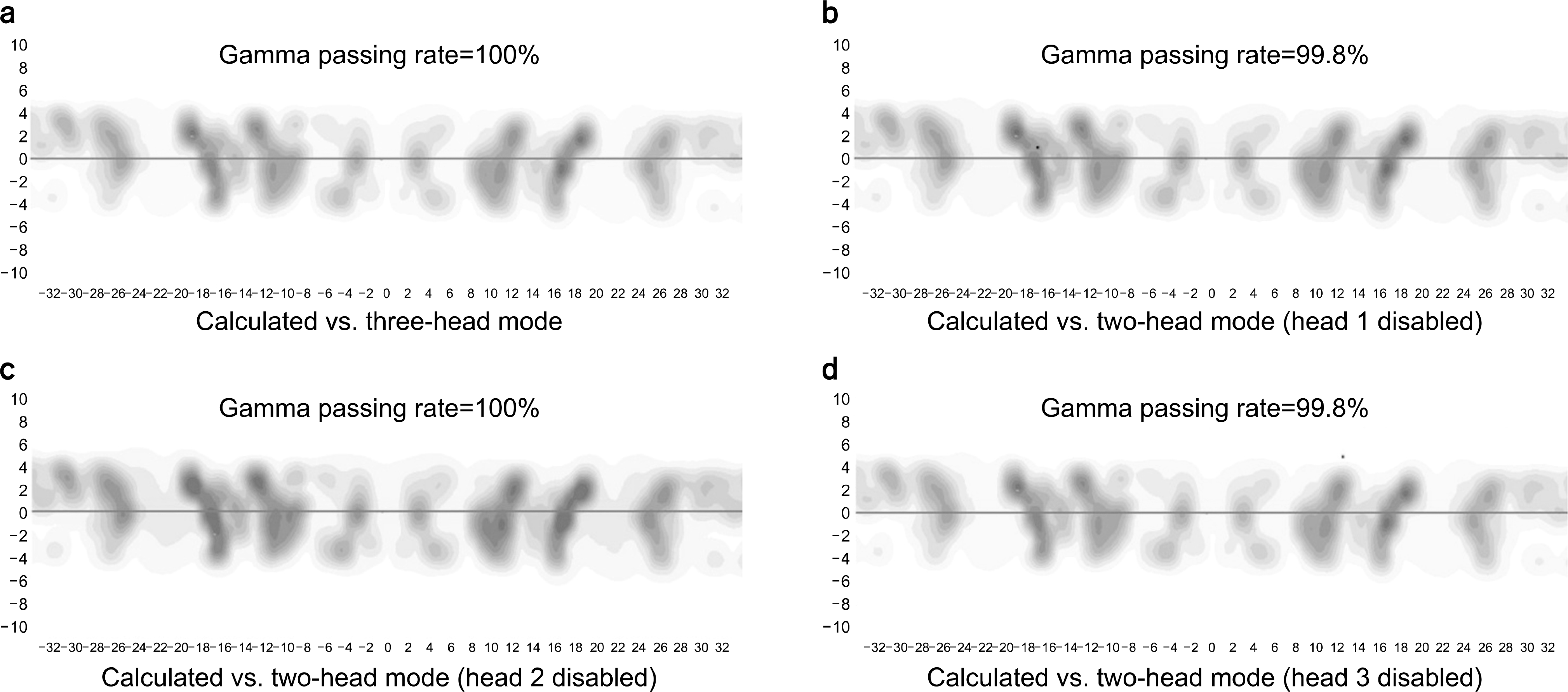
 | Fig. 2Measured dose distributions with the ArcCHECKTM of the intensity modulated radiation therapy (IMRT) plan for the treatment of head and neck cancer. The failed points by the gamma-index method with a 3%/3 mm gamma criterion are shown in red (measured value>calculated value) and blue (measured value<calculated value) colors. The distributions of the three-headed mode (a), two-headed mode with head 1 disabled (b), two-headed mode with head 2 disabled (c) and two-headed mode with head 3 disabled (d) are shown. |
Table 1.
Gamma passing rates of three-headed mode and two-headed mode.


 PDF
PDF ePub
ePub Citation
 Print
Print


 XML Download