

 PDF
PDF ePub
ePub Citation
 Print
Print


Abstract
Dual energy cone-beam CT can distinguish two materials with different atomic compositions. The principle of dual energy cone-beam CT based on modulation layer is that higher energy spectrum can be acquired at blocked x-ray window. To evaluate the possibility of modulation layer based dual energy cone-beam CT, we analyzed x-ray spectrum for various thicknesses of modulation layers by Monte Carlo simulation. To compare with the results of simulation, the experiment was performed on prototype cone-beam CT for 50∼100 kVp with CdTe XR-100T detector. As the result of comparing, the mean energy of energy spectrum for 80 kVp are well matched with that of simulation. The mean energy of energy spectrum for 80 and 120 kVp were increased as 1.67 and 1.52 times by 2.0 mm modulation layer, respectively. We realized that the virtual dual energy x-ray source can be generated by modulation layer.
Go to : 
References
1. Johnson Thorsten R. C., Schonberg Christian Fink Stefan O., Reiser Maximilian F.DualenergyCTinclinicalpractice. Springer;(. 2011. ), pp.p. 3–9.
2. Szczykutowicz T. P., Chen G. H.DualenergyCT usingslowkVpswitchingacquisitionandpriorimagecon-strainedcompressedsensing. PhysMedBiol. 55(21):6411–6429. 2010.
3. Altman A., Carmi R.ADouble‐LayerDetector,Dual‐EnergyCT—Principles,AdvantagesandApplications. Med. Phys. 36(6):2750–2750. 2009.
4. Faby Sebastian, Kuchenbecker Stefan, Sawall Stefan, Simons David, Heinz-Peter Schlemmer, Lell Michael, et al. Performance of today's dual energy CT and future multienergy CT invirtual non-contrast imaging and in iodine quantification: Asimulation study. Med.Phys. 42(7):4349–4366. 2015.
5. Yu Lifeng, Christner Jodie A., Leng Shuai, Wang Jia, Fletcher Joel G., McCollough Cynthia H.Virtual monochromaticimagingindual-sourcedual-energyCT: Radiationdoseandimagequality. Med. Phy. 38(12):6371–6379. 2011.
6. Ahn SH, Choi JH, Lee KC, Kim SY, Lee R, Shin SY. Developmentofabeamstoparraysystemwithdualscan modeforscattercorrectionofcone-beamCT. JKoreanPhys Soc. 64(8):),(. 2014.
7. Cranley K., Gilmore B.J., Fogarty G.W.A., Desponds L.CatalogueofDiagnosticX-raySpectraandOther Data, ReportNo. 78. TheInstituteofPhysicsandEngineering in Medicine. 1997.
8. Liu Xin, Yu Lifeng, Primak Andrew N., McCollough Cynthia H.Quantitativeimagingofelementcompositionand massfractionusingdual-energyCT:Three-materialdecom-position, Med. Phys. 36(5):1602–1609. 2009.
9. Hunemohr Nora, Paganetti Harald, Greilich Steffen, Jakel Oliver, Seco Joao. Tissuedecompositionfromdual energyCTdataforMC baseddosecalculationinparticle therapy. Med.Phys. 41(6):061714. 2014.
10. Goodsitt Mitchell M., Shenoy Apeksha, Shen Jincheng, Howard David, Schipper Matthew J., Wilderman Scott, et al. :. EvaluationofdualenergyquantitativeCTfordetermining thespatialdistributionsofredmarrowandbonefordosimetry ininternalemitterradiationtherapy. Med.Phys. 41(5):05190. 2014.
Go to :
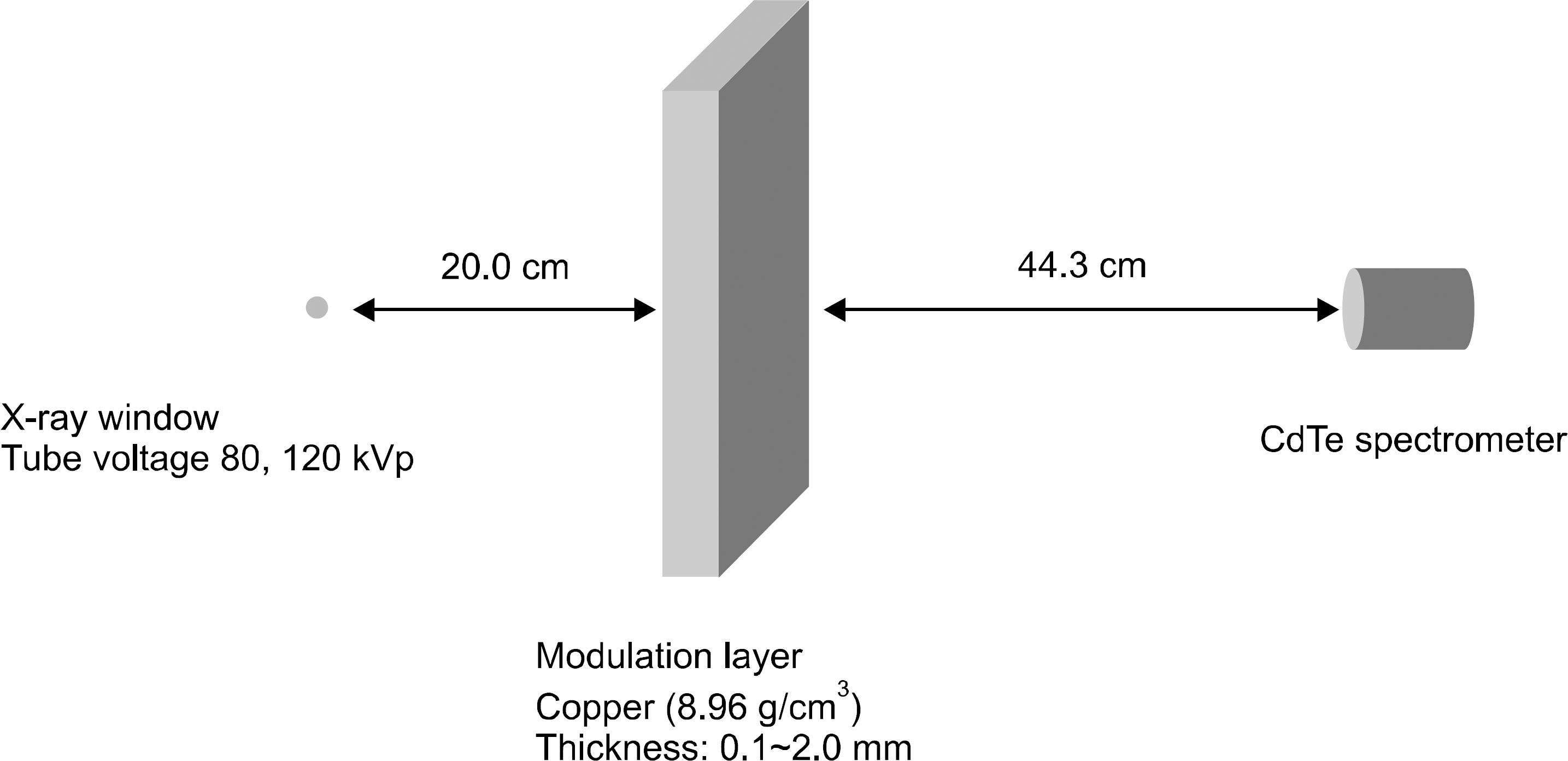 | Fig. 1.Geometrical modeling of Monte Carlo simulation. Modulation layer and CdTe spectrometer are located at 20.0 cm and 64.3 cm distances from x-ray window, respectively. |
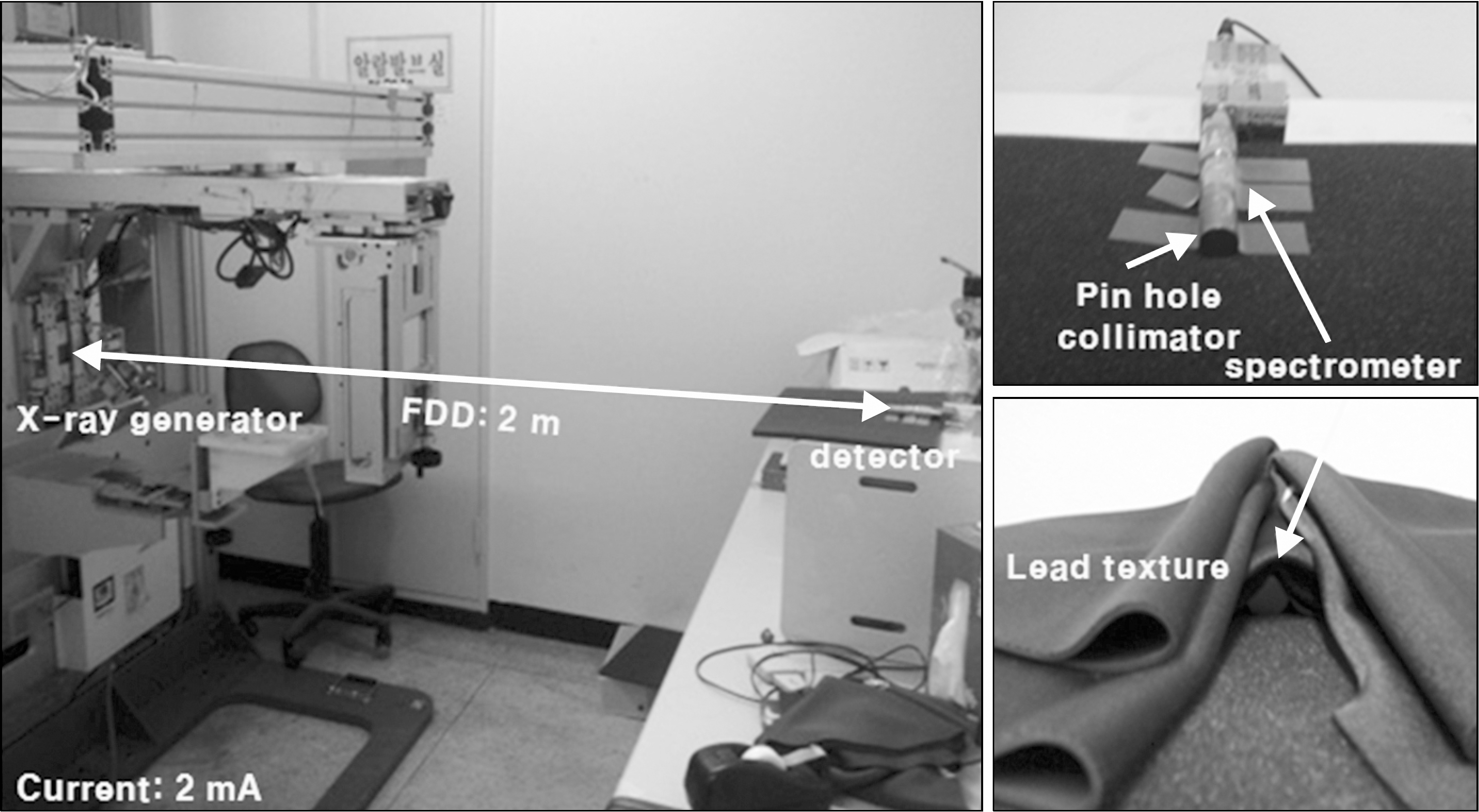 | Fig. 2.Experimental setup for measuring the energy spectrum with CdTe XR-100T detector. Pin hole collimator and lead texture were used to prevent saturation. Pin hole collimator has narrow hole with 0.3 mm diameter. |
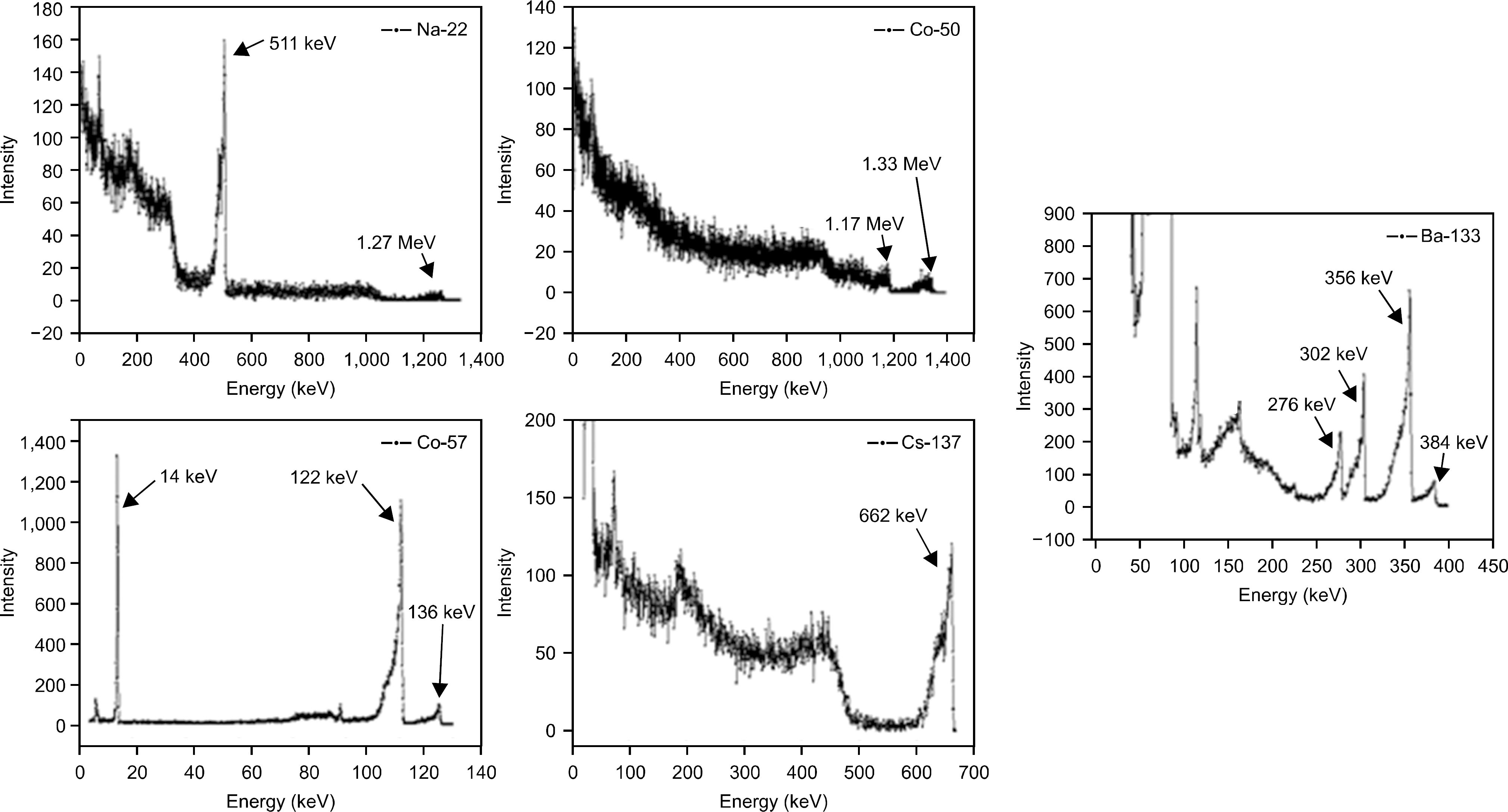
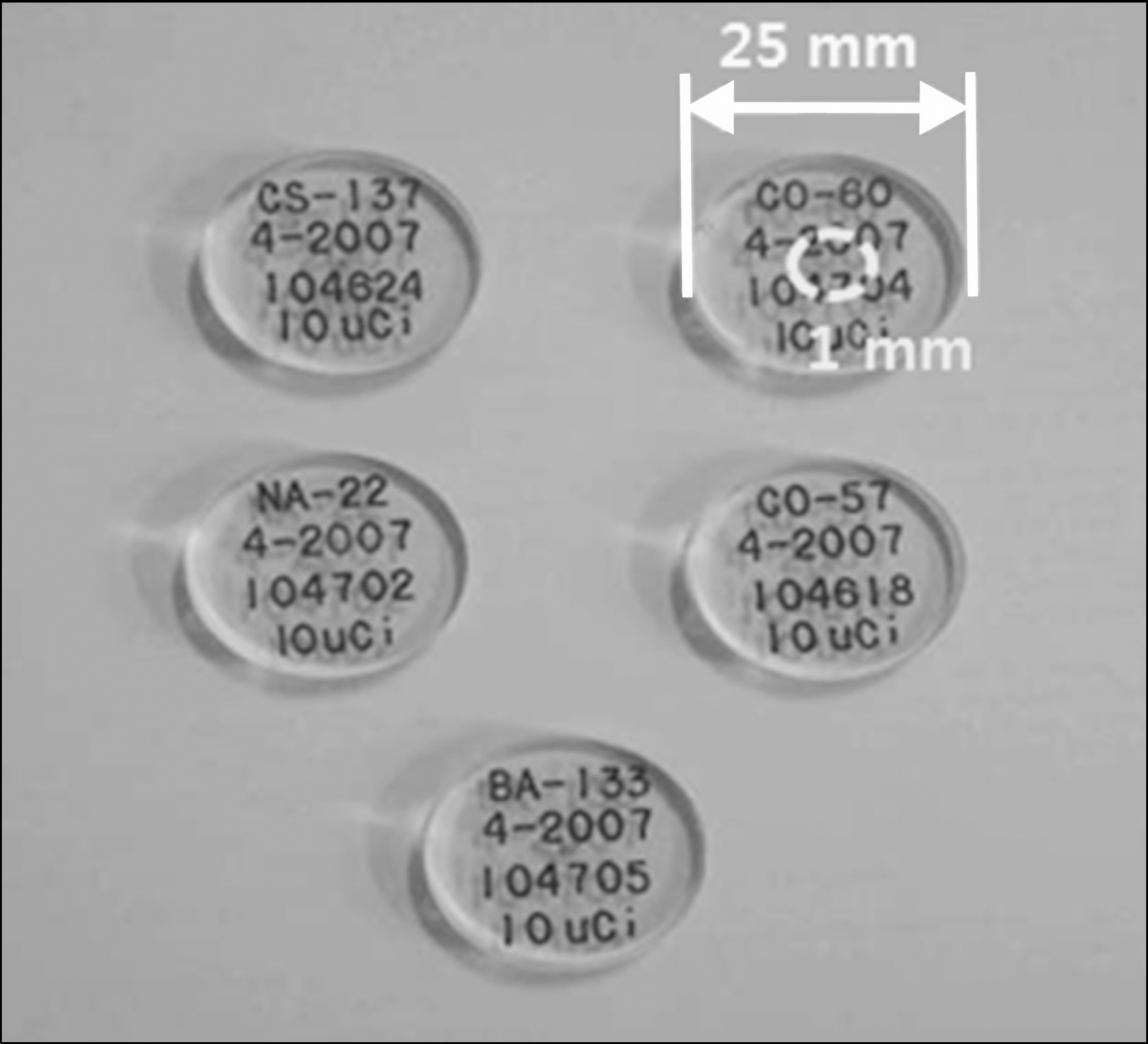 | Fig. 3.Photography of five isotopes used to calibrate CdTe XR-100T detector. The diameter of isotope is 1.0 mm and is enveloped in 25.0 mm diameter plastic protector. |
 | Fig. 4.Five energy spectra for energy calibration of CdTe XR-100T detector with five isotopes such as Na22, Ba133, Cs137, Co60, and Co57. |
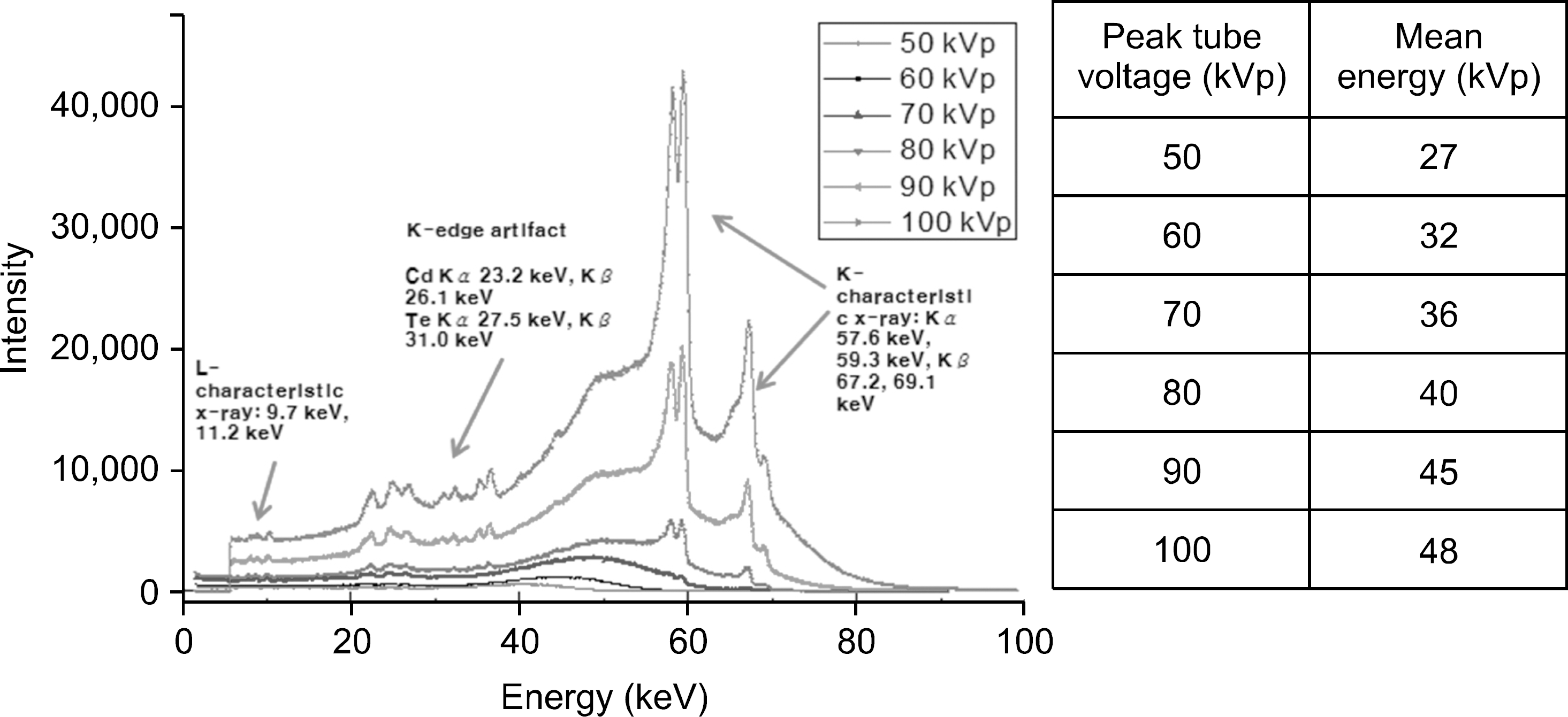
 | Fig. 5.Energy spectra acquired by CdTe XR-100T detector and their mean energy. The characteristic x-rays of detector materials, Cadmium and Telluride, are showed for peak tube voltage above 80 kVp. |
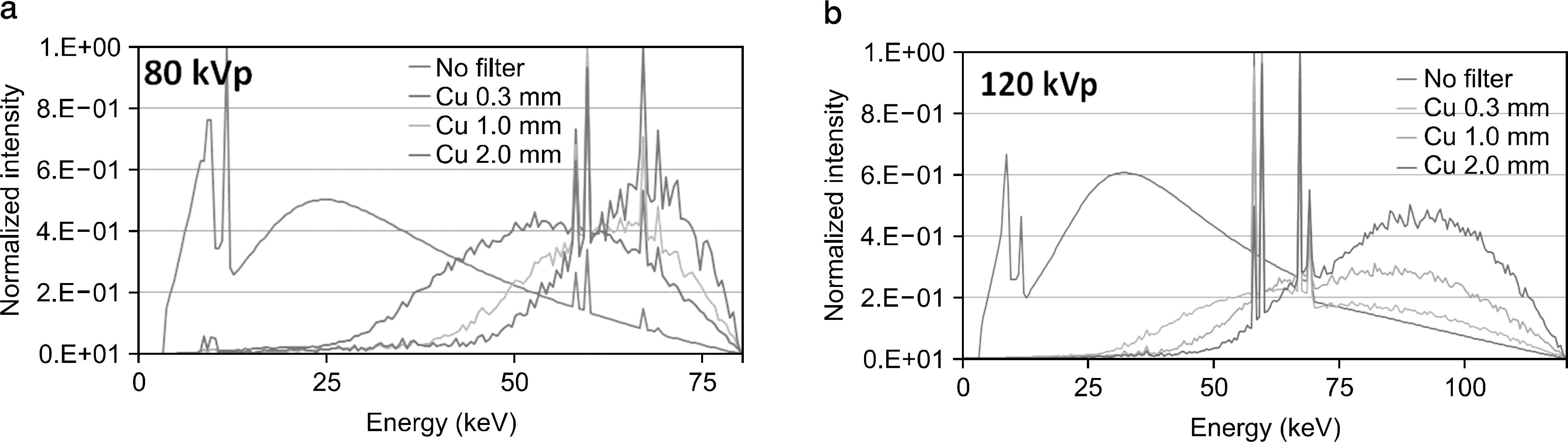 | Fig. 6.Energy spectra acquired by Monte Carlo simulation without modulation filter and with modulation filters for (a) 80 kVp and (b) 120 kVp. |
Table 1.
The energies of emitted gamma-rays from the selected five isotopes.
 XML Download