

 PDF
PDF ePub
ePub Citation
 Print
Print


Abstract
A CCD camera and an LED light source were combined to fabricate a compact optical CT scanner for the therapeutic radiation dose evaluation of a polymer gel dosimeter. After the collimated beam emitted by the LED passed through aquarium, gel phantom, and telecentric lens, an image was collected by the CCD camera and reconstructed using MATLAB. By using a stepping motor and LabVIEW, the gel dosimeter was rotated at every 0.72o, and the time for collecting 500 slice images per a revolution was within 20 min. At a spatial frequency of 4.5 lp/mm of the optical CT scanner, the modulation transfer function value was 72%. The linear correlation coefficient of the optical CT scanner for the polymer gel dosimeter was 0.987.
Go to : 
References
1. M. Humphreys, M. Teressa, G. Urbano, et al: Assessment of a customised immobilisation system for head and neck IMRT using electronic portal imaging. Radiother. Oncol. 77(1):39–74. 2005.
2. L. N. McDermott, M. Wendling, J. J. Sonke, M. Herk, B. J. Mijnheer: Replacing pretreatment verification with in vivo EPID dosimetry for prostate IMRT. Int. J. Radiat. Oncol. Biol. Phys. 67(5):1568–1577. 2007.
3. M. Oldham, J. H. Siewerdsen, S. Kumar, J. Wong, D. A. Jaffray: Optical-CT gel-dosimetry I: basic investigations. Med. Phys. 30(4):623–634. 2003.
4. Nikola Krstaji´c. Simon J. Doran: Focusing optics of a parallel beam CCD optical tomography apparatus for 3D radiation gel dosimetry. Phys. Med. Biol. 51:2055–2075. 2006.
5. A. Jirasek, D. Rudko, D. Wells, Journal of Physics: A prototype fan-beam optical CT scanner for polymer gel dosimetry. Conference Series. 164:1–6. 2009.
6. Nikola Krstaji´c, Simon J Doran: Fast laser scanning optical-CT apparatus for 3D radiation dosimetry. Phys. Med. Biol. 52:257–263. 2007.
7. S. J. Doran, N. Krstaji: The history and principles of optical computed tomography for scanning 3-D radiation dosimeters. Journal of Physics: Conference Series. 56:45–57. 2006.
8. Timothy Olding, Oliver Holmes, L John Schreiner: Cone beam optical computed tomography for gel dosimetry I. Scanner characterization. Phys. Med. Biol. 55:2819–2840. 2010.
9. M. Hilts, A. Jirasek, C. Duzenli: Technical considerations for implementation of X-ray CT polymer gel dosimetry. Phys. Med. Biol. 50:1727–1745. 2005.
10. K. H. Cho, S. J. Cho, S. Lee, S. H. Lee, C. K. Min: Dose responses in a normoxic polymethacrylic acid gel dosimeter using optimal CT scanning parameters. Nucl. Instr. and Meth. A. 675:112–117. 2012.
11. Y. R. Cho, H. W. Park, A. R. Kim, et. al: Fabrication of a Normoxic Polymer Gel Dosimeter and its Dose Distribution Characteristics. J. Korean Phys. Soc. 59(1):169–175. 2011.
12. A. R. Kim: Optical computed tomography for 3D gel dosimetry. MS thesis, Kyonggi University (. 2011.
13. J. C. Lee: Image characteristic of optical computed tomography. MS thesis, Kyonggi University (. 2011.
14. S. J. Doran, K. K. Koerkamp, M. A. Bero, et al: A CCD-based optical CT scanner for high-resolution 3D imaging of radiation dose distributions: equipment specifications, optical simulations and preliminary results. Phys. Med. Biol. 46(12):3191–213. 2001.
15. Corey Clift, Andrew Thomas, John Adamovics, Zheng Chang, Indra Das: Toward acquiring comprehensive radiosurgery field commissioning data using the PRESAGEⓇ/optical-CT 3D dosimetry system. Phys. Med. Biol. 55(5):1279–1293. 2010.
16. Gustavsson H, Back S A J, Lepage M, Rintoul L, Baldock C. Development and optimization of a 2-hydroxyethylacrylate MRI polymer gel dosimeter. Phys. Med. Biol. 49:227–241. 2004.
Go to :
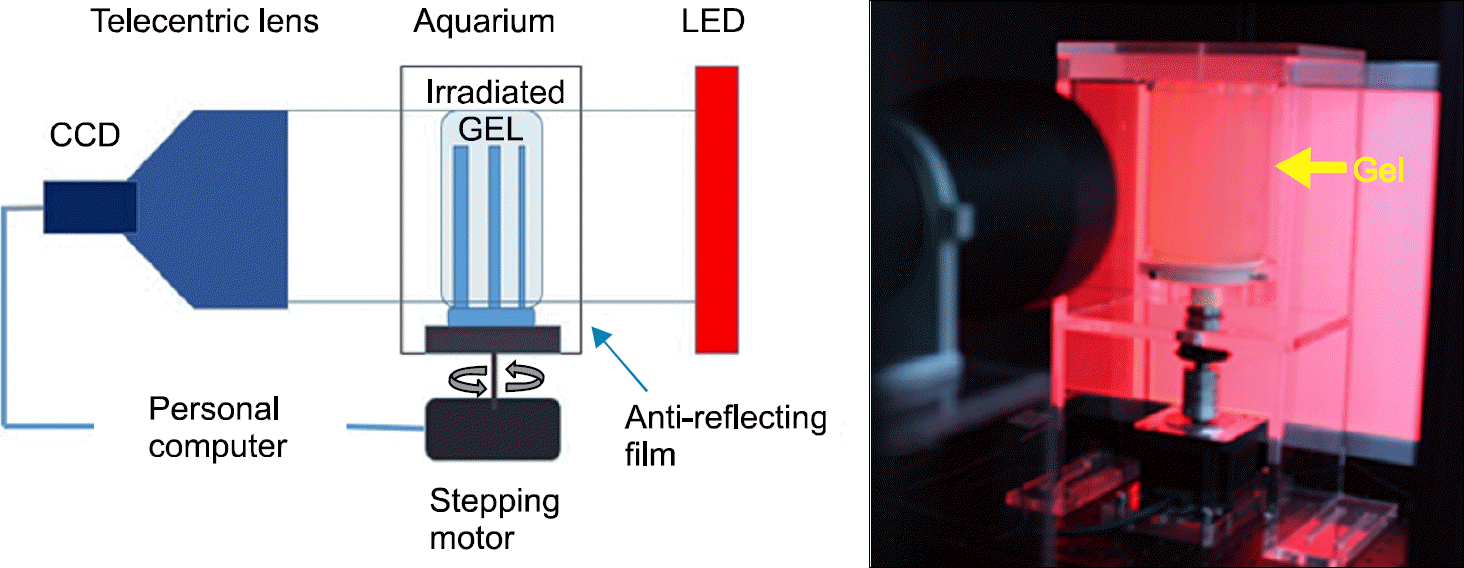 | Fig. 1.A schematic diagram of OCT Scanner (left) and a picture of OCT on the experiment (right). |
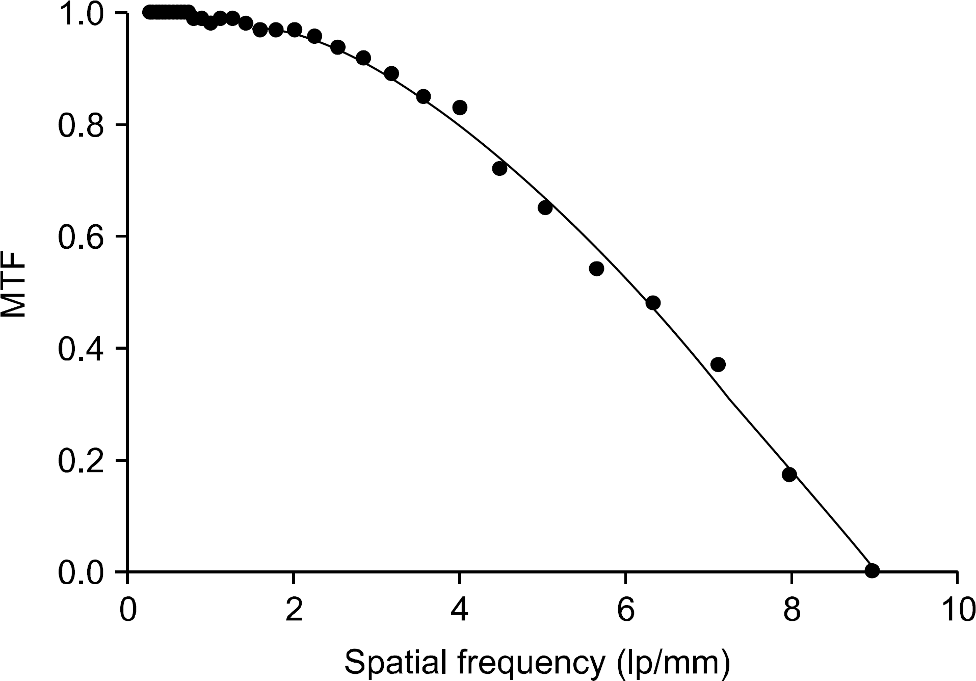 | Fig. 2.Modulation Transfer Function curve. Spatial resolution for a optical CT system is measured to be 72% out to 4.5 lp/mm. |
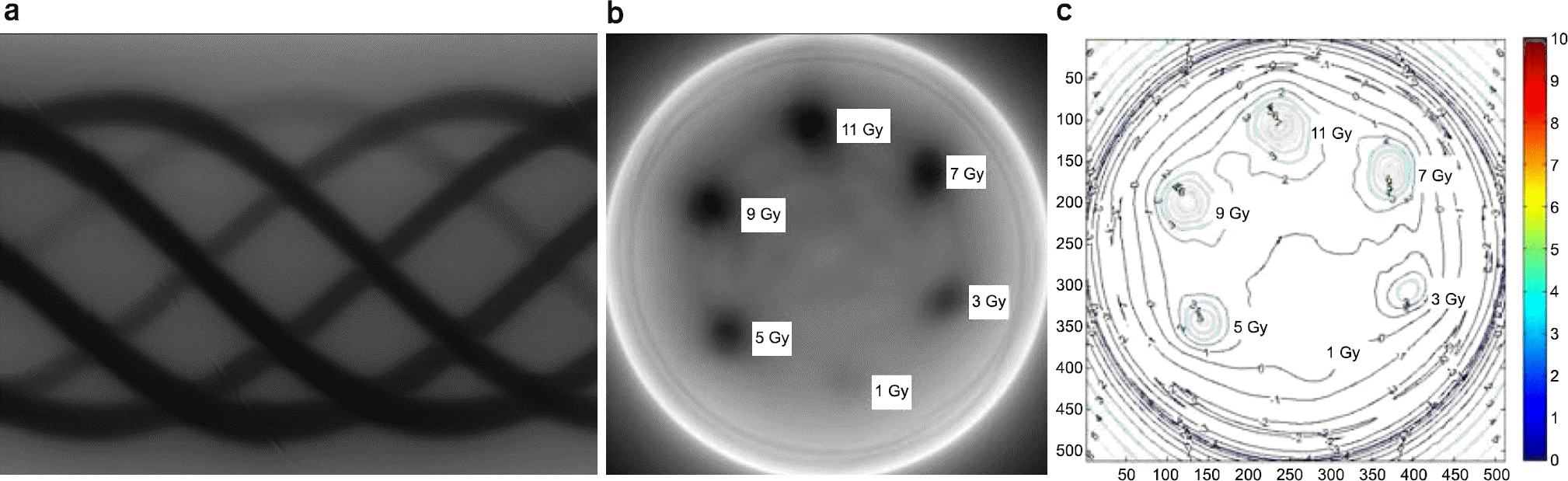
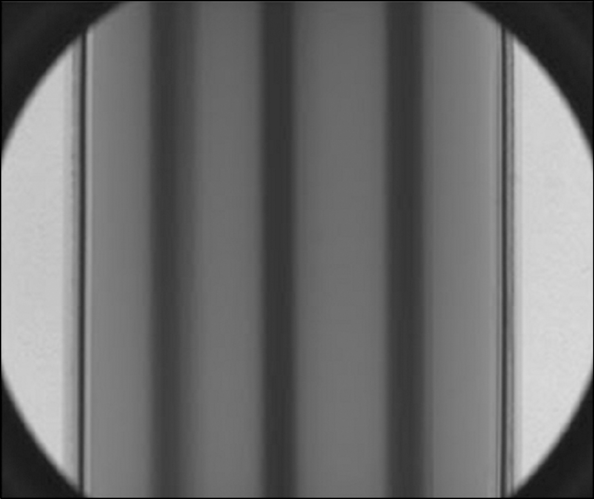 | Fig. 3.Projection image of a gel dosimeter irradiated with Cyberknife (1 Gy, 2 Gy and 3 Gy). |
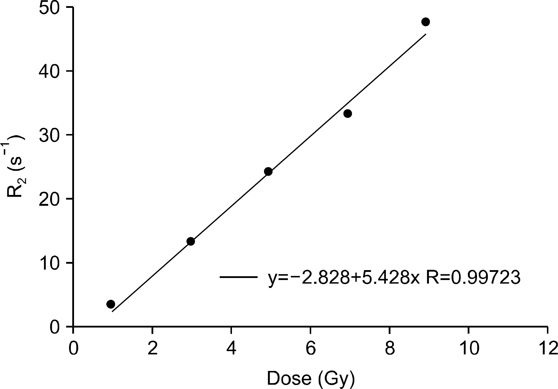
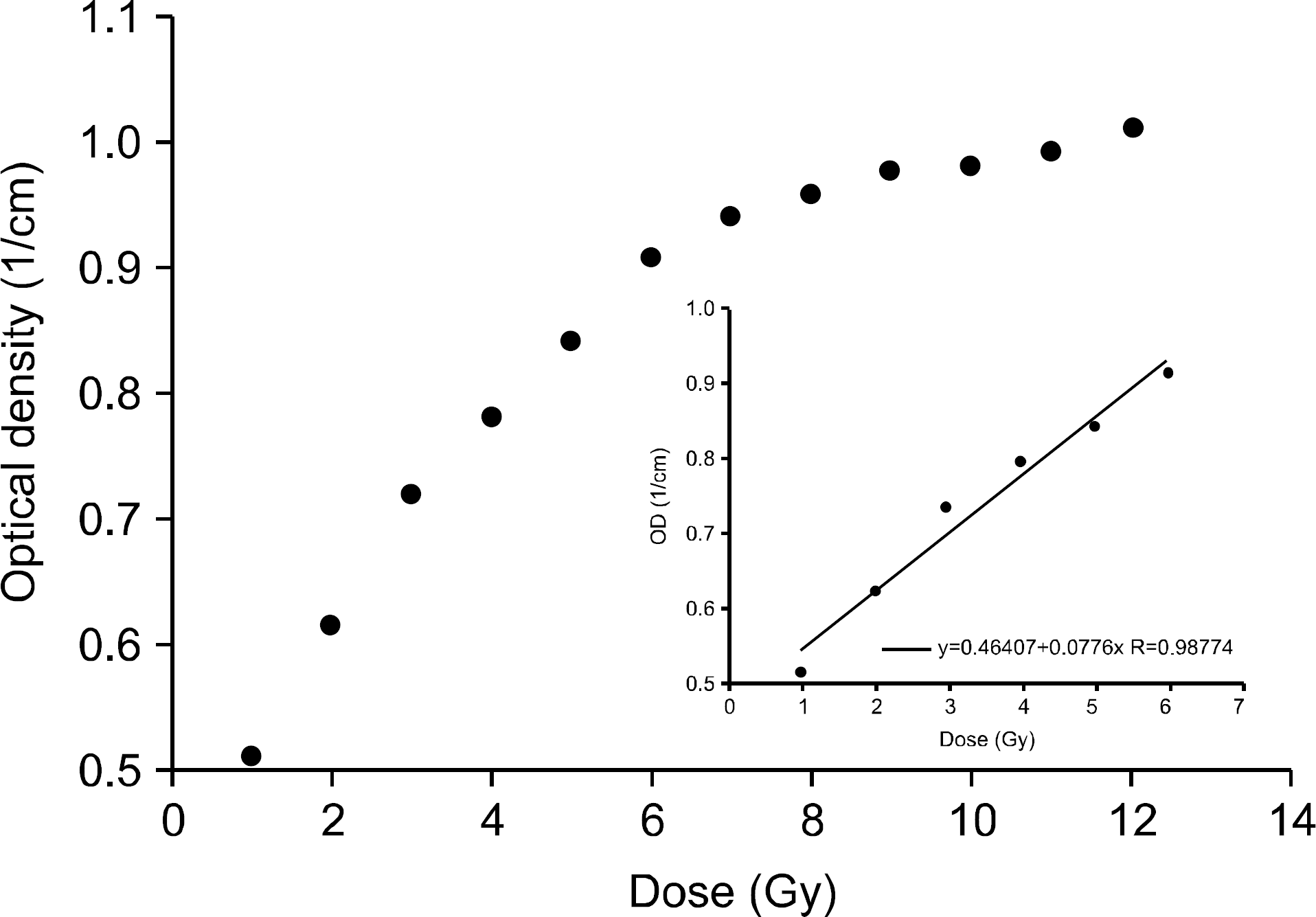 | Fig. 4.Dose response curve of a gel dosimeter measured by OCT scanner. The inset shows the linearity to 6 Gy and the linear correlation coefficient of r2=0.987. |
 | Fig. 5.Dose response curve of a gel dosimeter using MRI. The linear correlation coefficient is r2=0.997. |
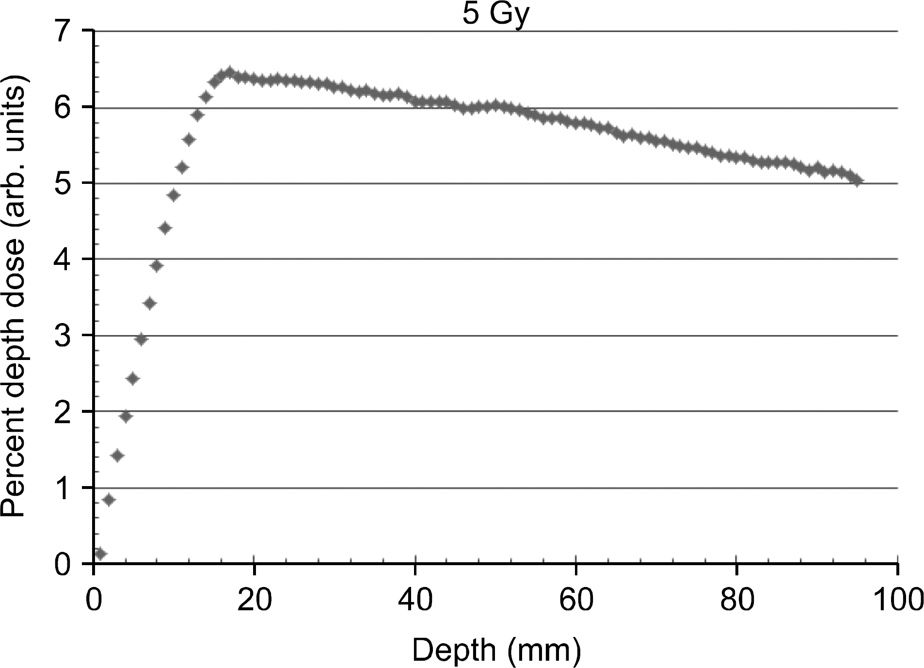 | Fig. 6.Percent Depth Dose distribution measured on the depth of central axis of a polymer gel dosimeter using 6 MV X-ray photon beam. |
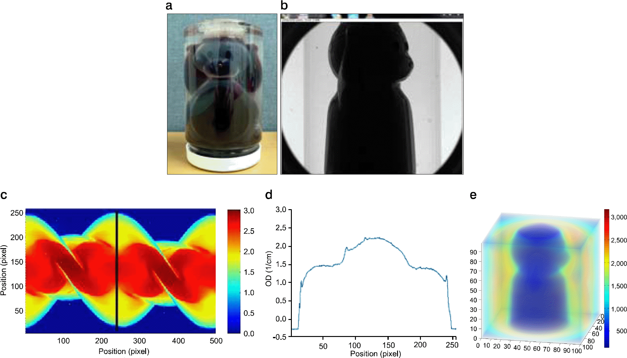 | Fig. 8.(a) A bear doll installed in PET container, (b) sagittal projection image (2,456×2,058 pixels, 16 bit), (c) sinogram, (d) line profile at the vertical black line of sinogram and (e) 3-dimensional rendering image. |
Table 1.
Timing characteristics of OCT scanner.
Table 2.
A comparison of the dosimetric characteristics for OCT, X-ray CT, and MRI.
 XML Download