Abstract
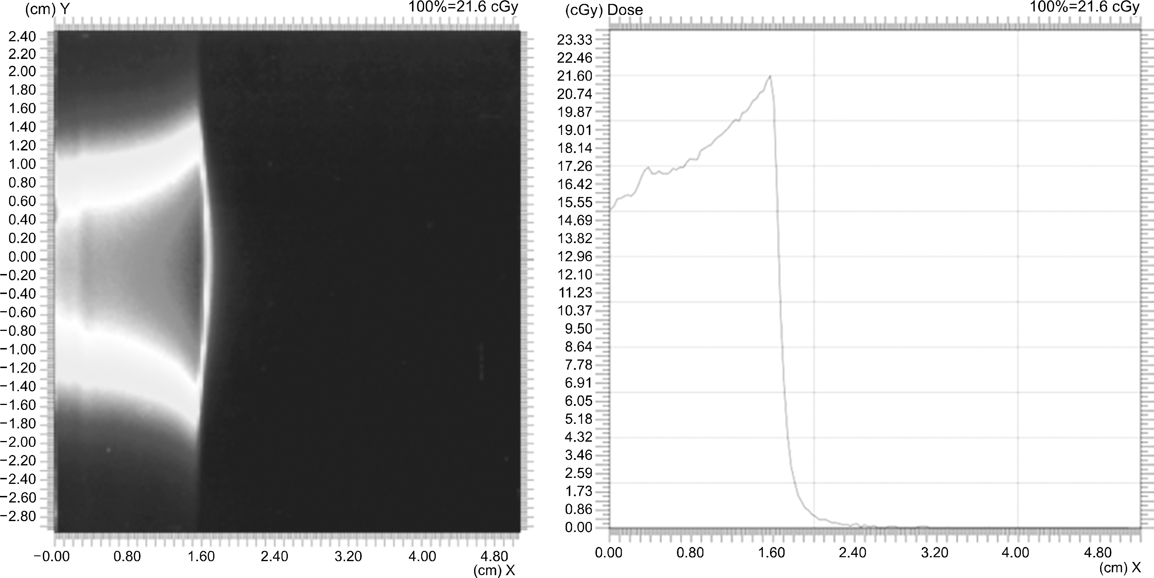
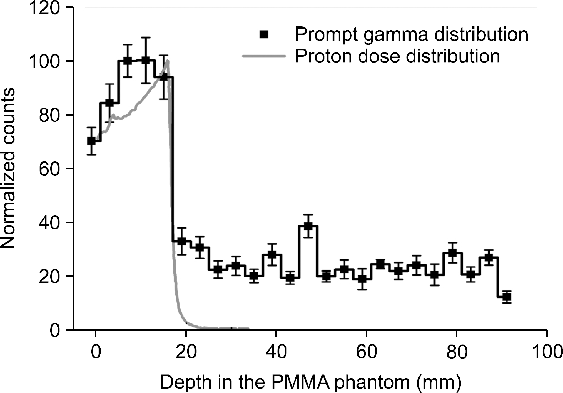
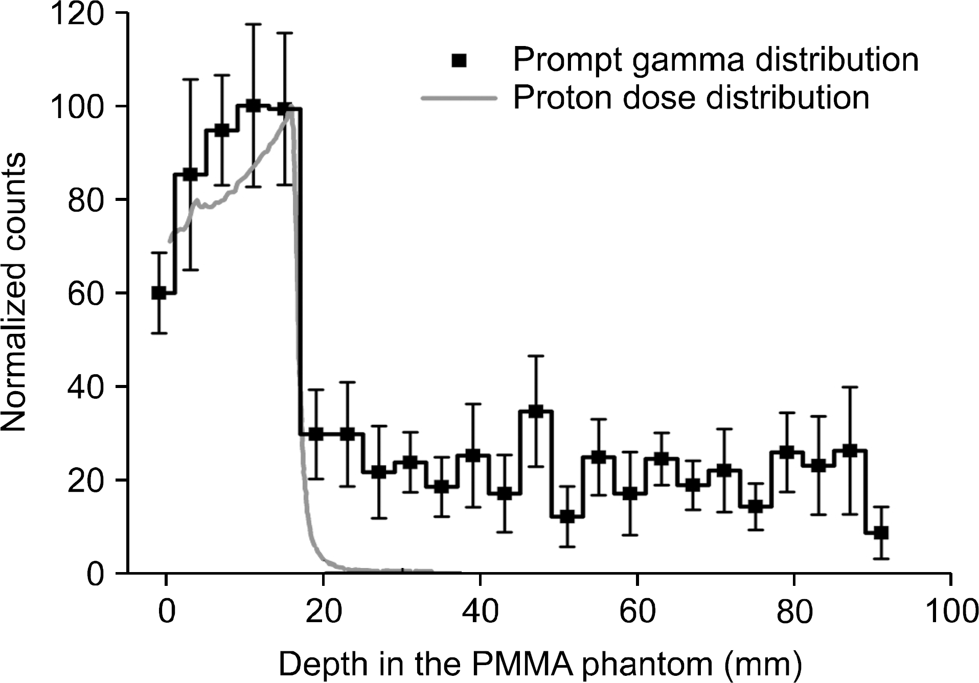
In proton therapy, in vivo proton beam range verification is very important to deliver conformal dose to the target volume and minimize unnecessary dose to normal tissue. For this purpose, a multi-slit prompt-gamma camera module made of 24 scintillation detectors and 24-channel signal processing system is under development. In the present study, we have developed and tested a dual-mode signal processing system, which can operate in the energy calibration mode and the fast data acquisition mode, to process the signals from the 24 scintillation detectors. As a result of performance test, using the energy calibration mode, we were able to perform energy calibration for the 24 scintillation detectors at the same time and determine the discrimination levels for the detector channels. Further, using the fast data acquisition mode, we were able to measure a prompt-gamma distribution induced by a 45 MeV proton beam. The measured prompt gamma distribution was found similar to the proton dose distribution at the distal fall-off region, and the estimated beam range was 17.13±0.76 mm, which is close to the proton beam range of 16.15 mm measured by an EBT film.
Go to : 
References
1. Paganetti H. Rangeuncertaintiesinprotontherapyandthe roleofMonteCarlosimulations.Phys.Med.Biol. 57:R99–R117. 2012.
2. Knopf AC, Lomax A. Invivoprotonrangeverification: a review.Phys.Med.Biol. 58:R131–R160. 2012.
3. Stichelbaut F, Jongen Y. Verificationoftheprotonbeam positioninthepatientbythedetectionofpromptgamma-rays emission. Meetingof39thParticleTherapyCo-OperativeGroup. (San Francisco,. 2003.
4. Min CH, Kim CH, Youn MY, Kim JW. Promptgamma measurementsforlocatingthedosefall-offregionintheproton therapy.Appl.Phys.Lett. 89:183517. 2006.
5. Smeets J, Roellinghoff F, Prieels D, Stichelbaut F, Benilov A, Busca P, Fiorini C, Peloso R, Basilavecchia M, Frizzi T, Dehaes JC Dubus A. Promptgammaimaging withaslitcameraforreal-timerangecontrolinprotontherapy. Phys.Med.Biol. 57:3371–3405. 2012.
6. Bom V, Joulaeizadeh L, Beekman F. Real-timeprompt gammamonitoringinspot-scanningprotontherapyusingimagingthroughaknife-edge-shapedslit.Phys.Med.Biol. 57:297–308. 2012.
7. Perali I, Celani A, Bombelli L, Fiorini C, Camera F, Clementel E, Henrotin S, Janssens G, Prieels D, Roellinghoff F, Smeets J, Stichelbaut F, Stappen FV. Promptgammaimagingofprotonpencilbeamsatclinicaldose rate.Phys.Med.Biol. 59:5849–5871. 2014.
8. Priegnitz M, Helmbrecht S, Janssens G, Perali I, Smeets J, Stappen FV, Sterpin E, Fiedler F. Detection ofmixed-rangeprotonpencilbeamswithapromptgammaslit camera.Phys.Med.Biol. 61:855–871. 2016.
9. Frandes M, Zoglauer A, Maxim V, Prost R. Atracking Compton-scatteringimagingsystemforhadrontherapy monitoring.IEEETrans. Nuc.Sci. 57:144–150. 2010.
10. Richard MH, Chevallier M, Dauvergne D, Freud N, Henriquet P, Le Foulher, Letang JM, Montarou G, Ray C, Roellinghoff F, Testa E, Testa M, Walenta AH. DesignguidelinesforadoublescatteringComptoncamerafor prompt-gammaimagingduringionbeamtherapy: aMonte Carlosimulationstudy.IEEETrans.Nuc.Sci. 58:87–94. 2011.
11. Polf JC, Avery S, Mackin DS, Beddar S. Evaluationofa stochasticreconstructionalgorithmforuseinComptoncamera imagingandbeamrangeverificationfromsecondarygamma emissionduringprotontherapy.Phys.Med.Biol. 57:3537–3553. 2012.
12. Krimmer J, Ley JL, Abellan C, Cachemiche JP, Caponetto L, Chen X, Dahoumane M, Dauvergne D, Freud N, Joly B, Lambert D, Lestand L, Letang JM, Magne M, Mathez H, Maxim V, Montarou G, Morel C, Pinto M, Ray C, Reithinger V, Testa E, Zoccarato Y. DevelopmentofaComptoncameraformedicalapplications basedonsiliconstripandscintillationdetectors.Nucl.Instrum. Meth.A. 787:98–101. 2014.
13. Mackin D, Peterson S, Beddar S, Polf JC. Imagingof promptgammaraysemittedduringdeliveryofclinicalproton beamswithaComptoncamera: Feasibilitystudiesforrange verification.Phys.Med.Biol. 60:7085–7099. 2015.
14. Kim CH, Park JH, Seo H, Lee HR. Gammaelectronvertex imaingandapplicationtobeamrangeverificationinproton therapy.Med.Phys. 39:1001–1005. 2012.
15. Lee HR, Park JH, Kim JH, Jung WG, Kim CH. Developmentofsignalprocessingmodulesfordouble-sidedsil-iconstripdetectorofgammaverteximagingforprotonbeam doseverification.J.Radiat.Prot. 39(2):81–88. 2014.
16. Golnik C, Hueso-Gonzalez F, Muller A, Dendooven P, Enghardt W, Fiedler F, Kormoll T, Roemer K, Petzoldt J, Wagner A, Pausch G. Rangeassessmentinparticletherapybasedonpromptγ-raytimingmeasurements.Phys.Med. Biol. 59:5399–5422. 2014.
17. Hueso-Gonzalez F, Enghardt W, Fiedler F, Golnik C, Janssens G, Petzoldt J, Prieels D, Priegnitz M, Romer K, Smeets J. Firsttestofthepromptgammaraytimingmeth-odwithheterogeneoustargetsataclinicalrotontherapyfacility. Phys.Med.Biol. 60:6247–6272. 2015.
18. Min CH, Lee HR, Kim CH, Lee SB. Developmentofar-ray-typepromptgammameasurementsystemforinvivorange verificationinprotontherapy.Med.Phys. 39:2100–2107. 2012.
19. 이한림, 박종훈, 김한성, 김찬형. 다채널 방사선 측정 장치의 데이터 획득 채널수 저감을 위한 멀티플렉싱 시스템 개발.2013춘계학술발표회 논문요약집 대한방사선방어학회. 140-141:2013.
Go to : 
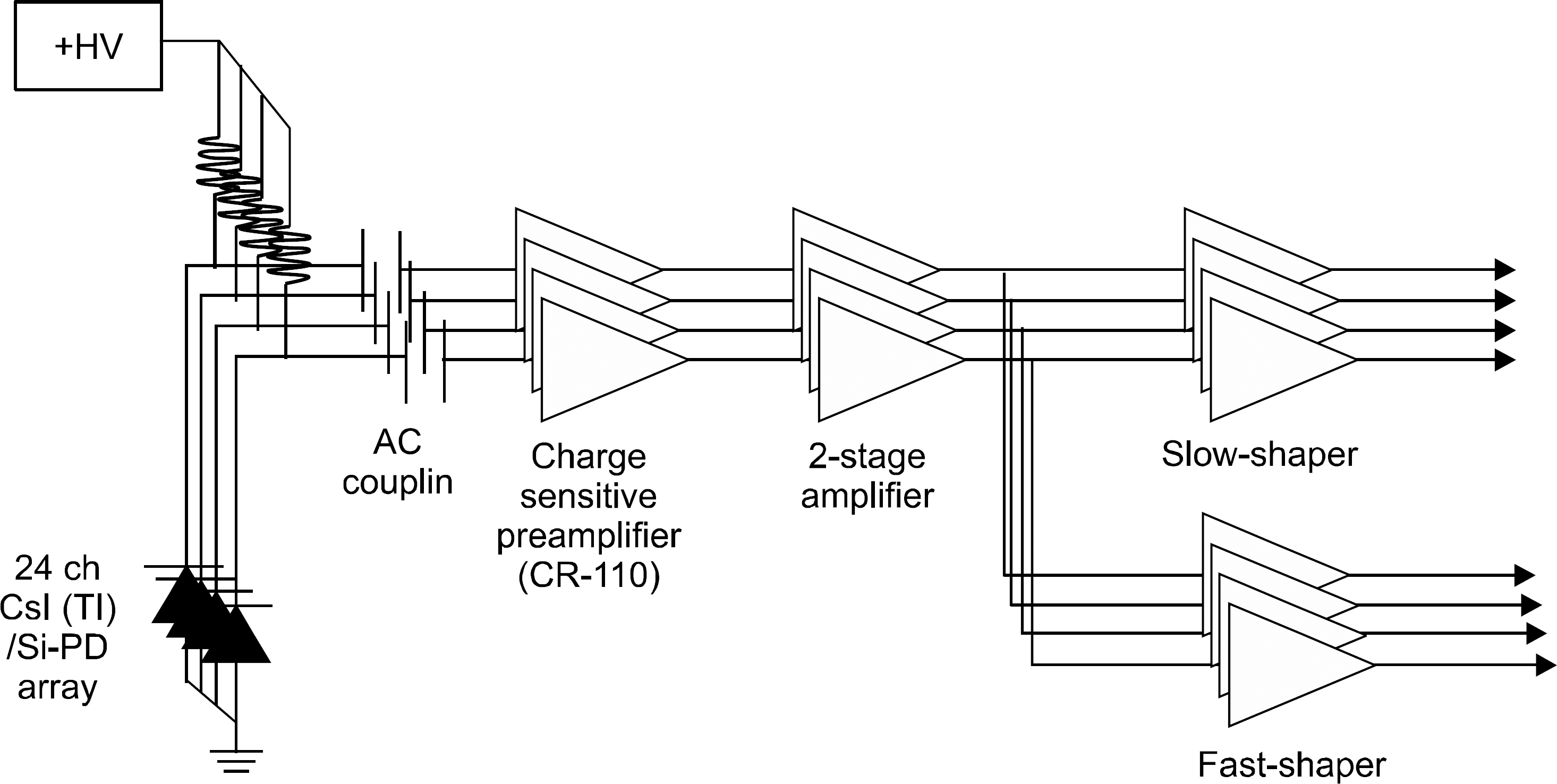 | Fig. 1.Schematic diagram of 24- channel front-end electronics system including charge sensitive amplifiers and slow and fast shaping amplifiers. |
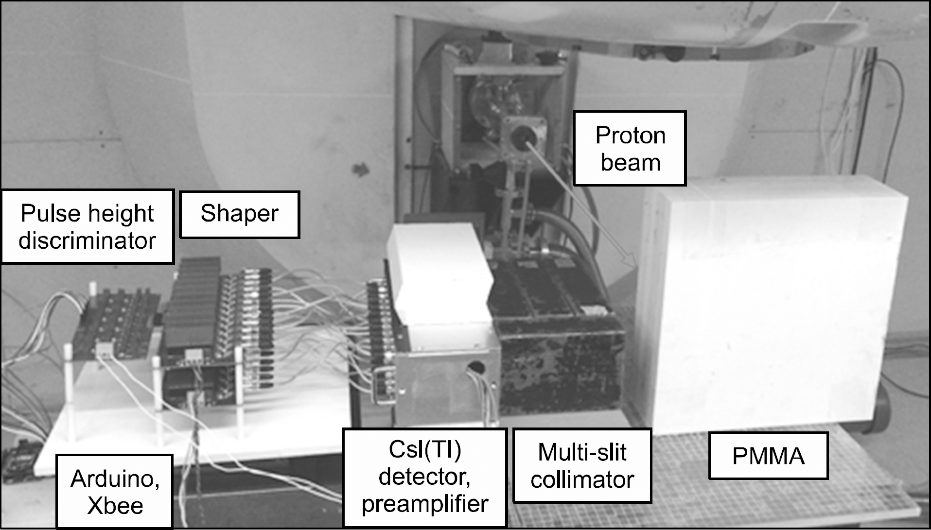
 | Fig. 4.Assembled dual-mode signal processing system including pream-plifiers, shapers, a multiplexing system, pulse height discrimina-tors, a microcontroller (Arduino), an Xbee. |
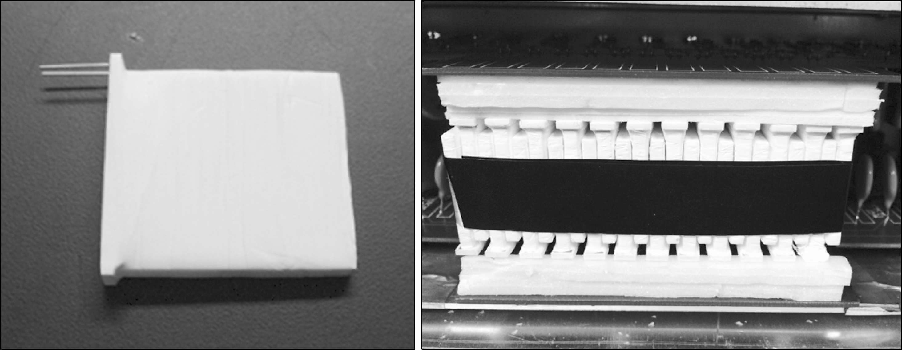 | Fig. 5.A scintillation detector (left) and an array of 24-channel scintillation detectors (right). |
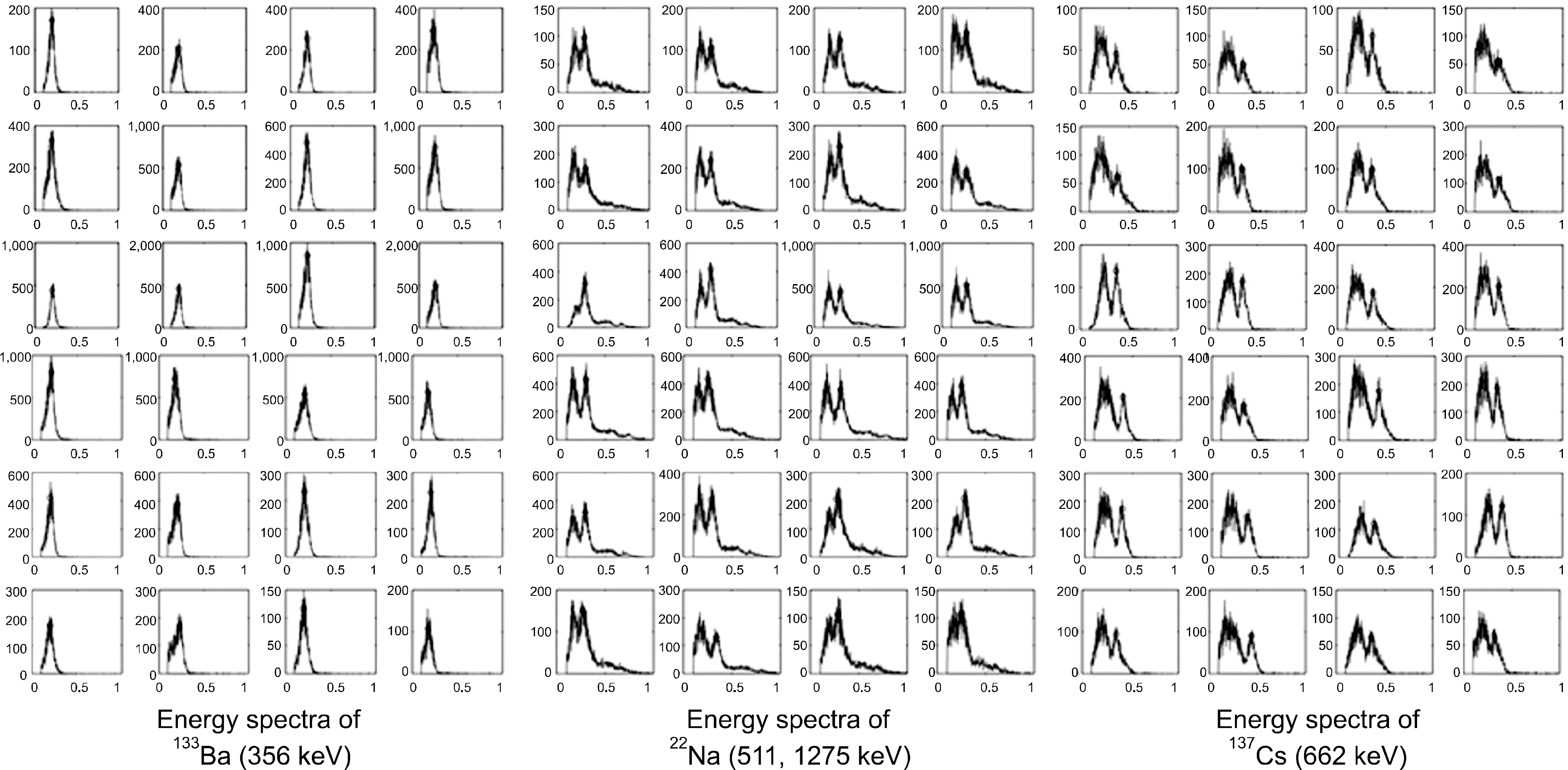
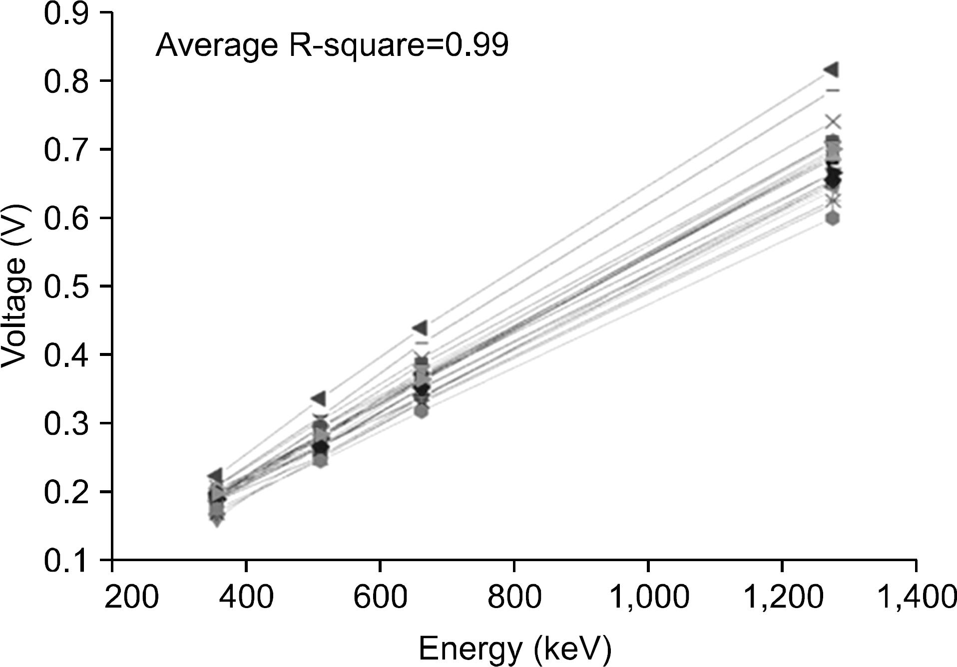 | Fig. 7.Energy spectra of mode of the dual-mode signal processing system developed in the present study.133 Ba (356 keV), 22 Na (511 keV, 1,275 keV), 137 Cs (662 keV) sources measured by the energy calibration |


 PDF
PDF ePub
ePub Citation
 Print
Print


 XML Download