Abstract
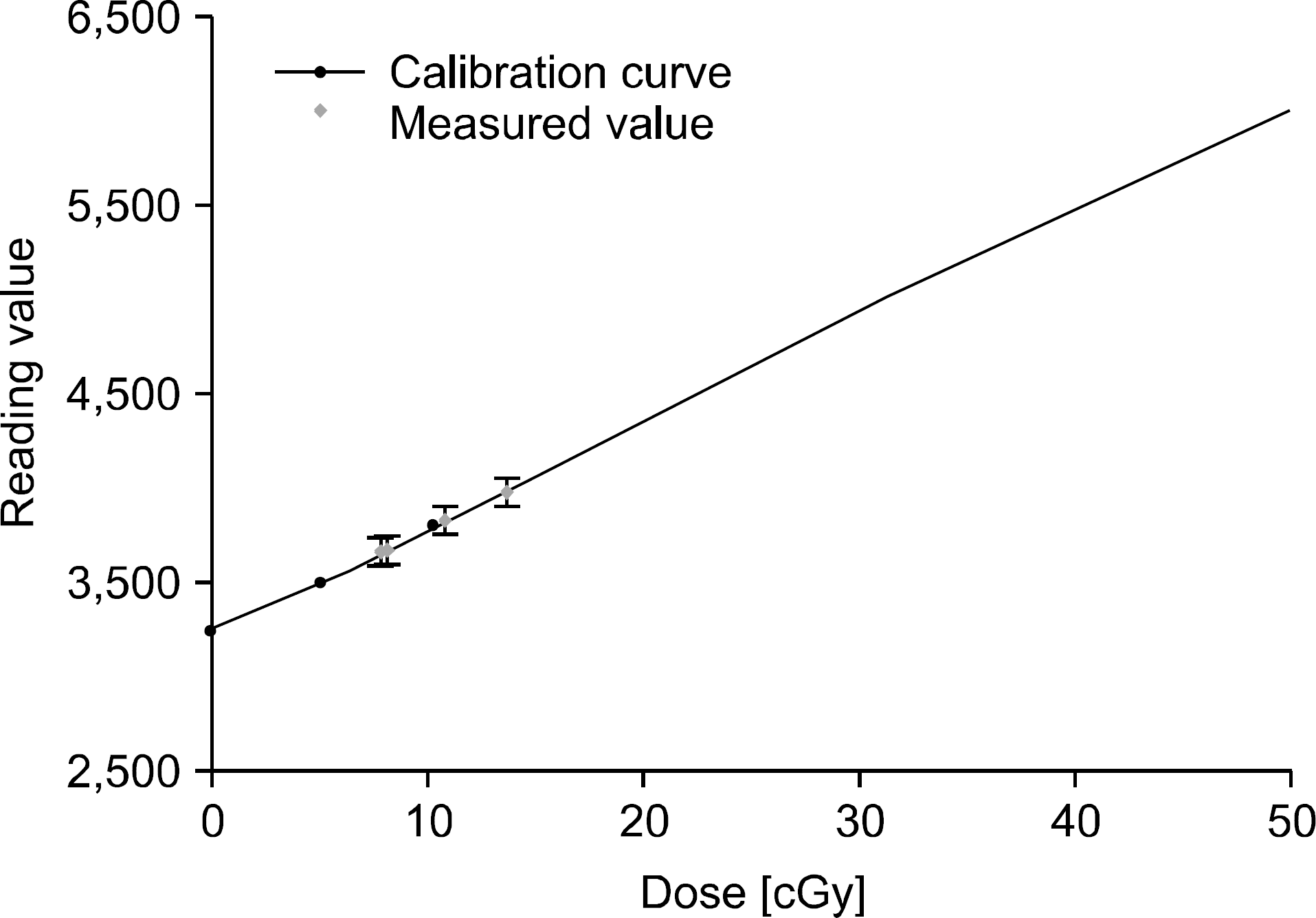
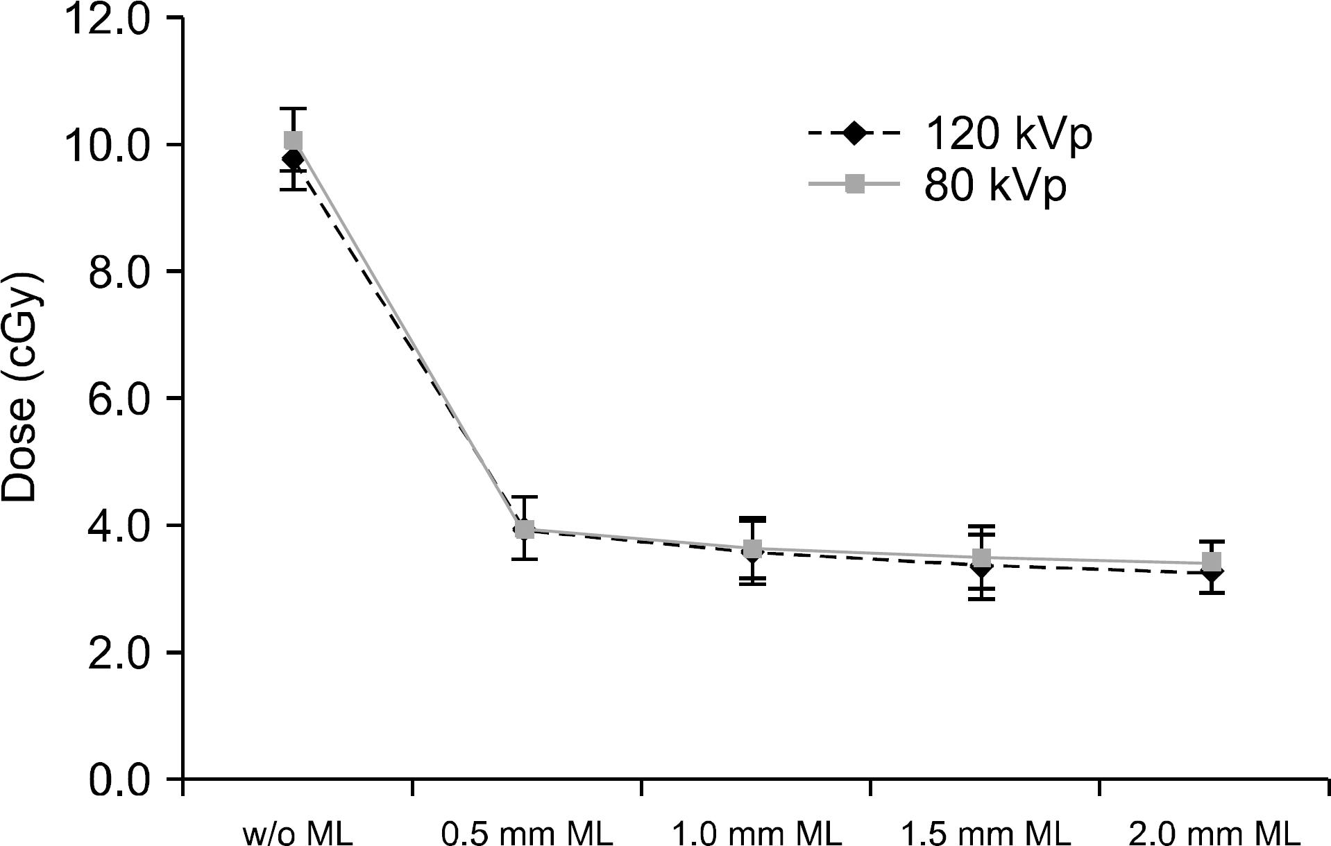
The paper discusses radiation dose of dual energy CT on which copper modulation layer, is mounted in order to improve diagnostic performance of the dual energy CT. The radiation dose is estimated using MCNPX and its results are compared with that of the conventional dual energy CT system. CT X-ray spectra of 80 and 120 kVp, which are usually used for thorax, abdominal, head, and neck CT scans, were generated by the SPEC78 code and were used for the source specification ‘SDEF' card for MCNPX dose modeling. The copper modulation layer was located 20 cm away from a source covering half of the X-ray window. The radiation dose was measured as changing its thickness from 0.5 to 2.0 mm at intervals of 0.5 mm. Since the MCNPX tally provides only normalized values to a single particle, the dose conversion coefficients of F6 tally for the modulation layer-based dual energy CBCT should be calculated for matching the modeling results into the actual dose. The dose conversion coefficient is 7.2*104 cGy/output that is obtained from dose calibration curve between F6 tally and experimental results in which GAFCHORMIC EBT3 films were exposed by an already known source. Consequently, the dose of the modulation layer-based dual energy cone beam CT is 33∼40% less than that of the single energy CT system. On the basis of the results, it is considered that scattered dose produced by the copper modulation layer is very small. It shows that the modulation layer-based dual energy CBCT system can effectively reduce radiation dose, which is the major disadvantage of established dual energy CT.
References
1. Ding GX, Duggan DM, Coffey CW, et al. Astudyonadaptive IMRTtreatmentplanningusingkVcone-beamCT. RadiotherapyandOncology. 85(1):116–125. 2007.
2. AAPM Task Group No. 142: Qualityassuranceofmedical accelerators. American Association of Physicists in Medicine;(. 2009.
3. Jaffray DA, Siewerdsen JH, Wong JW, Martinez AA. Flat-panelcone-beamcomputedtomographyforimage-guided radiationtherapy.InternationalJournalofRadiationOncology BiologyPhysics. 53(5):1337–1349. 2002.
4. Danad I, Fayad ZA, Willemink MJ, Min JK. Newapplications of cardiaccomputedtomography: dual-energy,spectral, andmolecularCTimaging.CardiovascularImaging. 8(6):710–723. 2015.
5. Johnson T. DualenergyCTinclinicalpractice. Springer Science & Business Media;2011. p. 3–8.
6. Primak AN, Giraldo JR, Liu X, Yu L, McCollough CH. Improveddual-energymaterialdiscriminationfordual-source CTbymeansofadditionalspectralfiltration.Medicalphysics. 36(4):1359–1369. 2009.
7. Petersilka M, Bruder H, Krauss B, Stierstorfer K, Flohr TG. TechnicalprinciplesofdualsourceCT.Europeanjournal ofradiology. 68(3):362–368. 2008.
8. Johnson TR, Krauss B, Sedlmair M, et al. Materialdiffer-entiationbydualenergyCT:initialexperience.Europeanradiol-ogy. 17(6):1510–7. 2007.
9. Bauer RW, Kramer S, Renker M, et al. Doseandimage qualityatCTpulmonaryangiography—comparisonoffirstand secondgenerationdual-energyCTand64-sliceCT.European radiology. 21(10):2139–2147. 2011.
10. Schenzle JC, Sommer WH, Neumaier K, et al. Dualen-ergyCTofthechest: howaboutthedose?Investigativeradiol-ogy. 45(6):347–353. 2010.
11. Ho LM, Yoshizumi TT, Hurwitz LM, et al. Dualenergyver-sussingleenergyMDCT:measurementofradiationdoseusing adultabdominalimagingprotocols.Academicradiology. 16(11):1400–1407. 2009.
12. Matsumoto K, Jinzaki M, Tanami Y, et al. Virtualmono-chromaticspectralimagingwithfastkilovoltageswitching:: im-provedimagequalityascomparedwiththatobtainedwithcon-ventional120-kVpCT.Radiology. 259(1):257–262. 2011.
13. Kalender WA, Perman WH, Vetter JR, Klotz E. Evaluationofaprototypedual‐energycomputedtomographic apparatus.I.Phantomstudies.Medicalphysics. 13(3):334–339. 1986.
14. Hao J, Kang K, Zhang L, Chen Z. Anovelimageopti-mizationmethodfordual-energycomputedtomography. NuclearInstrumentsandMethodsinPhysicsResearch Section A:Accelerators,Spectrometers,DetectorsandAssociated Equipment. 722:34–42. 2013.
15. Altman A, Carmi R. ADouble‐LayerDetector,Dual‐Energy CT—Principles,AdvantagesandApplications.MedicalPhysics. 36(6):2750–2750. 2009.
16. Virginia T, John E, Raju S, et al. DoseReductioninCT whileMaintainingDiagnosticConfidence: DiagnosticReference LevelsatRoutineHead,Chest,andAbdominalCT—IAEA-coor-dinatedResearchProject.Radiology. 240(3):828–834. 2006.
17. Song WY, Kamath S, Ozawa S, et al. Adosecomparison studybetweenXVIⓇ andOBIⓇ CBCTsystems.Medphy. 35(2):480–486. 2008.
18. Brown TA, Hogstrom KR, Alvarez D, et al. Dose-responsecurveofEBT,EBT2,andEBT3radiochromicfilmsto synchrotron-producedmonochromaticx-raybeams.Medical physics. 39(12):7412–7417. 2012.
19. Cho YS, Jeong WK, Kim Y, Heo JN. RadiationDosesof Dual-EnergyCTforAbdominopelvicCT:Comparisonwith Single-EnergyCT.JournaloftheKoreanSocietyofRadiology. 65(5):505–512. 2011.
20. Raju R, Thompson AG, Lee K, et al. Reducediodineload withCTcoronaryangiographyusingdual-energyimaging: a prospectiverandomizedtrialcomparedwithstandardcoronary CTangiography.Journalofcardiovascularcomputedtomography. 8(4):282–288. 2014.
21. Kerl JM, Bauer RW, Maurer TB, et al. DoselevelsatcoronaryCTangiography—acomparisonofdualenergy-,dual source-and16-sliceCT.Europeanradiology. 21(3):530–537. 2011.
22. Halliburton SS, Sola S, Kuzmiak SA, et al. Effectofdu-al-sourcecardiaccomputedtomographyonpatientradiation doseinaclinicalsetting: comparisontosingle-sourceimaging. Journalofcardiovascularcomputedtomography. 2(6):392–400. 2008.


 PDF
PDF ePub
ePub Citation
 Print
Print


 XML Download