Abstract
The purpose of this study is to see the usefulness of lead apron for critical organs near the breast under examining. For clinical experiment, 30 female volunteers who agreed to their participation in the experiments, were chosen and divided into two groups, 15 in group A and 15 in group B respectively. group A is to see whether each side of breast under mammography affects to other side glandular on the critical organs is same, because it is not allowed to scan the both breast for same person or to scan repeatedly. Group B is to see the effectiveness of lead apron during the mammography of right breast. Glass dosimeters were placed on the thyroid, the contralateral breast, and lower abdomen where near the breast during examining. The average glandular doses on the surface in mammography of the thyroid gland, the contralateral breast, the lower abdomen were 0.0692 mGy, 0.6790 mGy, and 0.0122 mGy, respectively, which was an extremely low level of glandular dose. In group B, as to the thyroid gland, average dose was decreased from 0.0922 mGy to 0.0158 mGy. The average dose of contralateral breast was decreased from 0.8575 mGy to 0.0286 mGy. The average doses of lower abdomen was decrease 0.0150 mGy to 0.0173 mGy. As to the lower abdomen, dose decreased from 0.0150 mGy before the use of an apron down to 0.0173 mGy after the use. As p-value was under 0.05, statistically significant difference was observed between the two groups. Wearing an apron can have the protective effects on the thyroid gland up to 20 times lower than not wearing one. Besides, it is also necessary to protect the other breast during the examination by wearing one.
References
1. A. D. Wrixon, New ICRP recommendations. J. Radiol. Prot. 28:161. 2008.
2. M. John and P. Boone, Glandual Breast Dose for Monoenergetic and High-Energy X-ray Beams: Monte Carlo Assessment. Med. Phys. 213:23. 1999.
3. ICRP, Recommendations of the International Commission on Radiological Protection. Publication 60, 1991. 21.
4. IAEA, International Basic Safety Standards for Protection against Ionizing Radiation and for the Safety of Radiation Sources. DS379 Paragraph Tracking from SS115. 2009. p.209.
5. Ministry for health, w.a.f.a., Diagnostic reference level guide line for mammography. 2008. 16: p.23.
6. Lee M.H., Dong K. R., Park S. J., Song Y. S., Cho C. H., Sanchez and M. I.The Effect of Scattering Dose on the Thyroid During Mammography. Journal of the Korean Institute of Electrical and Electronic Material Engineers. 23:826. 2010.

7. I. Sechopoulos, S. Suryanarayanan, S. Vedantham, C. J. D'Orsi, and A. Karellas, Radiation Dose to Organs and Tissues from Mammography: Monte Carlo and Phantom Study. Radiology. 246:434. 2008.
8. Hong E-H, Study on the usefulness and aluminum Face Block developed for dose reduction of the lens and thyroid mammography. KOREA UNIVERSITY. (. 2012.
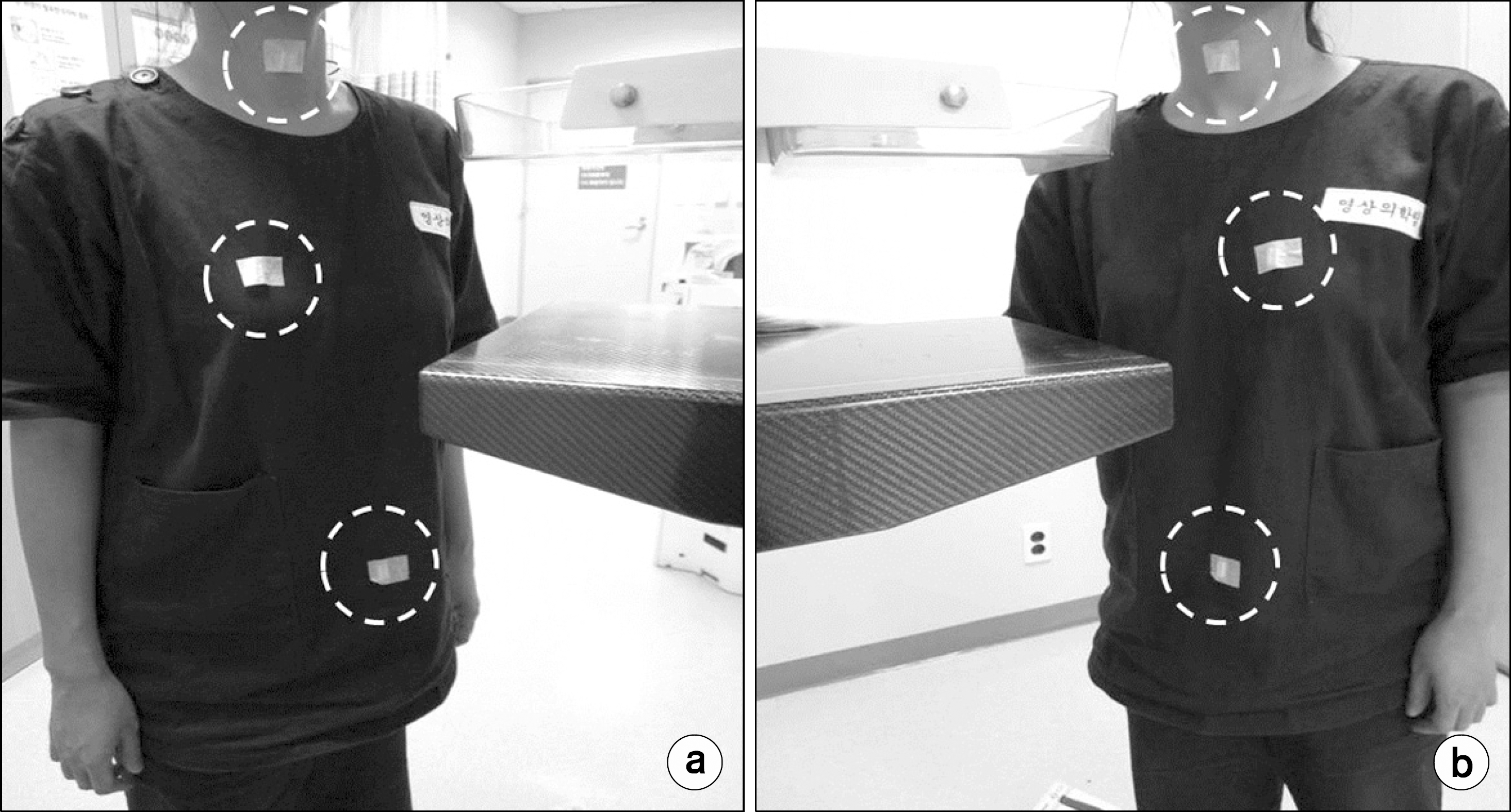
Fig. 1.
Positions of GDs applied on the volunteers to see the bias of left and right mammography. Dotted circles indicate the positions of GDs. (a) GD positions of group A for mammography on the left cc view, (b) GD positions of group A for mammography on the right cc view.

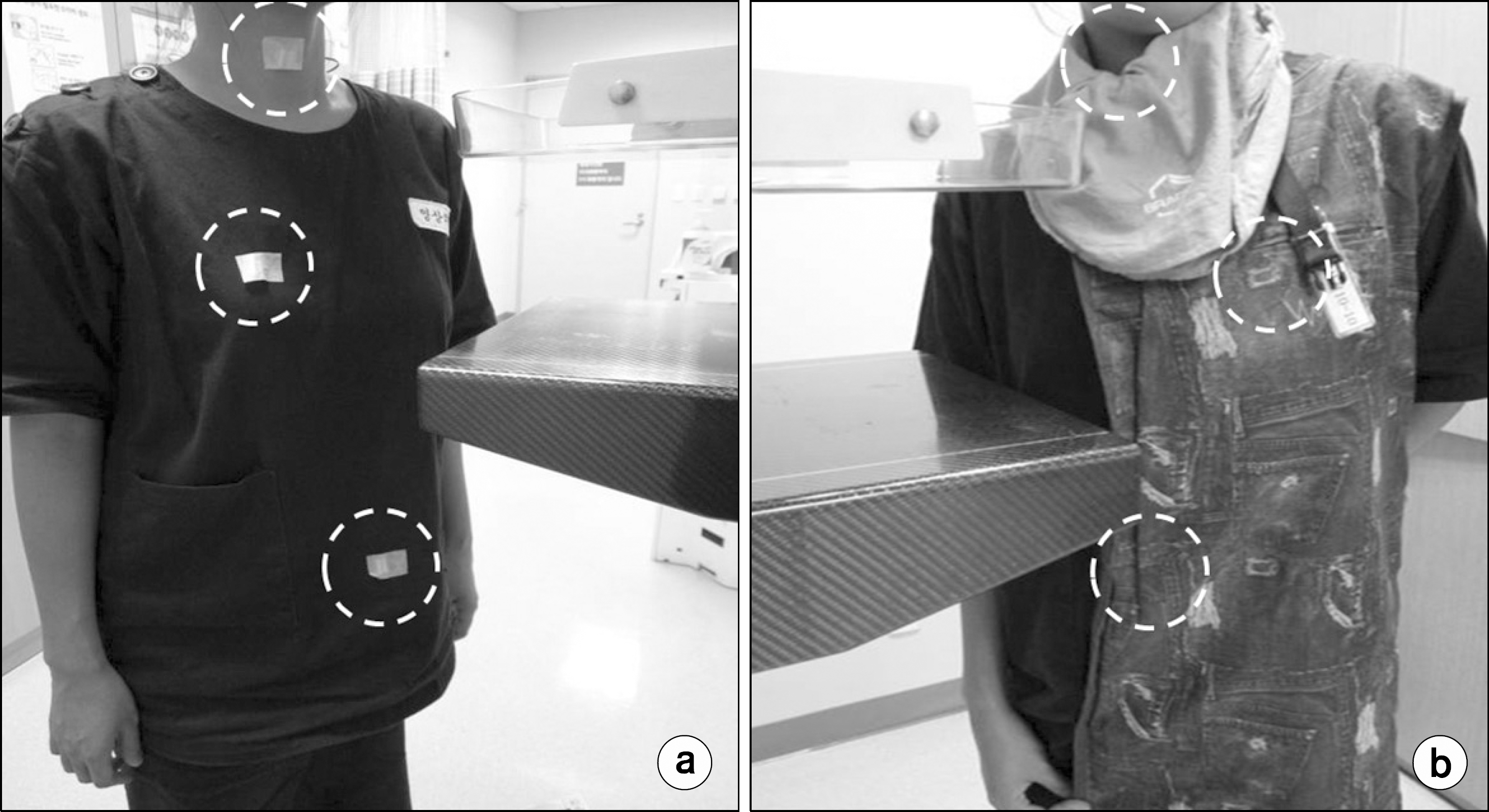
Fig. 2.
Positions of GDs applied on the volunteers to see the effectiveness of apron. Dotted circles indicate the positions of GDs. (a) GD positions of group B for mammography on the left cc view, (b) GD positions of group B for mammography on the right cc view.

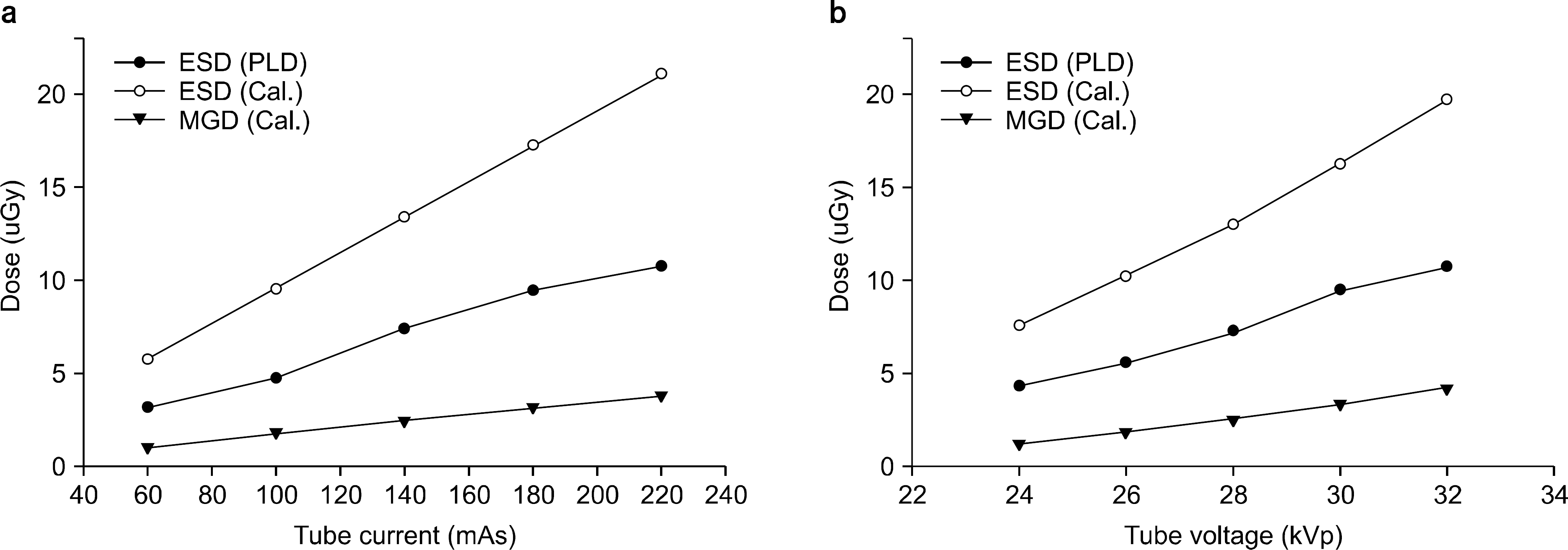
Fig. 3.
Absorbed dose calculated from mammogram and measurements using glass dosimeter. (a) Tube current dependency when tube voltage at 27 kVp. (b) Tube voltage dependency when tube current at 120 mAs.

Table 1.
Characteristics of volunteers between Group A and B.
| Group | Mean age (Y) | Rt. Breastmean thickness (cm) | Lt. Breastmean thickness (cm) | Mean tube peak voltage (kVp) | Mean tube current (mAs) |
|---|---|---|---|---|---|
| A | 52.8 | 4.2 | 4.2 | 27 | 121.2 |
| B | 54.3 | 4.5 | 4.5 | 27.3 | 124.2 |
Table 2.
Bias of left and right mammography; there are no differences between left and right.
Table 3.
Difference between entrance surface doses with and without lead apron (average).


 PDF
PDF ePub
ePub Citation
 Print
Print


 XML Download