Abstract
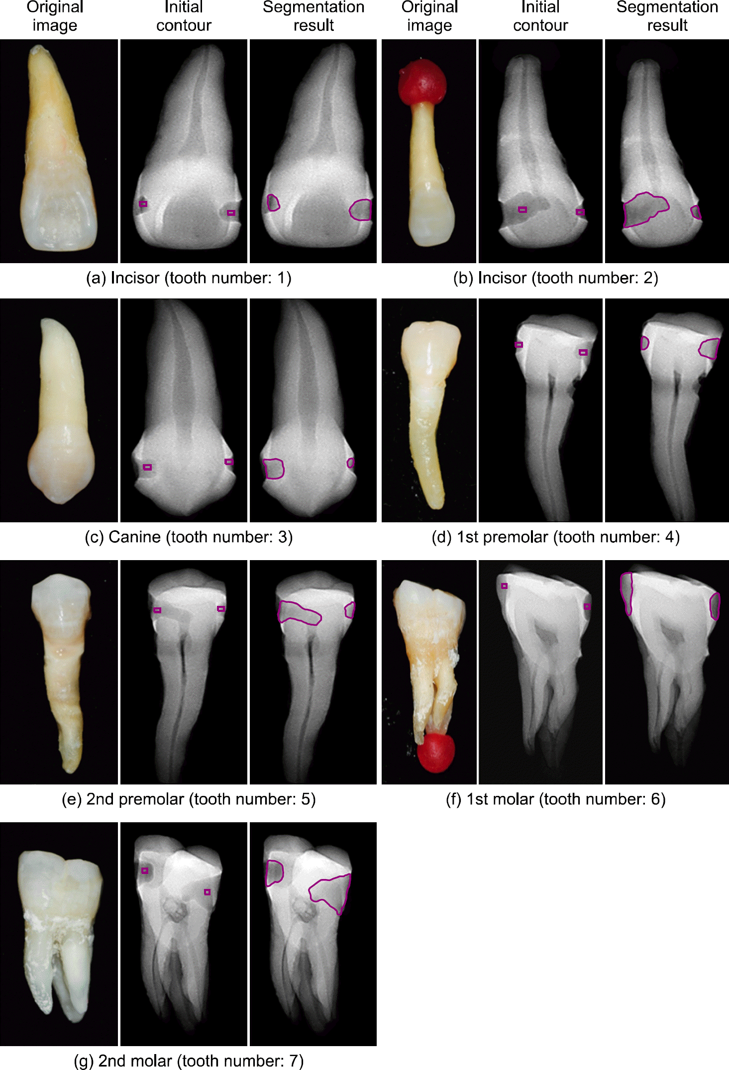
The automated dental cavity detection program for a new concept intra-oral dental x-ray imaging device, an auxiliary diagnosis system, which is able to assist a dentist to identify dental caries in an early stage and to make an accurate diagnosis, was to be developed. The primary theory of the automatic dental cavity detection program is divided into two algorithms; one is an image segmentation skill to discriminate between a dental cavity and a normal tooth and the other is a computational method to analyze feature of an tooth image and take an advantage of it for detection of dental cavities. In the present study, it is, first, evaluated how accurately the DRLSE (Direct Regularized Level Set Evolution) method extracts demarcation surrounding the dental cavity. In order to evaluate the ability of the developed algorithm to automatically detect dental cavities, 7 tooth phantoms from incisor to molar were fabricated which contained a various form of cavities. Then, dental cavities in the tooth phantom images were analyzed with the developed algorithm. Except for two cavities whose contours were identified partially, the contours of 12 cavities were correctly discriminated by the automated dental caries detection program, which, consequently, proved the practical feasibility of the automatic dental lesion detection algorithm. However, an efficient and enhanced algorithm is required for its application to the actual dental diagnosis since shapes or conditions of the dental caries are different between individuals and complicated. In the future, the automatic dental cavity detection system will be improved adding pattern recognition or machine learning based algorithm which can deal with information of tooth status.
References
1. Bennett T. Amaechi: Emerging technologies for diagnosis of dental caries: The road so far, J. Appl. Phys. VOL 105 (. 2009. ) 102047–1 – 102047–9.
2. Iain A. Pretty: Caries detection and diagnosis: Novel technologies, Journal of Dentistry, VOL 34 (. 2006. ) 727–739.
3. Nigel B. Pitts et al: Clinical Diagnosis of Dental Caries: A European Perspective, J. Dent. Educ., VOL 65, NO. 10, (. 2001. ) 972–978.
4. Juliana Gomez, et al. Non-cavitated carious lesions detection methods: a systematic review, Community Dent. Oral. Epidemiol., VOL 41, (. 2013. ) 55–66.
5. Sungho Cho, et al. Introduction of Dental X-ray Imaging with New Concept – intra Oral x-ray Tube. J. Inst. Electron Eng. Korea SC. VOL 48:NO.(4):(. 2011; 94–101.
6. Nigel B. Pitts and Kim R. Ekstrand: International Caries Detection and Assessment System (ICDAS) and its International Caries Classification and Management System (ICCMS) – methods for staging of the caries process and enabling dentists to manage caries, Community Dent. Oral. Epidemiol., VOL 41, (. 2013. ) e41-e52.
7. Shuo Li, et al. An automatic variational level set segmentation framework for computer aided dental X-rays analysis in clinical environments, Comput. Med. Imaging Graphics, VOL 30. (. 2006. ) 65–74.
8. Stanley Osher and James A. Sethian: Fronts propagating with curvature-dependent speed: algorithms based on Hamilton-Jacobi formulations, J. Comput. Phys. VOL 79, (. 1988. ) 12–49.
9. Stanley Osher and Ronald P. Fedkiw: Level set methods: an overview and some recent results. J. Comput. Phys. VOL 169, (. 2001. ) 463–502.
10. C. Li, et al: Distance regularized level set evolution and its application to image segmentation. IEEE Transactions on Image Processing. Vol. 19:No.12 (. 2010; ) 3243–3254.
11. Michael Kass, et al. Snake: Active Contour Models, Int. J. Compter Vision, VOL 1, NO. 2 (. 1988. ) 321–331.
12. Tony F. Chan and Luminita A. Vese: Active contours without edges, IEEE Trans. Image, VOL 10, NO. 2 (. 2001. ) 266–277.
13. Caselles Vincent, et al. Geodesic active contours, Int. J. Compter Vision, VOL 22, NO. 2 (. 1997. ) 61–79.
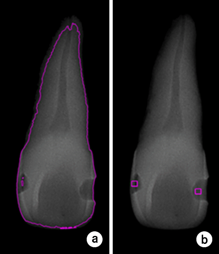
Fig. 2.
(a) Contour of the tooth. (b) Initial points in the cavities where the rectangular contour starts to evolve outside of the cavities.

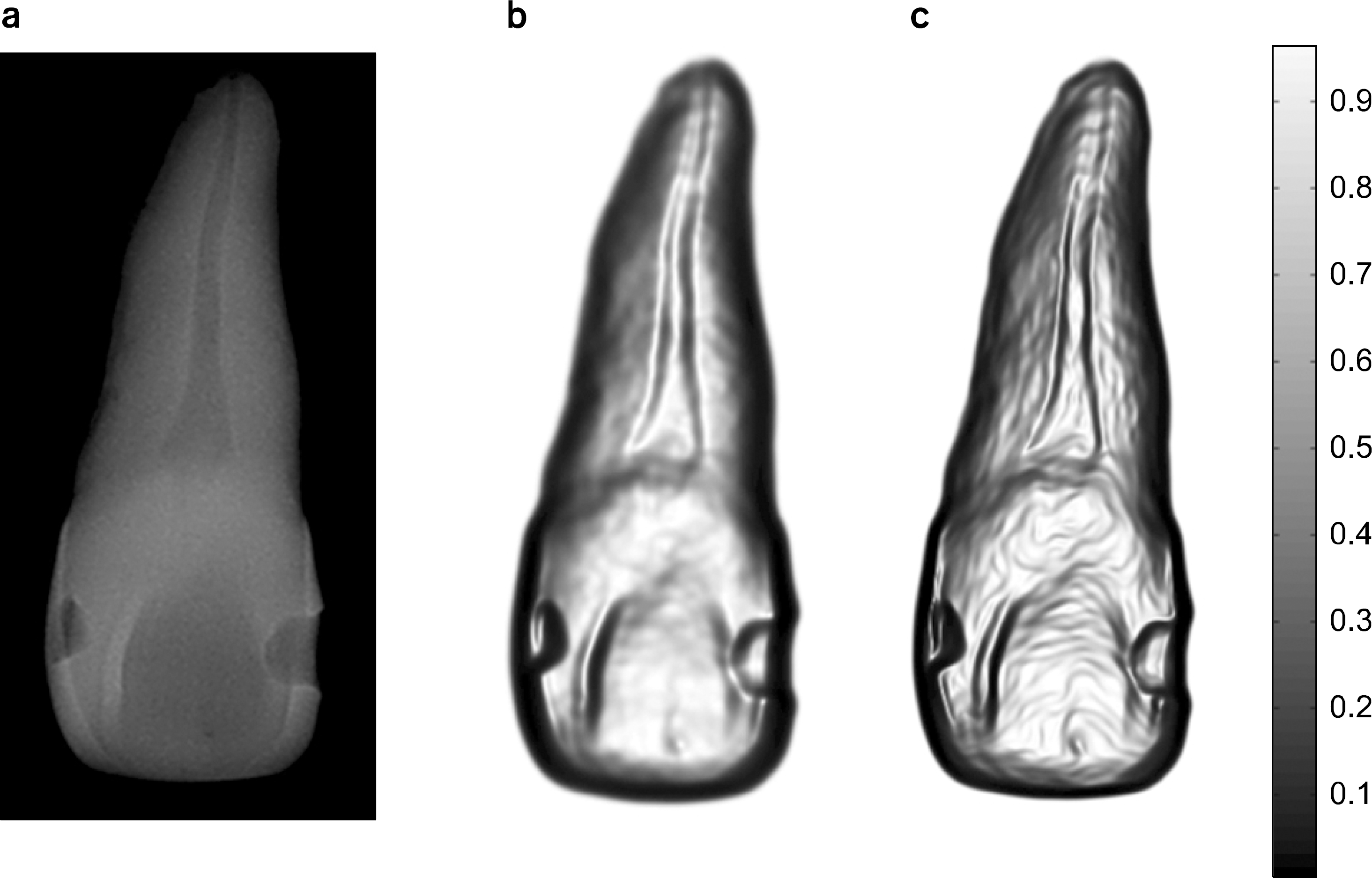
Fig. 3.
(a) Dental X-ray image, (b) Edge function by Gaussian smoothing, and (c) Edge function by coherent filter.


 PDF
PDF ePub
ePub Citation
 Print
Print


 XML Download