Abstract
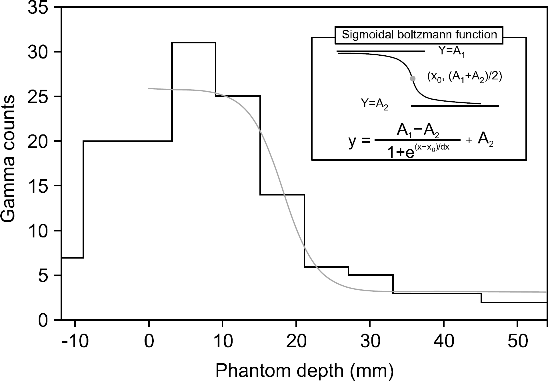
In proton therapy, verification of proton dose distribution is important to treat cancer precisely and to enhance patients' safety. To verify proton dose distribution, in a previous study, our team incorporated a vertically-aligned one-dimensional array detection system. We measured 2D prompt-gamma distribution moving the developed detection system in the longitudinal direction and verified similarity between 2D prompt-gamma distribution and 2D proton dose distribution. In the present, we have developed two-dimension prompt-gamma measurement system consisted of a 2D parallel-hole collimator, 2D array-type NaI(Tl) scintillators, and multi-anode PMT (MA-PMT) to measure 2D prompt-gamma distribution in real time. The developed measurement system was tested with22Na (0.511 and 1.275 MeV) and137Cs (0.662 MeV) gamma sources, and the energy resolutions of 0.511, 0.662 and 1.275 MeV were 10.9%±0.23p%, 9.8%±0.18p% and 6.4%±0.24p%, respectively. Further, the energy resolution of the high gamma energy (3.416 MeV) of double escape peak from Am-Be source was 11.4%±3.6p%. To estimate the performance of the developed measurement system, we measured 2D prompt-gamma distribution generated by PMMA phantom irradiated with 45 MeV proton beam of 0.5 nA. As a result of comparing a EBT film result, 2D prompt-gamma distribution measured for 9×10⁹ protons is similar to 2D proton dose distribution. In addition, the 45 MeV estimated beam range by profile distribution of 2D prompt gamma distribution was 17.0±0.4 mm and was intimately related with the proton beam range of 17.4 mm.
Go to : 
References
1. Schardt D, Elsässer T, Schulz-Ertner D. Heavy-ion tumor therapy: Physical and radiobiological benefits. Rev. Mod. Phys. 82:383–425. 2010.

3. Paganetti H. Range uncertainties in proton therapy and the role of Monte Carlo simulations. Phys. Med. Biol. 57:R99–R117. 2012.
4. Knopf AC, Lomax A. In vivo proton range verification: a review. Phys. Med. Biol. 58:R131–R160. 2012.
5. Oelfke U, Lam G K Y, Atkins M S. Proton dose monitoring with PET: quantitative studies in Lucite. Phys. Med. Biol. 41:177–196. 1996.
6. Moteabbed M, Espana S, Paganetti H. Monte Carlo patient study on the comparison of prompt gamma and PET imaging for range verification in proton therapy. Phys. Med. Biol. 56:1063–1082. 2011.
7. Min CH, Kim CH, Youn MY, Kim JW. Prompt gamma measurements for locating the dose fall-off region in the proton therapy. Appl. Phys. Lett. 89:183517. 2006.

8. Bom V, Joulaeizadeh L, Beekman F. Realtime prompt gamma monitoring in spot-scanning proton therapy using imaging through a knife-edge-shaped slit. Phys. Med. Biol. 57:297–308. 2012.
9. Smeets J, Roellinghoff F, Prieels D, Stichelbaut F, Benilov A, Busca P, Fiorini C, Peloso R, Basilavecchia M, Frizzi T, Dehaes JC, Dubus A. Prompt gamma imaging with a slit camera for real-time range control in proton therapy. Phys. Med. Biol. 57:3371–3405. 2012.
10. Mackin D, Peterson S, Beddar S, Polf J. Evaluation of a stochastic reconstruction algorithm for use in Compton camera imaging and beam range verification from secondary gamma emission during proton therapy. Phys. Med. Biol. 57:3537–3553. 2012.
11. Kurosawa S, Kubo H, Ueno K, Kabuki S, Iwaki S, Takahashi M, Taniue K, Higashi N, Miuchi K, Tanimori T, Kim D, Kim J. Prompt gamma detection for range verification in proton therapy. Curr. Appl. Phys. 12:364–368. 2012.

12. Kim CH, Park JH, Seo H, Lee HR. Gamma electron vertex imaing and application to beam range verification in proton therapy. Med. Phys. 39:1001–1005. 2012.
13. Lee HR, Park JH, Kim HS, Kim CH, Kim SH. Two-dimensional measurement of the prompt-gamma distribution for proton dose distribution monitoring. J. Korean Phys. Soc. 63(7):1385–1389. 2013.
14. Lee HR, Min CH, Park JH, Kim SH, Kim CH. Study on optimization of detection system of prompt gamma distribution for proton dose verification. Prog. Med. Phys. 23(3):162–168. 2012.
Go to : 
 | Fig. 1.Development of two-dimension prompt-gamma measurement system consisted of multichannel signal processing device, 2D array-type NaI(Tl) scintillators, 2D photo sensors, pallel-holes collimator and data acquisition system. |
 | Fig. 3.Energy spectra and position of 22Na gamma source measured by two-dimension prompt-gamma measurement system. |
 | Fig. 4.Energy spectra and position of 137Cs gamma source measured by two-dimension prompt-gamma measurement system. |
 | Fig. 5.Energy spectra of Am-Be source measured by two-dimension prompt-gamma measurement system. |
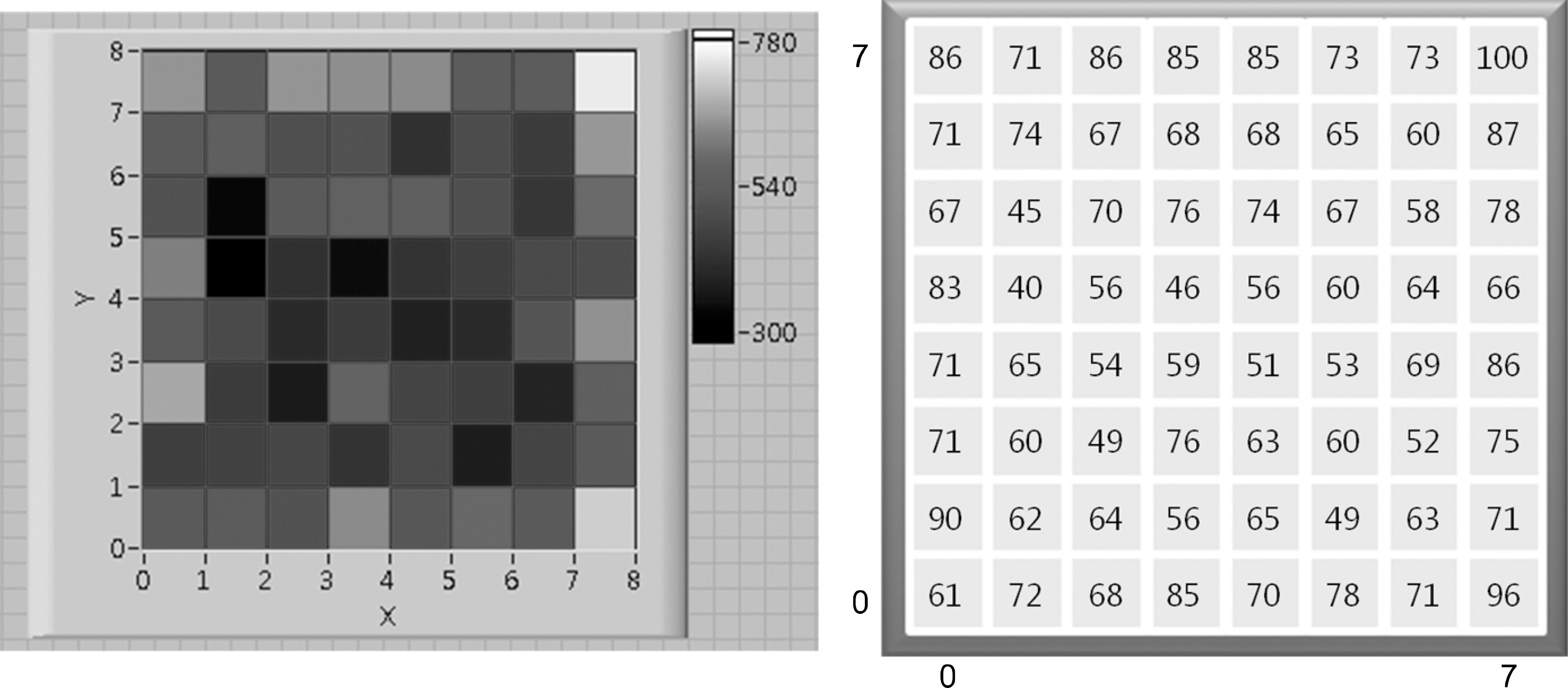 | Fig. 6.Efficiency map of two-dimension prompt-gamma measurement system with 137Cs gamma source. |
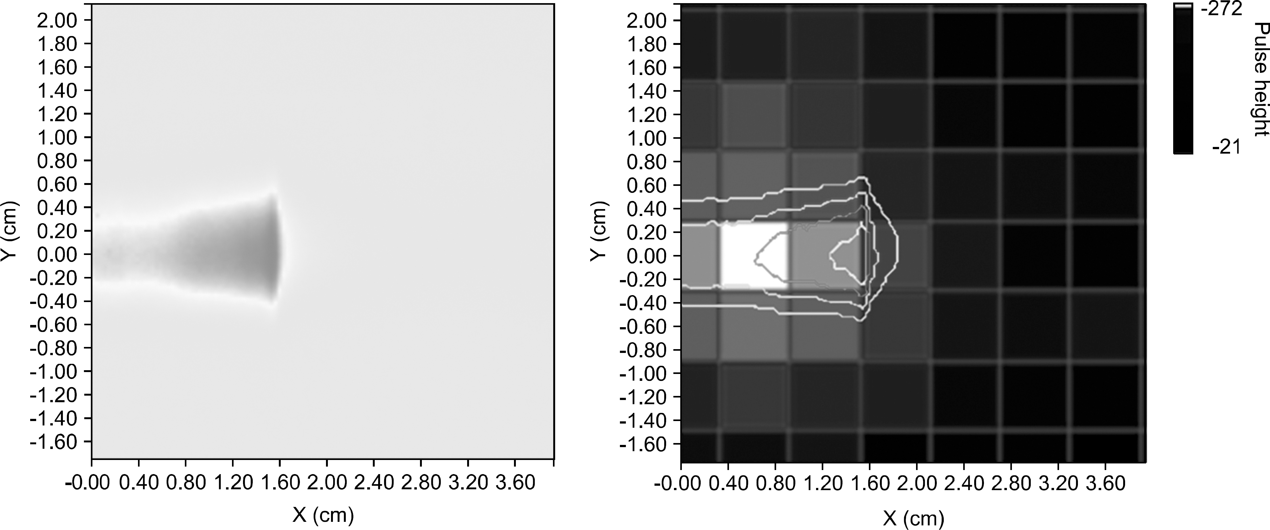 | Fig. 7.Proton dose distribution with EBT film (left) and image registration between EBT film result and prompt gamma 2D distribution (right). |


 PDF
PDF ePub
ePub Citation
 Print
Print


 XML Download